. 2008 Feb;172(2):288-98.
doi: 10.2353/ajpath.2008.070726. Epub 2008 Jan 17.

Galectin-3 expression and secretion links macrophages to the promotion of renal fibrosis

Affiliations

Galectin-3 expression and secretion links macrophages to the promotion of renal fibrosis

Neil C Henderson et al. Am J Pathol. 2008 Feb.

Abstract

Macrophages have been proposed as a key cell type in the pathogenesis of renal fibrosis; however, the mechanism by which macrophages drive fibrosis is still unclear. We show that expression of galectin-3, a beta-galactoside-binding lectin, is up-regulated in a mouse model of progressive renal fibrosis (unilateral ureteric obstruction, UUO), and absence of galectin-3 protects against renal myofibroblast accumulation/activation and fibrosis. Furthermore, specific depletion of macrophages using CD11b-DTR mice reduces fibrosis severity after UUO demonstrating that macrophages are key cells in the pathogenesis of renal fibrosis. Disruption of the galectin-3 gene does not affect macrophage recruitment after UUO, or macrophage proinflammatory cytokine profiles in response to interferon-gamma/lipopolysaccharide. In addition, absence of galectin-3 does not affect transforming growth factor-beta expression or Smad 2/3 phosphorylation in obstructed kidneys. Adoptive transfer of wild-type but not galectin-3(-/-) macrophages did, however, restore the fibrotic phenotype in galectin-3(-/-) mice. Cross-over experiments using wild-type and galectin-3(-/-) macrophage supernatants and renal fibroblasts confirmed that secretion of galectin-3 by macrophages is critical in the activation of renal fibroblasts to a profibrotic phenotype. Therefore, we demonstrate for the first time that galectin-3 expression and secretion by macrophages is a major mechanism linking macrophages to the promotion of renal fibrosis.


Figures

Figure 1
Figure 1
Galectin-3 expression is up-regulated in a mouse model of progressive renal fibrosis (UUO). Galectin-3 expression in tubular epithelium in sham-operated mouse kidney (a) and after 14 days of UUO (b). c: Real-time PCR quantification of galectin-3 expression in whole kidney homogenates from control sham-operated (day 0) and at 3, 7, and 14 days after UUO. ***P < 0.0001 compared with control. Scale bar = 100 μm.
Figure 2
Figure 2
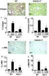
Absence of galectin-3 protects against renal fibrosis. Mice underwent control (sham operation) or UUO and were sacrificed at 7 days (n = 6 mice in each group). a–c: Renal collagen deposition was examined by picrosirius red staining (a, b) and quantified using digital image analysis (c). c: Significantly reduced collagen deposition was observed in the galectin-3−/− mice compared with WT 7 days after ureteral ligation (*P < 0.05). d: Furthermore, transcripts for procollagen (I) were also reduced in the galectin-3−/− group compared with WT animals (*P < 0.05). e and f: Immunohistochemistry revealed markedly reduced α-SMA positivity (a marker of myofibroblast activation) in galectin-3−/− compared with WT mice 7 days after ureteral ligation. g: α-SMA was quantified using digital image analysis, and significantly less α-SMA staining occurred in the galectin-3−/− mice compared with WT (*P < 0.05). h: α-SMA mRNA transcripts, as assessed by real-time RT-PCR, were significantly decreased in the galectin-3−/− mice compared with WT animals (*P < 0.01). Scale bars = 100 μm.
Figure 3
Figure 3
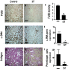
Macrophage ablation by DT reduces renal fibrosis after UUO. Mice underwent UUO and received DT or control vehicle after ureteric ligation (n = 6 mice in each group) as described in Materials and Methods. Kidneys were harvested at day 7, and immunostaining was performed for the specific macrophage marker F4/80 (a, b), α-SMA (d, e), and collagen (g, h) in vehicle control-treated (left) and DT-treated (right) mice. c: Quantification of F4/80 staining (macrophage infiltration) by digital image analysis. Real-time RT-PCR quantitation of α-SMA (f) and procollagen (I) expression (i) in vehicle- and DT-treated mice. *P < 0.05 compared to vehicle treated mice. Scale bars = 100 μm.
Figure 4
Figure 4
Macrophage ablation by DT does not affect recruitment of CD34+/ColI+ and CD45+/ColI+ fibrocytes after UUO. a: CD34; b: ColI; c: merged. Arrows indicate CD34+/ColI+ fibrocytes. d: The number of infiltrating fibrocytes (CD34+/ColI+ and CD45+/ColI+) increased at day 7 after UUO, with no significant difference in infiltrating fibrocyte numbers between non-DT-treated (white bars) and DT-treated (black bars) mice. Day 7 UUO DTCD34+/ColI+, 26.33 ± 2.8 per mm2; day 7 UUO DT+CD34+/ColI+, 29.83 ± 4.7 per mm2 (n = 6 mice in each group, P = NS). Day 7 UUO DTCD45+/ColI+, 35.67 ± 3.2 per mm2 (n = 6), day 7 UUO DT+ CD45+/ColI+, 37.3 ± 4.1 per mm2 (n = 6 mice in each group, P = NS). Values are the mean ± SEM. Scale bars = 100 μm.
Figure 5
Figure 5
Disruption of the galectin-3 gene does not affect macrophage recruitment after UUO or macrophage proinflammatory cytokine profiles in response to IFN-γ/LPS. a–d: H&E staining of kidneys from WT and galectin-3−/− mice 3 days after control sham operation or UUO. Renal macrophages were stained with F4/80 (e–h) and quantitated by digital image analysis (i). Macrophage recruitment was similar in WT and galectin-3−/− mice at all time points studied after UUO (days 0, 3, 7, and 14) (n = 6 mice in each group, P = NS). BMDMs and peritoneal macrophages were stimulated with IFN-γ (100 U/ml) for 48 hours, and LPS (100 ng/ml) was added for the last 24 hours. BMDM supernatants were assayed for IL-6 (j) and TNF-α (k) (galectin-3−/−, open bars; WT, filled bars). Peritoneal macrophage supernatants were assayed for IL-6 (l) and TNF-α (m) (galectin-3−/−, open bars; WT, filled bars). The results represent the mean ± SEM of three experiments (P = NS). Scale bars = 100 μm.
Figure 6
Figure 6
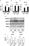
Disruption of the galectin-3 gene does not affect TGF-β expression or Smad 2/3 phosphorylation in obstructed kidneys. WT and galectin-3−/− mice underwent UUO, and kidneys were harvested at days 3, 7, and 14. a–c: RNA was extracted, and TGF-β gene expression was measured as described in Materials and Methods by real-time RT-PCR. TGF-β mRNA transcripts were markedly elevated compared to control after UUO at days 3, 7, and 14 (P < 0.01 compared to control kidney). However, there was no significant difference in renal TGF-β mRNA expression between WT and galectin-3−/− mice after UUO at any of the time points studied. P = NS. d: Western blot of pSMAD2 and pSMAD3 expression in lysates from control and UUO kidneys at day 7 (each lane represents a different mouse). e: Quantification by optical densitometry using Image J.
Figure 7
Figure 7
Macrophage-derived galectin-3 drives myofibroblast accumulation/activation in the kidney after UUO. a: Galectin-3 immunofluorescence staining of WT BMDMs. Nuclei are labeled with 4,6-diamidino-2-phenylindole. b: Representative Western blot of galectin-3 expression in WT BMDM lysate and supernatant. c–m: Galectin-3−/− mice underwent UUO, and mature WT or galectin-3−/− BMDMs (5 × 10⁶) were adoptively transferred at days 1, 3, and 5 after ureteric ligation (n = 6 in each group) and kidneys were harvested at day 7 after UUO. c: Staining of WT macrophages with galectin-3 antibody at day 7 after UUO demonstrating recruitment and engraftment of adoptively transferred WT macrophages. d: Control staining of galectin-3−/− macrophages with galectin-3 antibody. e and f: Fluorescence microscopy demonstrating Cell Tracker Orange-labeled WT (e) and galectin-3−/− (f) macrophages recruited and engrafted at day 7 after UUO. g and h: α-SMA staining of obstructed kidney after adoptive transfer of WT (g) or galectin-3−/− (h) macrophages (arrows indicate α-SMA staining of blood vessels). i and j: Collagen staining (picrosirius red stain) of obstructed kidney after adoptive transfer of WT (i) or galectin-3−/− (j) macrophages. k: Quantitation of recruitment and engraftment of WT and galectin-3−/− macrophages by digital image analysis (P = NS). l: Quantitation of α-SMA staining using digital image analysis. m: Quantitation of collagen (picrosirius red) staining using digital image analysis. n: Quantitation of procollagen (I) gene expression by real-time RT-PCR. *P < 0.01 compared to WT adoptively transferred macrophages. Scale bars = 100 μm.
Figure 8
Figure 8
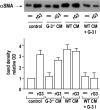
Galectin-3-positive macrophages promote renal fibroblast activation in vitro. Mature BMDMs (1 × 10⁶ cells) were plated in six-well tissue culture dishes, incubated in serum-free media, and conditioned media (0.5 ml) was collected after 48 hours. Galectin-3−/− renal fibroblasts were isolated by trypsin digestion as described in Materials and Methods. rG3 refers to recombinant mouse galectin-3 (30 μg/ml). Conditioned media (CM) from BMDMs (0.5 ml) or control media were added to 0.5 ml of renal fibroblasts (4 × 10⁴ cells). WT BMDM-conditioned media were also added in the presence of 5 μmol/L galectin-3 inhibitor (G-3I) [bis-(3-deoxy-3-{3-methoxybenzamido}-β-d-galactopyranosyl)-sulfane]. Cells were incubated for a further 48 hours (final fetal calf serum concentration, 7.5%) before lysis. α-SMA expression in galectin-3−/− renal fibroblast lysates was measured by Western analysis and quantified by optical densitometry using Image J. Results represent the mean ± SEM of three to four independent experiments.

Similar articles

Cited by

References

    1. El Nahas AM, Bello AK. Chronic kidney disease: the global challenge. Lancet. 2005;365:331–340. - PubMed
    1. Sayegh MH, Carpenter CB. Transplantation 50 years later—progress, challenges, and promises. N Engl J Med. 2004;351:2761–2766. - PubMed
    1. Leibovich SJ, Ross R. The role of the macrophage in wound repair. A study with hydrocortisone and antimacrophage serum. Am J Pathol. 1975;78:71–100. - PMC - PubMed
    1. Leibovich SJ, Ross R. A macrophage-dependent factor that stimulates the proliferation of fibroblasts in vitro. Am J Pathol. 1976;84:501–514. - PMC - PubMed
    1. Duffield JS, Forbes SJ, Constandinou CM, Clay S, Partolina M, Vuthoori S, Wu S, Lang R, Iredale JP. Selective depletion of macrophages reveals distinct, opposing roles during liver injury and repair. J Clin Invest. 2005;115:56–65. - PMC - PubMed