. 1991 May;1(2):149-67.

CTD kinase large subunit is encoded by CTK1, a gene required for normal growth of Saccharomyces cerevisiae

Affiliations

CTD kinase large subunit is encoded by CTK1, a gene required for normal growth of Saccharomyces cerevisiae

J M Lee et al. Gene Expr. 1991 May.

Abstract

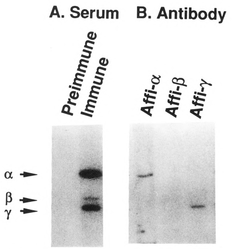
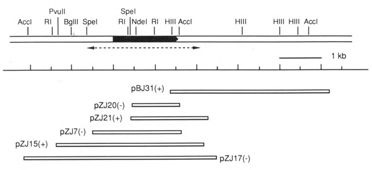
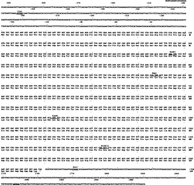
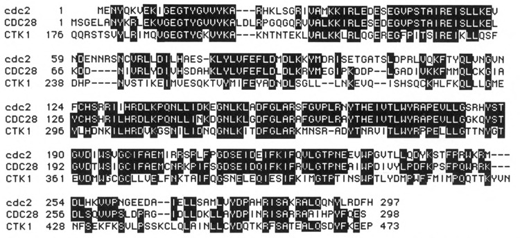
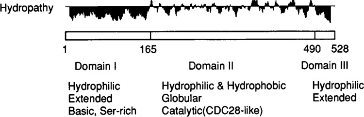
We previously purified a yeast protein kinase that specifically hyperphosphorylates the carboxyl-terminal repeat domain (CTD) of RNA polymerase II largest subunit and showed that this CTD kinase consists of three subunits of 58, 38, and 32 kDa. We have now cloned, sequenced, and characterized CTK1, the gene encoding the 58 kDa alpha subunit. The CTK1 gene product contains a central domain homologous to catalytic subunits of other protein kinases, notably yeast CDC28, suggesting that the 58 kDa subunit is catalytic. Cells that carry a disrupted version of the CTK1 gene lack the characterized CTD kinase activity, grow slowly and are cold-sensitive, demonstrating that the CTK1 gene product is essential for CTD kinase activity and normal growth. While ctk1 mutant cells do contain phosphorylated forms of the RNA polymerase II largest subunit, these forms differ from those found in wild type cells, implicating CTK1 as a component of the physiologically significant CTD phosphorylating machinery. As befitting an enzyme with a nuclear function, the N-terminal region of the CTK1 protein contains a nuclear targeting signal.


Figures

Figure 1
Figure 1
Immunoblots of CTD kinase. Mono S purified CTD kinase was run in a 12% polyacrylamide SDS gel and analyzed by Western blotting as described in Materials and Methods (2 μl [37 ng protein] per lane; see Lee and Greenleaf, 1989). [125I]Protein A was used to reveal the bound antibody. A. Lanes were reacted with preimmune serum (lane 1) or immune serum (lane 2) at 1:100 dilution. B. Each lane was reacted with antibodies affinity purified against a single subunit of CTD kinase (1:20 dilution). Lane 1, anti-α subunit (58 kDa); lane 2, anti-β subunit (38 kDa); lane 3, anti-γ subunit (32 kDa). The positions of the three subunits of the CTD kinase are denoted at left.
Figure 2
Figure 2
Restriction map of CTK1 gene and surrounding region. The black box represents the open reading frame, pointing in the 5′ to 3′ direction. The sequenced region is indicated as a dotted arrow underneath the map. The sizes of inserts subcloned in recombinant plasmids and their corresponding positions are drawn at the bottom. + / – indicates the orientation of the subcloned insert with respect to the β-galactosidase gene in the vector pSK-. Plasmid pBJ31 contains the insert DNA from phage λYJ31 subcloned into pBluescript SK-. All other plasmids, designated pZJ, were obtained from recombinant λZAP phage by in vivo excision procedures. RI: EcoRI; HIII: HindIII.
Figure 3
Figure 3
Sequence of CTK1 gene. The nucleotide and amino acid sequences of the CTK1 gene and its open reading frame are shown. The 5′ region upstream of start ATG codon is expressed by - numbers. The total sequenced region covers from −654 to +1991. Every 30 nucleotides and every 10 amino acid residues are indicated by dots. Several restriction enzyme sites are also noted on the sequence. The polyadenylation signal is underlined.
Figure 4
Figure 4
Sequence homologies among cdc2 of S. pombe, CDC28 of S. cerevisiae, and CTK1. Identical sequences are shown as white letters in the dark background; amino acid numbers are denoted at left.
Figure 5
Figure 5
Structural features of CTK1 protein. Hydropathy values (Kyte and Doolittle, 1982) were calculated and plotted using MacGene II Plus computer program. The three tentatively assigned domains, as described in text, are indicated at the bottom. For hydropathy the + values (above the horizontal lines) indicate hydrophobicity, and the - values (below the line) the hydrophilicity. The numbers underneath the box indicate the amino acid number.
Figure 6
Figure 6
Southern blot analysis of gene disruptions. Total genomic DNAs were digested with restriction enzymes AccI and BglII and analyzed by Southern blotting as described in Materials and Methods (1% agarose gel). A. Schematic diagram of wild type and mutant CTK1 genes. The top line of each is a restriction map. Below that is shown the probe DNA fragments used for the Southern blotting. At the bottom are the expected sizes of DNA bands which will hybridize with the labeled DNA probes. B: BglII; C: ClaI; R: EcoRI; H: HindlII; A: AccI. B. Southern blot of genomic DNA from wild type diploid strain DBY1091 (lane 1), from two independent transformants of DBY 1091 (lanes 2 and 3), and from four haploid mutant ctk1 strains (lanes 4 to 7) derived meiotically from the diploids of lanes 2 and 3. The sizes of hybridized DNA bands are denoted at left. (The sibling wild type CTK1 haploids contained only the 2.6 kb band).
Figure 7
Figure 7
Phosphocellulose column profiles of CTD kinase activity in extracts prepared from CTK1 and ctk1 strains. Crude extracts from wild type CTK1 and mutant ctk1 haploid strains (30 ml culture at A600 = 1.0) were loaded directly onto two identical columns of 3 ml phosphocellulose (P11) at 0.2 M KCl in buffer H (Lee and Greenleaf, 1989) and eluted with a 15 ml gradient of 0.2–0.8 M KCl in buffer H. Fractions of 0.5 ml were collected and dialyzed against 25 mM KCl in buffer H, then 1 μl of each fraction was assayed for CTD kinase activity under standard conditions with yeast CTD fusion protein as substrate (Lee and Greenleaf, 1989). Reaction products were analyzed by 6% SDS polyacrylamide gel electrophoresis, and an autoradiogram was taken from dried gel. The position of the intact fusion protein is indicated. A. Extract of wild type CTK1 strain. B. Extract of mutant ctk1 strain.
Figure 8
Figure 8
Western blot analysis of subunit IIa/IIo in crude extracts from CTK1 and ctk1 strains using different antibodies. Crude extracts were prepared from different yeast strains and samples containing similar amounts of total protein were analyzed by Western blotting using different largest subunit-specific affinity-purified antibodies (all described in Materials and Methods). Lanes 1 and 2 of each blot contained purified yeast RNA polymerase II (Pol IIA) and the same polymerase II after phosphorylation in vitro by CTD kinase (Pol IIO), respectively. Bound antibody was detected with [125I]protein A for A–C, or with alkaline phosphatase-conjugated goat anti-rabbit antibody followed by reaction with chemiluminescent substrate AMPPD for D. The positions of subunit IIa and IIo are denoted as solid and dotted arrows respectively. A. Blot was reacted with anti-DmE2, directed against determinants outside the CTD (major bands in lanes 1 and 2 represent IIb, the proteolyzed form of IIa). Lane 3: wild type diploid strain (DBY 1091). Lane 4: diploid transformant. Lanes 5 and 6: haploid wild type strains. Lanes 7 and 8: haploid mutant strains. (Strains in lanes 5–8 were siblings from one tetrad derived from the strain of lane 4). Lane 9: a diploid mutant strain constructed by crossing two haploid mutant strains. The low IIo/IIa ratio in this blot (lane 2 vs lane 1) is presumably an artifact (compare blot in D). B. Blot was reacted with anti CTD. Lanes 3 and 4 contained extracts from wild type and mutant strains, respectively (replicate samples of those used in A, lanes 5 and 7). C. Blot was reacted with anti-PCTD, directed against phosphorylated CTD. The lanes contain samples as in B. D. Crude extract prepared from a wild type strain (lane 5 in A) was immuno-precipitated with anti-DmE2 antibody and the immuno precipitate analyzed after no treatment (lane 3), treatment with alkaline phosphatase (lane 4), or treatment with yeast CTD kinase (lane 5). (See Materials and Methods.) Blot was reacted with anti-DmE2 antibody.
Figure 9
Figure 9
Intracellular localization of CTKl- β-galactosidase fusion proteins. Cells were stained with 4′, 6-diamidino-2-phenylindole (DAPI) (panels A, C) and rabbit anti- β-galactosidase antibody followed by rhodamine-labeled second antibody (panels B, D). The strain containing plasmid pCTKl(118)-LacZ is shown in panels A and B. The strain containing pCTKl (463, Δ3 – 181)LacZ is shown in panels C and D.
Figure 10
Figure 10
Immunoblot of CTD kinase with anti-CTD kinase and anti-CDC28 antibodies. Mono-S purified CTD kinase (lanes 1 and 3, 0.1 μl) and crude extract (lanes 2 and 4, 10 μl) were run in a 12% polyacrylamide SDS gel and blotted onto nitrocellulose membrane. A. Blots were reacted with a mixture of all three affinity-purified antibodies against the subunits of CTD kinase (lanes 1 and 2), or with these antibodies preincubated with an 18 amino acid oligopeptide representing the C-terminus of CDC28 (lanes 3 and 4). B. Blots were reacted with affinity purified anti-CDC28 antibody (lanes 1 and 2), or this antibody preincubated with the 18 amino acid oligopeptide (lanes 3 and 4) (see Materials and Methods). Bound antibodies were detected with alkaline phosphatase conjugated goat anti-rabbit IgG followed by reaction with NBT and BCIP. Positions of the CTD kinase subunits are indicated at left.

Similar articles

Cited by

References

    1. Allison L. A., Moyle M., Shales M., and Ingles C. J. (1985), Cell 42, 599–610. - PubMed
    1. Allison L. A., Wong J. K., Fitzpatrick V. D., Moyle M., and Ingles C. J. (1988), Mol Cell Biol 8, 321–329. - PMC - PubMed
    1. Allison L. A. and Ingles C. J. (1989), Proc Natl Acad Sci USA 86, 2794–2798. - PMC - PubMed
    1. Arion D., Meijer L., Brizuela L., and Beach D. (1988), Cell 55, 371–378. - PubMed
    1. Arndt K. T., Styles C. A., and Fink G. R. (1989), Cell 56, 527–537. - PubMed

Publication types

Associated data