

Visualizing cold spots: TRPM8-expressing sensory neurons and their projections
- PMID: 18199758
- PMCID: PMC6670358
- DOI: 10.1523/JNEUROSCI.3976-07.2008
Visualizing cold spots: TRPM8-expressing sensory neurons and their projections
Abstract
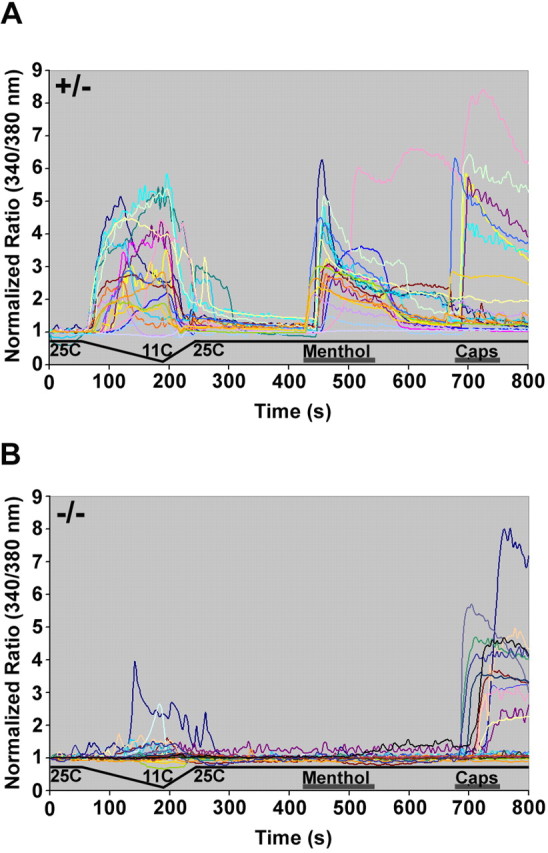
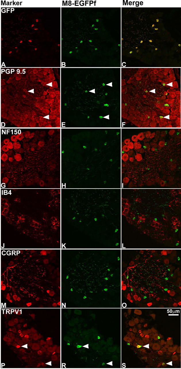
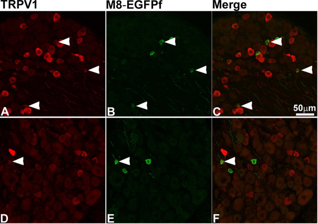
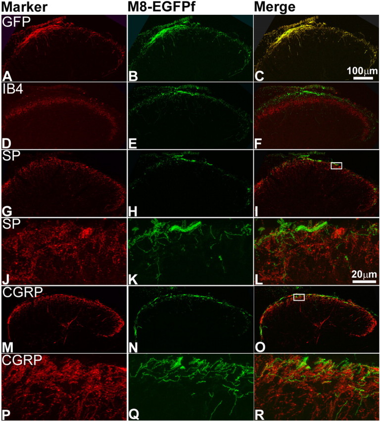
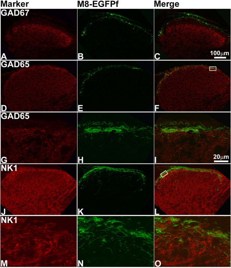
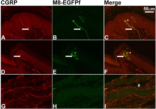
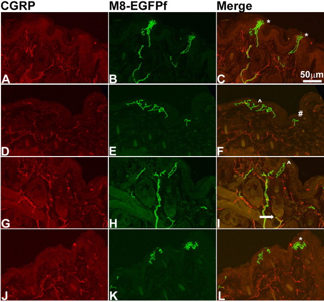
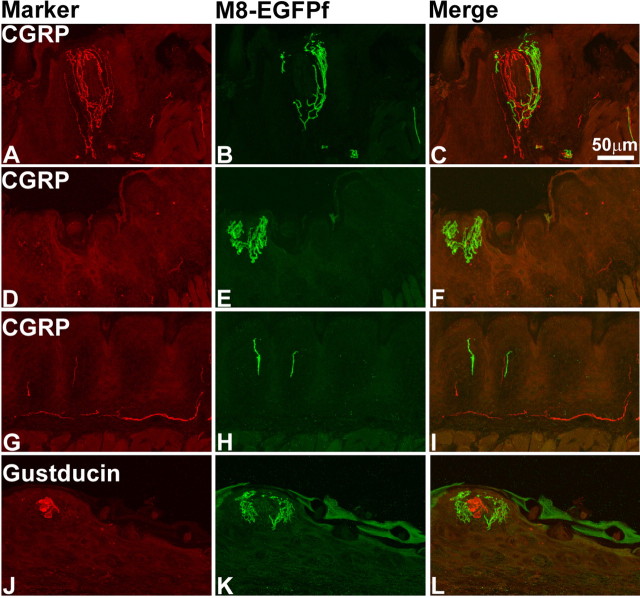
Environmental stimuli such as temperature and pressure are sensed by dorsal root ganglion (DRG) neurons. DRG neurons are heterogeneous, but molecular markers that identify unique functional subpopulations are mainly lacking. ThermoTRPs are members of the transient receptor potential family of ion channels and are gated by shifts in temperature. TRPM8 is activated by cooling, and TRPM8-deficient mice have severe deficits in cool thermosensation. The anatomical and functional properties of TRPM8-expressing fibers have not been not comprehensively investigated. We use mice engineered to express the farnesylated enhanced green fluorescent protein (EGFPf) from the TRPM8 locus (TRPM8(EGFPf)) to explore this issue. Virtually all EGFPf-positive cultured DRG neurons from hemizygous mice (TRPM8(EGFPf/+)) responded to cold and menthol. In contrast, EGFPf-positive DRGs from homozygous mice (TRPM8(EGFPf/EGFPf)) had drastically reduced cold responses and no menthol responses. In vivo, EGFPf-positive neurons marked a unique population of DRG neurons, a majority of which do not coexpress nociceptive markers. The fraction of DRG neurons expressing EGFPf was not altered under an inflammatory condition, although an increase in TRPV1-coexpressing neurons was observed. TRPM8(EGFPf) neurons project to the superficial layer I of the spinal cord, making distinct contacts when compared with peptidergic projections. At the periphery, TRPM8(EGFPf) projections mark unique endings in the most superficial layers of epidermis, including bush/cluster endings of the mystacial pads. We show that TRPM8 expression functionally associates with cold sensitivity in cultured DRGs, and provide the first glimpses of the unique anatomical architecture of cold fibers in vivo.
Figures
Similar articles
-
CGRPα-expressing sensory neurons respond to stimuli that evoke sensations of pain and itch.PLoS One. 2012;7(5):e36355. doi: 10.1371/journal.pone.0036355. Epub 2012 May 1. PLoS One. 2012. PMID: 22563493 Free PMC article.
-
Expression of the transient receptor potential channels TRPV1, TRPA1 and TRPM8 in mouse trigeminal primary afferent neurons innervating the dura.Mol Pain. 2012 Sep 12;8:66. doi: 10.1186/1744-8069-8-66. Mol Pain. 2012. PMID: 22971321 Free PMC article.
-
Emergence of functional sensory subtypes as defined by transient receptor potential channel expression.J Neurosci. 2007 Mar 7;27(10):2435-43. doi: 10.1523/JNEUROSCI.5614-06.2007. J Neurosci. 2007. PMID: 17344381 Free PMC article.
-
Regulation of TRPM8 channel activity.Mol Cell Endocrinol. 2012 Apr 28;353(1-2):68-74. doi: 10.1016/j.mce.2011.10.023. Epub 2011 Oct 28. Mol Cell Endocrinol. 2012. PMID: 22061619 Free PMC article. Review.
-
The TRPM8 channel as a potential therapeutic target for bladder hypersensitive disorders.J Smooth Muscle Res. 2022;58(0):11-21. doi: 10.1540/jsmr.58.11. J Smooth Muscle Res. 2022. PMID: 35354708 Free PMC article. Review.
Cited by
-
Protein kinase C modulation of thermo-sensitive transient receptor potential channels: Implications for pain signaling.J Nat Sci Biol Med. 2011 Jan;2(1):13-25. doi: 10.4103/0976-9668.82311. J Nat Sci Biol Med. 2011. PMID: 22470230 Free PMC article.
-
The odyssey of the TR(i)P journey to the cellular membrane.Front Cell Dev Biol. 2024 Jul 23;12:1414935. doi: 10.3389/fcell.2024.1414935. eCollection 2024. Front Cell Dev Biol. 2024. PMID: 39108834 Free PMC article. Review.
-
TRPM8 and Migraine.Headache. 2016 Oct;56(9):1406-1417. doi: 10.1111/head.12948. Epub 2016 Sep 16. Headache. 2016. PMID: 27634619 Free PMC article. Review.
-
Quantitative characterization reveals three types of dry-sensitive corneal afferents: pattern of discharge, receptive field, and thermal and chemical sensitivity.J Neurophysiol. 2012 Nov;108(9):2481-93. doi: 10.1152/jn.00523.2012. Epub 2012 Aug 22. J Neurophysiol. 2012. PMID: 22914652 Free PMC article.
-
Expression of vesicular glutamate transporters in transient receptor potential melastatin 8 (TRPM8)-positive dental afferents in the mouse.Neuroscience. 2015 Sep 10;303:378-88. doi: 10.1016/j.neuroscience.2015.07.013. Epub 2015 Jul 9. Neuroscience. 2015. PMID: 26166724 Free PMC article.
References
-
- Abe J, Hosokawa H, Okazawa M, Kandachi M, Sawada Y, Yamanaka K, Matsumura K, Kobayashi S. TRPM8 protein localization in trigeminal ganglion and taste papillae. Brain Res Mol Brain Res. 2005;136:91–98. - PubMed
-
- Amaya F, Oh-hashi K, Naruse Y, Iijima N, Ueda M, Shimosato G, Tominaga M, Tanaka Y, Tanaka M. Local inflammation increases vanilloid receptor 1 expression within distinct subgroups of DRG neurons. Brain Res. 2003;963:190–196. - PubMed
-
- Babes A, Zorzon D, Reid G. Two populations of cold-sensitive neurons in rat dorsal root ganglia and their modulation by nerve growth factor. Eur J Neurosci. 2004;20:2276–2282. - PubMed
-
- Barber RP, Vaughn JE, Saito K, McLaughlin BJ, Roberts E. GABAergic terminals are presynaptic to primary afferent terminals in the substantia gelatinosa of the rat spinal cord. Brain Res. 1978;141:35–55. - PubMed
-
- Bautista DM, Siemens J, Glazer JM, Tsuruda PR, Basbaum AI, Stucky CL, Jordt SE, Julius D. The menthol receptor TRPM8 is the principal detector of environmental cold. Nature. 2007;448:204–208. - PubMed
Publication types
MeSH terms
Substances
Grants and funding
LinkOut - more resources
Full Text Sources
Other Literature Sources
Molecular Biology Databases
Miscellaneous
