. 2008 Apr;19(4):1317-27.
doi: 10.1091/mbc.e07-11-1099. Epub 2008 Jan 16.

Roles of Greatwall kinase in the regulation of cdc25 phosphatase

Affiliations

Roles of Greatwall kinase in the regulation of cdc25 phosphatase

Yong Zhao et al. Mol Biol Cell. 2008 Apr.

Abstract

We previously reported that immunodepletion of Greatwall kinase prevents Xenopus egg extracts from entering or maintaining M phase due to the accumulation of inhibitory phosphorylations on Thr14 and Tyr15 of Cdc2. M phase-promoting factor (MPF) in turn activates Greatwall, implying that Greatwall participates in an MPF autoregulatory loop. We show here that activated Greatwall both accelerates the mitotic G2/M transition in cycling egg extracts and induces meiotic maturation in G2-arrested Xenopus oocytes in the absence of progesterone. Activated Greatwall can induce phosphorylations of Cdc25 in the absence of the activity of Cdc2, Plx1 (Xenopus Polo-like kinase) or mitogen-activated protein kinase, or in the presence of an activator of protein kinase A that normally blocks mitotic entry. The effects of active Greatwall mimic in many respects those associated with addition of the phosphatase inhibitor okadaic acid (OA); moreover, OA allows cycling extracts to enter M phase in the absence of Greatwall. Taken together, these findings support a model in which Greatwall negatively regulates a crucial phosphatase that inhibits Cdc25 activation and M phase induction.


Figures
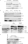
Figure 1.
Figure 1.
Greatwall regulates Cdc25. (A) Depletion of Greatwall from cycling extracts prevents Cdc25 activation and mitotic entry. Mock-depleted control extracts enter M phase within 80 min, as shown by M phase–specific upshifts of Cdc25 and Greatwall, the loss of the inhibitory Tyr15 phosphorylation of Cdc2, and the rapid degradation of cyclin B. Greatwall-depleted extracts fail to enter mitosis as judged by all of these criteria; similar experiments published previously (Yu et al., 2006) show that the block to mitotic entry continues for at least 60 min beyond the latest time point shown here. (B) Maintenance of mitotic phosphorylations on Cdc25C in CSF extracts requires Greatwall but not Plx1 or Cdc2 activity. Removal of Plx1 by immunodepletion and/or inhibition of Cdc2 activity by 250 μM roscovitine have no detectable impact on Cdc25C mobility (groups of Plx1Δ/−Ros and MockΔ/+Ros). Depletion of Greatwall completely eliminates the migration shift of Cdc25C (groups of Plx1Δ/GwlΔ and GwlΔ). The electrophoretic mobility of Greatwall itself is unaffected by Plx1 depletion, but roscovitine appears to cause the removal of some phosphorylations on Greatwall; this effect is only partial because Greatwall's migration is still significantly retarded (the arrow at left indicates the mobility of fully dephosphorylated Greatwall during interphase). (C) Premature mitotic entry induced by active Greatwall. Freshly made cycling extracts were incubated at 23°C starting at t = 0. Untreated cycling extracts enter mitosis at 70 min. Addition at 20 min of excess (five times the endogenous level) active wild-type Greatwall [Gwl(BV)] purified from OA-treated Sf9 cells promotes premature mitotic entry at t = 30 min, as indicated by the mobility shifts of Cdc25C and endogenous (endo) Greatwall and the dephosphorylation of Tyr15 of Cdc2. Within 20 min of mitotic entry, the extracts exit mitosis (note the degradation of cyclins A1 and B1), indicating that excess active Greatwall does not block mitotic exit. Addition of an even larger excess (7.5 times the endogenous level) of kinase-dead Greatwall (G41S) does not accelerate mitotic entry. Throughout this article, the relative amounts of recombinant and endogenous proteins were determined by Coomassie Blue staining (data not shown); the intensities of the exogenous Greatwall bands on the Western blots in B–D are distorted by the immunoglobulin Z domain in these recombinant proteins as well as some nonlinearity in the relationship between the protein amount and Western blot signal intensity (see Supplementary Figure S1E). The interaction between the Z domain and secondary antibodies accounts for ∼50% of the total signal on Western blots (Supplementary Figure S1E). (D) Greatwall purified from OA-treated Sf9 cells is less active than endogenous Greatwall. CSF egg extracts were subjected to Greatwall depletion or mock depletion (see Yu et al., 2006 for details). Greatwall-depleted egg extract was then supplemented with various amounts of mock-depleted CSF egg extract or with active purified Greatwall. Extracts were prepared for analysis 30 min after supplementation. Purified Greatwall preparations have only about one-third the activity of endogenous mitotic Greatwall in maintaining the M phase status of the extracts.
Figure 2.
Figure 2.
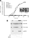
Active Greatwall induces oocyte maturation in the absence of progesterone in a protein synthesis–dependent manner. (A) Oocytes were injected with active (WT) or kinase-dead (KD) Greatwall protein. Control oocytes were stimulated by progesterone (Pg). In some cases as indicated, cycloheximide (CHX) was added 1 h before Greatwall injection or progesterone addition. The graph shows the time course of germinal vesicle breakdown (GVBD). Photographs illustrate the external morphology of Greatwall-injected oocytes (Gwl) or progesterone-stimulated oocytes (Pg). (B) Oocytes were homogenized at GVBD, or 7 h after injection or stimulation when GVBD did not occur. Lysates were immunoblotted with the indicated antibodies. Cyclin B1 is expressed at a low level in immature oocytes and its synthesis is stimulated during meiotic maturation; Cyclin B2 migrates as a doublet when MPF is inactive and as a single band when MPF is active (Karaiskou et al., 2004).
Figure 3.
Figure 3.
Greatwall can influence Cdc25 phosphorylation independent of MPF activity. (A) Greatwall promotes mitotic entry in the presence of the Cdc2 inhibitor roscovitine (Ros). Cycling extracts were incubated at 23°C (starting at t = 0) for 30 min before the addition of 250 μM Ros as indicated. Excess active Greatwall (5×) or an equal volume of buffer was added 10 min later. Extracts were processed every 10 min thereafter for Western blot analysis and Histone H1 kinase assay. (B) Greatwall promotes Cdc25 phosphorylation in the absence of MPF. To eliminate MPF activity, CSF extracts were first induced into interphase (at t = 0) by adding CaCl2 to a final concentration of 0.5 mM and then incubated at 23°C for 15 min. Cycloheximide (CHX) was then added (100 μg/ml final concentration) to inhibit protein synthesis as indicated. After another 15 min, exogenous cyclin B was supplemented to the indicated groups. At t = 40 min, active Greatwall was added as shown, and samples were collected every 10 min and processed for immunoblotting. In untreated control extracts, mitotic entry occurs at ∼120 min after CaCl2 addition (data not shown). Note that in the presence of active Greatwall, extracts enter mitosis prematurely at 50–60 min when cyclin B concentrations are low. In addition, a partial mobility shift of Cdc25 occurs at this time even when cyclin B is undetectable and (as shown in Figure 6 below) H1 kinase activity is extremely low due to CHX.
Figure 4.
Figure 4.
Plx1 is not required for Greatwall-induced mitotic entry. Cycling extracts were subjected to mock depletion or Plx1 depletion, and then incubated starting at t = 0 for 40 min at 23°C. The extract was then supplemented with buffer (PBS) or with WT or KD Greatwall purified from OA-treated Sf9 cells. One-microliter aliquots of extracts were subsequently collected every 10 min for immunoblot analysis. As expected, Plx1 depletion causes a defect in mitotic entry. However, Plx1 depletion does not interfere with Greatwall-induced mitotic entry even though Cdc25C does not obtain its full complement of M phase phosphorylations.
Figure 5.
Figure 5.
The MAPK pathway is not critical for Greatwall-induced activation of Cdc25. The involvement of the MAPK pathway in Greatwall-induced mitotic entry was tested in interphase extracts released from CSF arrest by the addition of CaCl2 at t = 0. Extracts were then incubated at 23°C for 30 min before U0126 was supplemented to a final concentration of 400 μM as shown. After 10 min of incubation, excess active Greatwall was added as indicated. Immunoblot analysis reveals that U0126 shuts down signaling through the MAPK pathway, but does not interfere with Greatwall's ability to induce Cdc25 phosphorylation and precocious mitotic entry.
Figure 6.
Figure 6.
Greatwall induces the phosphorylation of Cdc25 by several mitotic kinases. Interphase extract was prepared by addition of CaCl2 to CSF extract a t = 0 and incubated at 23°C for 20 min. The extract was then supplemented with the MEK inhibitor U0126 (400 μM) and/or the protein synthesis inhibitor CHX (100 μM) or DMSO, followed by another 20-min incubation at 23°C. Extracts were then subjected to Plx1 depletion or mock depletion as indicated. One-microliter aliquots were collected immediately after the depletion and every 10 min thereafter for immunoblotting and H1 kinase assay.
Figure 7.
Figure 7.
Greatwall overrides the effect of the PKA activator 8-Br-cAMP. Addition of 8-Br-cAMP (40 μM final concentration) effectively blocks mitotic entry in control cycling extracts; Cdc25 remains unphosphorylated, whereas inhibitory Tyr15 phosphorylations accumulate on Cdc2. The addition of excess (five times the endogenous level) active Greatwall at t = 40 min overcomes this effect of 8-Br-cAMP. In controls, the drug does not increase phosphorylation at the S287 residue of Cdc25C; the mechanism by which 8-Br-cAMP blocks mitotic entry remains unknown. The inset shows the result of a similar experiment that emphasizes the existence of Cdc25 species of retarded mobility that retain substantial Ser287 phosphorylation.
Figure 8.
Figure 8.
OA overcomes the Greatwall depletion phenotype in cycling extracts. (A) Mock-depleted or Greatwall-depleted cycling extracts were incubated at 23°C for 40 min before supplementation with 100 or 400 nM OA in DMSO or with an equal volume of DMSO alone (0). One-microliter aliquots were collected every 10 min thereafter and processed for immunoblotting. (B) OA rescues the Plx1 depletion phenotype in cycling extracts. Mock-depleted or Plx1-depleted cycling extracts were incubated at 23°C for 40 min before supplementation with 400 nM OA or DMSO.

Similar articles

Cited by

References

    1. Abrieu A., Brassac T., Galas S., Fisher D., Labbe J., Doree M. The Polo-like kinase Plx1 is a component of the MPF amplification loop at the G2/M-phase transition of the cell cycle in Xenopus eggs. J. Cell Sci. 1998;111:1751–1757. - PubMed
    1. Barr F. A., Sillje H.H.W., Nigg E. A. Polo-like kinases and the orchestration of cell division. Nat. Rev. Mol. Cell Biol. 2004;5:429–441. - PubMed
    1. Chun J., Chau A. S., Maingat F. G., Edmonds S. D., Ostergaard H. L., Shibuya E. K. Phosphorylation of Cdc25C by pp90Rsk contributes to a G2 cell cycle arrest in Xenopus cycling egg extracts. Cell Cycle. 2005;4:148–154. - PubMed
    1. Clarke P. R., Hoffmann I., Draetta G., Karsenti E. Dephosphorylation of cdc25-C by a type-2A protein phosphatase: specific regulation during the cell cycle in Xenopus egg extracts. Mol. Biol. Cell. 1993;4:397–411. - PMC - PubMed
    1. Descombes P., Nigg E. A. The polo-like kinase Plx1 is required for M phase exit and destruction of mitotic regulators in Xenopus egg extracts. EMBO J. 1998;17:1328–1335. - PMC - PubMed

Publication types

MeSH terms