. 2008 Mar;82(6):2715-26.
doi: 10.1128/JVI.02456-07. Epub 2008 Jan 16.

Mitochondrial and secretory human cytomegalovirus UL37 proteins traffic into mitochondrion-associated membranes of human cells

Affiliations

Mitochondrial and secretory human cytomegalovirus UL37 proteins traffic into mitochondrion-associated membranes of human cells

Petros Bozidis et al. J Virol. 2008 Mar.

Abstract

The human cytomegalovirus (HCMV) UL37 exon 1 protein (pUL37x1), also known as vMIA, is the predominant UL37 isoform during permissive infection. pUL37x1 is a potent antiapoptotic protein, which prevents cytochrome c release from mitochondria. The UL37x1 NH(2)-terminal bipartite localization signal, which remains uncleaved, targets UL37 proteins to the endoplasmic reticulum (ER) and then to mitochondria. Based upon our findings, we hypothesized that pUL37x1 traffics from the ER to mitochondria through direct contacts between the two organelles, provided by mitochondrion-associated membranes (MAMs). To facilitate its identification, we cloned and tagged the human phosphatidylserine synthase 1 (huPSS-1) cDNA, whose mouse homologue localizes almost exclusively in the MAM. Using subcellular fractionation of stable HeLa cell transfectants expressing mEGFP-huPSS-1, we found that HCMV pUL37x1 is present in purified microsomes, mitochondria, and MAM fractions. We further examined the trafficking of the full-length UL37 glycoprotein cleavage products, which divergently traffic either through the secretory apparatus or into mitochondria. Surprisingly, pUL37(NH2) and gpUL37(COOH) were both detected in the ER and MAM fraction, even though only pUL37(NH2) is preferentially imported into mitochondria but gpUL37(COOH) is not. To determine the sequences required for MAM importation, we examined pUL37x1 mutants that were partially defective for mitochondrial importation. Deletion mutants of the NH(2)-terminal UL37x1 mitochondrial localization signal were reduced in trafficking into the MAM, indicating partial overlap of MAM and mitochondrial targeting signals. Taken together, these results suggest that HCMV UL37 proteins traffic from the ER into the MAM, where they are sorted into either the secretory pathway or to mitochondrial importation.


Figures

FIG. 1.
FIG. 1.
HCMV UL37 proteins and ORFs. The UL37x1 hydrophobic leader (aa 1 to 22) and juxtaposed basic (aa 23 to 29) and acidic (aa 81 to 108) domains are represented. The downstream UL37x2 and UL37x3 N-glycosylation domain, basic residues, transmembrane, and cytosolic tail are present in gpUL37 and pUL37M. The unique internal cleavage site of gpUL37 is indicated by the arrow at aa 193/194. The epitope for Ab1064 and DC35 (aa 27 to 40) and the C-terminal Flag tag (flag) are shown. For the pUL37x1 mutants, the sequences in pUL37x1 Δ2-23 and pUL37x1 Δ23-34 (19, 25), as well as the C-terminal Myc tag (hexagon), are shown.
FIG. 2.
FIG. 2.
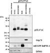
pUL37x1 traffics into microsomes, mitochondria, and MAM in transfected HeLa cells. PSS-120 cells that stably express the tagged MAM protein marker (mEGFP-huPSS-1) were transfected with either empty vector (p396) or vector expressing pUL37x1 (p327) and fractionated into microsomes, MAM, mitochondria, and cytosol by using Percoll gradient fractionation (10). A total of 10 μg of microsomes (0.7% of fraction), MAM (9.8%), mitochondria (9.7%), and cytosol (0.3%) was resolved by SDS-PAGE using 12% polyacrylamide, blotted, and reacted with a rabbit polyclonal antibody (DC35, 1:1,000) that recognizes UL37x1. The blots were stripped and reprobed with antibodies to specific protein markers for mitochondria (Grp75, mouse anti-Grp75 antibody, 1:1,000; Stressgen), MAM (mEGFP-huPSS-1, mouse anti-GFP antibody, 1:100; Santa Cruz), and ER (DPM1, goat anti-DPM1 antibody, 1:100; Santa Cruz) in order to verify the identity of purified fractions.
FIG. 3.
FIG. 3.
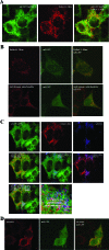
(A) Partial colocalization of mEGFP-huPSS-1 and dsRed-1-Mito markers. PSS-120 cells expressing mEGFP-huPSS-1 were transiently transfected with a plasmid expression vector for DsRed-1-Mito. After 24 h they were fixed and probed with mouse anti-GFP (1:200). The secondary antibody was FITC-goat anti-mouse (1:250, green). The cells were imaged for emissions at both 520 nm (FITC, left) and 615 nm (TR, middle) by using confocal microscopy. The overlaid image is on the right. (B) Diffuse mEGFP localization in transfected HeLa cells. HeLa cells were transfected with plasmids expressing mEGFP (pmEGFP-C1) and DsRed-1-Mito. Cells were fixed 24 h later and examined as in panel A for the presence of mEGFP and DsRed-1- mito. Fixed cells were examined for DsRed (top) or probed with human autoimmune anti-mitochondria antibodies (1:50) (bottom). Secondary antibody was TR-goat anti-human IgG (1:50). Confocal images were taken sequentially through a ×100 objective lens. Zeiss Lasersharp 2000 Acquisition software was used for additional magnifications. (C) Colocalization of pUL37x1 with mEGFP-huPSS-1 and with dsRed-1-Mito. PSS-120 cells expressing mEGFP-huPSS-1 were transiently transfected with DsRed-1-Mito expression vector and a pUL37x1 expression plasmid (p327). After 24 h they were fixed and probed with mouse anti-GFP (1:200) and rabbit anti-UL37x1 (Ab1064, 1:250). The secondary antibodies were FITC-goat anti-mouse (1:250, green) and Cy5-Goat anti-rabbit (1:50, blue). Cells were imaged by using confocal microscopy at 520 nm (FITC, top row, left panel), 615 nm (TR, top row, middle panel), and 670 nm (Cy5, top row, right panel). Panels also show the overlaid images of green-red (center row, left), green-cyan (center row, middle), red-cyan (center row, right) and green-red-cyan (bottom row, left). The bottom right panel shows detail of the boxed area from the triple overlay. (D) Lack of colocalization of mEGFP and pUL37x1 in transfected HeLa cells. HeLa cells were transfected with expression plasmid for mEGFP (pmEGFP-C1) and for pUL37x1 (p327). Cells were fixed 24 h later and examined as panel C for the presence of EGFP and for pUL37x1 by using Ab1064 (1:300) and TR-goat anti-rabbit IgG (1:300). Cells were imaged by using confocal microscopy at 520 nm (FITC, left panel) and 615 nm (TR, middle panel). The overlaid image is on the right.
FIG. 4.
FIG. 4.
pUL37x1 partially colocalizes with mEGFP-huPSS-1 in permissive cells. LE-HFFs were transiently transfected with vectors expressing mEGFP-huPSS-1. (A) Cells were HCMV infected (3 PFU/cell) 24 h later, harvested at 12 h postinfection, and stained with Ab1064 (anti-pUL37x1) and MAb810 (anti-IE1/IE2). Cy5-Goat anti-mouse IgG (1:100) or TR-goat anti-rabbit IgG (1:100) secondary antibodies were used for visualization. Green (upper left), red (upper right), cyan (lower left), and overlaid channels (lower right) are shown. (B) Uninfected LE-HFFs, transiently expressing mEGFP-huPSS-1, were stained with human autoimmune anti-mitochondria (1:50) or (C) with mouse anti-KDELR (1:100). Cy5-goat anti-human IgG (1:50) or TR-goat anti-mouse IgG (1:100) secondary antibodies were used for visualization. In panel B, the green channel is at the top, red channel is in the middle, and the overlay is at the bottom. In panel C, the green channel is on the left, the red channel is in the middle, and the overlay is on right.
FIG. 5.
FIG. 5.
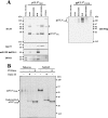
Both gpUL37 cleavage products traffic into microsomes, mitochondria, and MAM in transfected HeLa cells. PSS-120 cells that stably express the tagged MAM protein marker (mEGFP-huPSS-1) were transfected with either empty vector (p790) or vector expressing gpUL37-Flag (p816) and fractionated into microsomes, MAM, mitochondria, and cytosol by using Percoll gradient fractionation (10). A total of 10 μg of microsomes (0.6% of fraction), MAM (2.1%), mitochondria (1.6%), and cytosol (0.3%) were resolved by SDS-PAGE using 12% polyacrylamide and blotted. The blot was reacted with DC35 (anti-UL37x1, aa 27 to 40, 1:1,000) (left panel), stripped, and then reacted with anti-Flag antibody (M2, 1:2,000; Covance) (right panel) that recognizes the C-terminal tag of gpUL37. Blots were stripped and reprobed with antibodies against specific protein markers for mitochondria (Grp75, mouse anti-Grp75 antibody, 1:1,000; StressGen), MAM (mEGFP-huPSS-1, mouse anti-GFP antibody, sc-9996, 1:100; Santa Cruz), and ER (DPM1, goat anti-DPM1 antibody 1:100; Santa Cruz) to verify the identity of the purified fractions. In the left panel, the pUL37NH2 monomeric (arrowhead) and multimeric (filled circle) species are marked. The asterisk marks a background band. On the right, the multiple N-glycosylated gpUL37COOH species are indicated. (B) Endoglycosidase sensitivity of gpUL37COOH in MAM fraction. The microsomal (10 μg) and MAM (20 μg) proteins from panel A were treated with either PNGase (+) or EndoH (+). Control reactions were treated with buffer but lacked the added enzyme. Samples were then resolved by SDS-PAGE, blotted, and reacted with anti-Flag antibody as described above.
FIG. 6.
FIG. 6.
Inefficient trafficking of pUL37x1 NH2-terminal deletion mutants to both MAM and mitochondria subcellular fractions. PSS-120 cells were lipofected with empty vector (p796) or vector expressing (A) pUL37x1 Δ2-23-myc (p856) or (B) pUL37x1 Δ23-34-myc (p857) and fractionated into microsomes, MAM, mitochondria, and cytosol. A total of 10 μg of p856 transfected microsomes (2.1% of fraction), MAM (3.3%), mitochondria (8.7%), and cytosol (1.5%) and 10 μg of p857 transfected microsomes (2% of fraction), MAM (4%), mitochondria (7.9%), and cytosol (1.2%) were analyzed by Western blot analysis by using a mouse antibody to the C-terminal Myc tag (1:100, MAb9E10; BabCo). Membranes were stripped and reprobed with antibodies to organelle-specific markers as described in Fig. 2.
FIG. 7.
FIG. 7.
Model of UL37 protein sorting through the MAM to the Golgi or mitochondria. (Step 1) UL37 proteins are synthesized anchored to the ER membrane. The UL37x3 sequences are translocated into the ER lumen, where they are cleaved by signal peptidase I and N glycosylated. (Step 2) pUL37x1 and both cleavage products of gpUL37 traffic into the MAM. The presence of PSS-1 in the MAM is shown. Sorting of UL37 proteins from the MAM to the Golgi apparatus (step 3) or into mitochondria (step 4) is represented. Mitochondrial importation likely occurs through direct membrane contacts between the MAM and mitochondria, which are stabilized by PACS-2.

Similar articles

Cited by

References

    1. Adair, R., G. W. Liebisch, and A. M. Colberg-Poley. 2003. Complex alternative processing of human cytomegalovirus UL37 pre-mRNA. J. Gen. Virol. 843353-3358. - PubMed
    1. Al-Barazi, H. O., and A. M. Colberg-Poley. 1996. The human cytomegalovirus UL37 immediate-early regulatory protein is an integral membrane N-glycoprotein which traffics through the endoplasmic reticulum and Golgi apparatus. J. Virol. 707198-7208. - PMC - PubMed
    1. Anandatheerthavarada, H. K., G. Biswas, J. Mullick, N. B. Sepuri, L. Otvos, D. Pain, and N. G. Avadhani. 1999. Dual targeting of cytochrome P4502B1 to endoplasmic reticulum and mitochondria involves a novel signal activation by cyclic AMP-dependent phosphorylation at ser128. EMBO J. 185494-5504. - PMC - PubMed
    1. Ardail, D., I. Popa, J. Bodennec, P. Louisot, D. Schmitt, and J. Portoukalian. 2003. The mitochondria-associated endoplasmic-reticulum subcompartment (MAM fraction) of rat liver contains highly active sphingolipid-specific glycosyltransferases. Biochem. J. 3711013-1019. - PMC - PubMed
    1. Arnoult, D., L. M. Bartle, A. Skaletskaya, D. Poncet, N. Zamzami, P. U. Park, J. Sharpe, R. J. Youle, and V. S. Goldmacher. 2004. Cytomegalovirus cell death suppressor vMIA blocks Bax- but not Bak-mediated apoptosis by binding and sequestering Bax at mitochondria. Proc. Natl. Acad. Sci. USA 1017988-7993. - PMC - PubMed

Publication types

LinkOut - more resources