. 2008 Jan;14(1):86-96.
doi: 10.1016/j.devcel.2007.11.002.

Molecular integration of wingless, decapentaplegic, and autoregulatory inputs into Distalless during Drosophila leg development

Affiliations

Molecular integration of wingless, decapentaplegic, and autoregulatory inputs into Distalless during Drosophila leg development

Carlos Estella et al. Dev Cell. 2008 Jan.

Abstract

The development of the Drosophila leg requires both Decapentaplegic (Dpp) and Wingless (Wg), two signals that establish the proximo-distal (PD) axis by activating target genes such as Distalless (Dll). Dll expression in the leg depends on a Dpp- and Wg-dependent phase and a maintenance phase that is independent of these signals. Here, we show that accurate Dll expression in the leg results from the synergistic interaction between two cis-regulatory elements. The Leg Trigger (LT) element directly integrates Wg and Dpp inputs and is only active in cells receiving high levels of both signals. The Maintenance (M) element is able to maintain Wg- and Dpp-independent expression, but only when in cis to LT. M, which includes the native Dll promoter, functions as an autoregulatory element by directly binding Dll. The "trigger-maintenance" model describes a mechanism by which secreted morphogens act combinatorially to induce the stable expression of target genes.


Figures
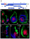
Figure 1
Figure 1. The LT enhancer
(A) The Dll 5′ cis-regulatory region. DNA fragments were cloned based on sequence conservation to other Drosophilids and assayed in transgenic reporter genes for expression in imaginal discs. E, Eco-R1, B, Bam-H1 and R, RSR II. LT is in red, 304 in yellow and M in light blue. Of the fragments tested in a standard reporter gene (using the minimal promoter from the hsp43 gene), only LT drove expression in discs. Although fragments LT (previously 215), 304, 208, and 179 were originally cloned by Vachon et al., (1992), no imaginal disc expression was reported. (B) Wild type leg discs at various stages of development stained for LT-lacZ (red), Dll (blue) and Homothorax (Hth) (green). The age of the larvae (+/− 6h) is indicated below each disc. Early in development (66h +/− 6h or before) LT was active in all the cells that express Dll. Later in development LT was active in a subset of Dll expressing cells. (C) Cross-section image of the 110h leg disc from (B). LT was only active in a subset of Dll expressing cells. (D) Wild type third instar leg disc stained for LT-lacZ (red), dpp-Gal4; UAS-GFP (green), and Wg (blue). LT was active in the center of the disc where the Wg and Dpp expression domains meet. (E) Wild type early third instar leg disc (~96h AEL) stained for LT-Gal4; UAS-GFP (red) and brk-lacZ (green). LT was active in cells that have no or very low Brk levels.
Figure 2
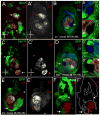
Figure 2. LT continuously requires the Wg and Dpp pathways
(A – A′) Clones expressing Arm* marked by GFP (green) activated LT-lacZ (red) in dorsal (arrow), but not ventral (arrowhead) regions of the disc. (B – B″) An arr clone induced between 48–72 hours after egg laying (h AEL), marked by absence of GFP (green), had no LT-lacZ expression (red), but maintained Dll expression (blue). (C – C′) Clones expressing TkvQD marked by GFP (green) activated LT-lacZ (red) in ventral (arrow), but not dorsal (arrowhead) regions of disc. (D – D″) A Mad clone induced between 48–72 hours AEL, marked by absence of GFP (green), had no LT-lacZ expression (red), but maintained Dll expression (blue). (E – E′) A brk clone induced 48–72h AEL, marked by absence of GFP (green), de-repressed LT-lacZ (red) in the ventral disc, close to the source of Wg (arrow), but not in dorsal regions (arrowhead). (F – F′) Clones expressing Tcf-RNAi, marked by GFP (green), did not de-repress LT-lacZ expression (red) in dorsal (arrowhead) or lateral regions of the leg disc. LT-lacZ was not expressed in Tcf-RNAi expressing cells (arrow).
Figure 3
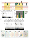
Figure 3. The regulation of LT by the Dpp and Wg pathways is direct
(A) Diagram of LT with transcription factor binding sites. Oval size indicates the relative affinities of these binding sites in EMSAs; ovals above and below the line indicate different binding site orientations. All of these binding sites, except for Brk2, are well conserved through D. virilis. The thin red lines summarize the results of a set of ~100 bp deletions tested in reporter genes. Regions shaded in yellow indicate deletions that had reduced or no reporter activity. Except for #2 and #7, all deletions that had an effect removed a Tcf or Mad binding site. Deletions #2 and #7 indicate that other inputs besides the mapped Tcf and Mad binding sites are required for LT activity. (B) EMSAs showing binding of Tcf to probes containing wild type or mutant binding sites (see Experimental Procedures for sequences). Arrows indicate protein-DNA complexes. (C) ChIP experiments demonstrating specific binding of Tcf to LT in imaginal discs. Anti-Tcf antibodies pulled down LT ~twice as efficiently from leg discs as from wing plus haltere discs. Similar results were seen for two independent PCR fragments, LT-1 and LT-2 (whose positions in LT are indicated in panel A). Each column shows the averages and standard error of the mean for four independent IPs and real time PCRs. (D) EMSAs showing binding of Mad and Brk to probes containing wild type or mutant binding sites (see Experimental Procedures for sequences). Arrows indicate protein-DNA complexes; M, Mad; B, Brk. Although Mad1 has a lower affinity for Mad than Mad2, its binding is sequence-specific. (E) X-Gal stains of leg discs from flies containing wild type LT-lacZ (i), LTTcf-lacZ (with all Tcf sites mutated; ii), LTMad1-lacZ (with the Mad1 site mutated; iii), LTMad2-lacZ (with the Mad2 site mutated; iv), LTBrk-lacZ (with both Brk sites mutated; v), and LTMadBrk-lacZ (with both Mad and Brk sites mutated; vi). Mutation of the Tcf sites or either Mad site resulted in loss of activity. Mutation of both Brk sites resulted in the ventral expansion of expression (arrow). The LTBrk-lacZ disc shown here has an intermediate amount of derepression; other transformant lines show stronger and more uniform ventral expression of lacZ. Mutation of both Mad and Brk sites resulted in no expression
Figure 4
Figure 4. The Dll promoter region has maintenance activity
(A) LT-lacZ was active in subset of Dll-expressing cells (with the hsp43 promoter, hsP). (B) M-lacZ is expressed nearly ubiquitously in leg discs at very low levels, with slightly higher levels in the Dll domain. M-lacZ is not expressed in wing discs (not shown). (C) LT+M-lacZ was expressed in all cells that express Dll in the leg imaginal disc, including low-level expression in the Dll-expressing trochanter ring. LT+M-lacZ was not expressed in wing discs (not shown). The inset shows the expression of Dll (green) and LT+M-lacZ (red) in combination with Hth (blue). (D and E) Genetic tests of LT+M-lacZ maintenance. (D-D‴) A Mad clone induced between 48–72h AEL, marked by absence of GFP, continues to express LT+M-lacZ (red) and Dll (blue). (E) An arr clone induced between 48–72h AEL, marked by absence of GFP, continues to express LT+M-lacZ (red) and Dll (blue). The insets (D′,D″,D‴,E,E″,E‴) show blow-ups of the clones, outlined in red.
Figure 5
Figure 5. Dll is required for Dll maintenance
(A) A Dll clone induced between 48–72h AEL, marked by absence of GFP, resulted in loss of LT+M-lacZ expression (red). The inset shows the clone outlined in red and the cell autonomous loss of LT+M-lacZ expression. (B) EMSAs showing Dll binding to each of the three Dll sites in the M element (WT and mutant; see Experimental Procedures for sequences). The arrow indicates the Dll-induced complexes. (C) The expression of a LT+M reporter gene with all three Dll binding sites mutated (LT+MDll-lacZ) was only weakly expressed in Dll-expressing cells of the leg disc. (D) Expression driven by the LT-5M-hsP-lacZ reporter gene. The level and pattern of expression indicates that the 5′M fragment confers partial maintenance activity. The insets and brackets compare β-gal and Dll expression. (E) When the Dll1 binding site is mutated in this reporter (LT-5MDll1-hsp-lacZ), expression resembles that driven by LT-lacZ (compare with Fig. 4A).
Figure 6
Figure 6. LT is required for maintenance
(A) Diagram of a LT+M reporter gene in which LT is flanked by FRT sites (black triangles). After expression of Flp, LT is deleted, leaving a single FRT site and the M element. (B) In the absence of Flp, >LT>M-lacZ generated a Dll-like expression pattern. This disc came from a larva of the same genotype as the one shown in (D) (hs-flp122; >LT>M-lacZ), but was not given a heat shock. (C) Deletion of the LT enhancer in the posterior compartment (green) early in development (prior to maintenance) using en-Gal4, UAS-flp resulted in the loss of lacZ expression in that compartment, while leaving expression in the anterior compartment intact. (D) Heat shock-induced expression of Flp during the maintenance stage of Dll expression resulted in the loss of reporter expression within the Dll domain. Due to the design of this experiment (see Experimental Procedures) only a subset of these heat-shock induced events were marked by GFP+; other, unmarked events are outlined. The inset shows a blow-up of the GFP-marked clone. In this experiment, Flp was provided 90 +/−6 h AEL via a 8 minute heat shock, significantly after maintenance begins.
Figure 7
Figure 7. The trigger-maintenance model
LT drives Dll expression early in larval development by directly integrating inputs from the Wg and Dpp signaling pathways. Tcf and Mad bind LT to activate, while Brk binds LT to repress, resulting in LT activity in the center of the young leg disc. Dll is also required for LT activity, although it is not known if this input is direct. We also suggest that other elements within the Dll locus may act redundantly with LT to integrate the Wg and Dpp signals (not indicated). As the disc grows, Dll becomes independent of Wg and Dpp signaling. During the maintenance phase, the composite LT+M element behaves as an autoregulatory element as it is directly activated by Dll binding to sequences close to the Dll promoter (M). Dll input into LT may also contribute to maintenance, as well as other currently unknown factors. Consistent with this model, a lineage tracing experiment using LT demonstrates that all Dll-expressing cells in a third instar leg disc are derived from LT-expressing cells (McKay et al, in preparation).

Similar articles

Cited by

References

    1. Abu-Shaar M, Mann RS. Generation of multiple antagonistic domains along the proximodistal axis during Drosophila leg development. Development. 1998;125:3821–3830. - PubMed
    1. Arnosti DN. Analysis and function of transcriptional regulatory elements: insights from Drosophila. Annu Rev Entomol. 2003;48:579–602. - PubMed
    1. Barolo S, Posakony JW. Three habits of highly effective signaling pathways: principles of transcriptional control by developmental cell signaling. Genes Dev. 2002;16:1167–1181. - PubMed
    1. Breiling A, Sessa L, Orlando V. Biology of polycomb and trithorax group proteins. Int Rev Cytol. 2007;258:83–136. - PubMed
    1. Brock HW, Fisher CL. Maintenance of gene expression patterns. Dev Dyn. 2005;232:633–655. - PubMed

Publication types

LinkOut - more resources