Review
. 2008 Feb;4(2):151-75.
doi: 10.4161/auto.5338. Epub 2007 Nov 21.

Guidelines for the use and interpretation of assays for monitoring autophagy in higher eukaryotes

Daniel J Klionsky  1 Hagai AbeliovichPatrizia AgostinisDevendra K AgrawalGjumrakch AlievDavid S AskewMisuzu BabaEric H BaehreckeBen A BahrAndrea BallabioBruce A BamberDiane C BasshamEttore BergaminiXiaoning BiMartine Biard-PiechaczykJanice S BlumDale E BredesenJeffrey L BrodskyJohn H BrumellUlf T BrunkWilfried BurschNadine CamougrandEduardo CebolleroFrancesco CecconiYingyu ChenLih-Shen ChinAugustine ChoiCharleen T ChuJongkyeong ChungPeter G H ClarkeRobert S B ClarkSteven G ClarkeCorinne ClavéJohn L ClevelandPatrice CodognoMaría I ColomboAna Coto-MontesJames M CreggAna Maria CuervoJayanta DebnathFrancesca DemarchiPatrick B DennisPhillip A DennisVojo DereticRodney J DevenishFederica Di SanoJ Fred DiceMarian DifigliaSavithramma Dinesh-KumarClark W DistelhorstMojgan Djavaheri-MergnyFrank C DorseyWulf DrögeMichel DronWilliam A Dunn JrMichael DuszenkoN Tony EissaZvulun ElazarAudrey EsclatineEeva-Liisa EskelinenLászló FésüsKim D FinleyJosé M FuentesJuan FueyoKozo FujisakiBrigitte GalliotFen-Biao GaoDavid A GewirtzSpencer B GibsonAntje GohlaAlfred L GoldbergRamon GonzalezCristina González-EstévezSharon GorskiRoberta A GottliebDieter HäussingerYou-Wen HeKim HeidenreichJoseph A HillMaria Høyer-HansenXun HuWei-Pang HuangAkiko IwasakiMarja JäätteläWilliam T JacksonXuejun JiangShengkan JinTerje JohansenJae U JungMotoni KadowakiChanhee KangAmeeta KelekarDavid H KesselJan A K W KielHong Pyo KimAdi KimchiTimothy J KinsellaKirill KiselyovKatsuhiko KitamotoErwin KnechtMasaaki KomatsuEiki KominamiSeiji KondoAttila L KovácsGuido KroemerChia-Yi KuanRakesh KumarMondira KunduJacques LandryMarianne LaporteWeidong LeHuan-Yao LeiMichael J LenardoBeth LevineAndrew LiebermanKah-Leong LimFu-Cheng LinWillisa LiouLeroy F LiuGabriel Lopez-BeresteinCarlos López-OtínBo LuKay F MacleodWalter MalorniWim MartinetKen MatsuokaJosef MautnerAlfred J MeijerAlicia MeléndezPaul MichelsGiovanni MiottoWilhelm P MistiaenNoboru MizushimaBaharia MograbiIryna MonastyrskaMichael N MoorePaula I MoreiraYuji MoriyasuTomasz MotylChristian MünzLeon O MurphyNaweed I NaqviThomas P NeufeldIchizo NishinoRalph A NixonTakeshi NodaBernd NürnbergMichinaga OgawaNancy L OleinickLaura J OlsenBulent OzpolatShoshana PaglinGlen E PalmerIssidora PapassideriMiles ParkesDavid H PerlmutterGeorge PerryMauro PiacentiniRonit Pinkas-KramarskiMark PrescottTassula Proikas-CezanneNina RabenAbdelhaq RamiFulvio ReggioriBärbel RohrerDavid C RubinszteinKevin M RyanJunichi SadoshimaHiroshi SakagamiYasuyoshi SakaiMarco SandriChihiro SasakawaMiklós SassClaudio SchneiderPer O SeglenOleksandr SeleverstovJeffrey SettlemanJohn J ShackaIrving M ShapiroAndrei SibirnyElaine C M Silva-ZacarinHans-Uwe SimonCristiano SimoneAnne SimonsenMark A SmithKatharina Spanel-BorowskiVickram SrinivasMeredith SteevesHarald StenmarkPer E StromhaugCarlos S SubausteSeiichiro SugimotoDavid SulzerToshihiko SuzukiMichele S SwansonIra TabasFumihiko TakeshitaNicholas J TalbotZsolt TallóczyKeiji TanakaKozo TanakaIsei TanidaGraham S TaylorJ Paul TaylorAlexei TermanGianluca TettamantiCraig B ThompsonMichael ThummAviva M TolkovskySharon A ToozeRay TruantLesya V TumanovskaYasuo UchiyamaTakashi UenoNéstor L UzcáteguiIda van der KleiEva C VaqueroTibor VellaiMichael W VogelHong-Gang WangPaul WebsterJohn W WileyZhijun XiGutian XiaoJoachim YahalomJin-Ming YangGeorge YapXiao-Ming YinTamotsu YoshimoriLi YuZhenyu YueMichisuke YuzakiOlga ZabirnykXiaoxiang ZhengXiongwei ZhuRussell L Deter
Affiliations
Review

Guidelines for the use and interpretation of assays for monitoring autophagy in higher eukaryotes

Daniel J Klionsky et al. Autophagy. 2008 Feb.

Abstract

Research in autophagy continues to accelerate,(1) and as a result many new scientists are entering the field. Accordingly, it is important to establish a standard set of criteria for monitoring macroautophagy in different organisms. Recent reviews have described the range of assays that have been used for this purpose.(2,3) There are many useful and convenient methods that can be used to monitor macroautophagy in yeast, but relatively few in other model systems, and there is much confusion regarding acceptable methods to measure macroautophagy in higher eukaryotes. A key point that needs to be emphasized is that there is a difference between measurements that monitor the numbers of autophagosomes versus those that measure flux through the autophagy pathway; thus, a block in macroautophagy that results in autophagosome accumulation needs to be differentiated from fully functional autophagy that includes delivery to, and degradation within, lysosomes (in most higher eukaryotes) or the vacuole (in plants and fungi). Here, we present a set of guidelines for the selection and interpretation of the methods that can be used by investigators who are attempting to examine macroautophagy and related processes, as well as by reviewers who need to provide realistic and reasonable critiques of papers that investigate these processes. This set of guidelines is not meant to be a formulaic set of rules, because the appropriate assays depend in part on the question being asked and the system being used. In addition, we emphasize that no individual assay is guaranteed to be the most appropriate one in every situation, and we strongly recommend the use of multiple assays to verify an autophagic response.


Figures
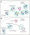
Figure 1
Figure 1
Schematic model demonstrating the induction of autophagosome formation when turnover is blocked versus normal autophagic flux. (A) Induction results in the initiation of autophagy including the formation of the phagophore, the initial sequestering compartment, which expands into an autophagosome. A defect in autophagosome turnover due, for example, to a block in fusion with lysosomes or disruption of lysosomal functions will result in an increased number of autophagosomes. In this scenario, autophagy has been induced, but there is no or limited autophagic flux. This is a different outcome than the situation shown in (B) where autophagosome formation is followed by fusion with lysosomes and degradation of the contents, allowing complete flux, or flow, through the entire pathway.
Figure 2
Figure 2
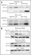
LC3-I conversion and LC3-II turnover. (A) HEK293 and HeLa cells were cultured in nutrient-rich medium (DMEM containing 10% FCS) or incubated for 4 h in starvation conditions (Krebs-Ringer medium) in the absence (−) or presence (+) of E64d and pepstatin at 10 μg/ml each (Inhibitors). Cells were then lysed and the proteins resolved by SDS-PAGE. Endogenous LC3 was detected by immunoblotting. Positions of LC3-I and LC3-II are indicated. In the absence of lysosomal protease inhibitors, starvation results in a modest increase (HEK293 cells) or even a decrease (HeLa cells) in the amount of LC3-II. The use of inhibitors reveals that this apparent decrease is due to lysosome-dependent degradation. This figure was modified from data previously published in reference , and is reproduced by permission of Landes Bioscience, copyright 2005. (B) Expression levels of LC3-I and LC3-II during starvation. Atg5+/+ (wild-type) and Atg5−/− MEFs were cultured in DMEM without amino acids and serum for the indicated times, and then subjected to immunoblot analysis using anti-LC3 antibody and anti-tubulin antibody. E64d (10 μg/ml) and pepstatin A (10 μg/ml) were added to the medium where indicated. Positions of LC3-I and LC3-II are indicated. Similar to the result in (A), the inclusion of lysosomal protease inhibitors reveals that the apparent decrease in LC3-II is due to lysosomal degradation as easily seen by comparing samples with and without inhibitors at the same time points (the overall decrease seen in the presence of inhibitors may reflect decreasing effectiveness of the inhibitors over time). Monitoring autophagy by following steady state amounts of LC3-II without including inhibitors in the analysis can result in an incorrect interpretation that autophagy is not taking place (due to the apparent absence of LC3-II). Conversely, if there are high levels of LC3-II but there is no change in the presence of inhibitors this may indicate that induction has occurred but that the final steps of autophagy are blocked, resulting in stabilization of this protein. This figure was modified from data previously published in reference , and is reproduced by permission of Landes Bioscience, copyright 2007.
Figure 3
Figure 3
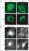
Changes in the localization of LC3 and GFP-LC3 upon the induction of autophagy. (A) Immunofluorescence in mouse fibroblasts and human T cells. The indicated cells were left untreated or were treated with 100 μM rapamycin for 4 h and were subjected to immunofluorescence with a selective antibody against LC3. LC3-stained autophagic compartments in T cells are indicated with arrows. Quantification of 20 cells similar to the ones shown here indicated that rapamycin-treated cells had 165 ± 8 vesicles per fibroblast and 6 ± 2 vesicles per T cell. Bar, 5 μm. This figure was previously published in reference , and is reproduced by permission of Landes Bioscience, copyright 2007. (B) Direct fluorescence in stable MEF transformants. GFP-LC3-expressing Atg5+/+ and Atg5−/− MEFs were cultured in DMEM with 10% FBS or DMEM without amino acids and serum for 1.5 h. Cells were fixed with 3% PFA and analyzed by fluorescence microscopy. Bar, 20 μm. This figure was previously published in reference , and is reproduced by permission of Landes Bioscience, copyright 2007.
Figure 4
Figure 4
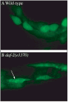
GFP::LGG-1 is an autophagy marker in C. elegans. GFP::LGG-1 expression in the hypodermal seam cells of (A) wild-type N2 animals and (B) daf-2(e1370) animals that have an increase in autophagy. The arrow shows representative GFP-positive punctate areas that label pre-autophagosomal and autophagosomal structures. This figure was modified from data previously published in Meléndez A, Tallóczy Z, Seaman M, Eskelinen E-L, Hall DH, Levine B. Autophagy genes are essential for dauer development and life-span extension in C. elegans. Science 2003; 301:1387–91. Reprinted with permission from AAAS.
Figure 5
Figure 5
GFP-LC3 processing can be used to monitor delivery of autophagosomal membranes. Atg5−/− MEFs engineered to express Atg5 under the control of the Tet-off promoter were grown in the presence of doxycyline (10 ng/ml) for one week to suppress autophagy. Cells were then cultured in the absence of drug for the indicated times, with or without a final 2 h starvation. Protein lysates were analyzed by western blot using anti-LC3 and anti-GFP antibodies. The positions of GFP-LC3-I, GFP-LC3-II and free GFP are indicated. This figure was modified from data previously published in reference , FEBS Letters, 580, Hosokawa N, Hara Y, Mizushima N, Generation of cell lines with tetracycline-regulated autophagy and a role for autophagy in controlling cell size, pp. 2623–9, copyright 2006, with permission from Elsevier.
Figure 6
Figure 6
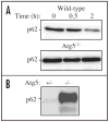
Regulation of the p62 protein during autophagy. (A) The level of p62 during starvation. Atg5+/+ and Atg5−/− MEFs were cultured in DMEM without amino acids and serum for the indicated times, and then subjected to immunoblot analysis using anti-p62 antibody (Progen Biotechnik). This figure was previously published in reference , and is reproduced by permission of Landes Bioscience, copyright 2007. (B) The level of p62 in the brain of neural-cell specific Atg5 knockout mice. This image was generously provided by Dr. Taichi Hara (Tokyo Medical and Dental University).
Figure 7
Figure 7
Detection of macroautophagy in tobacco BY-2 cells. (A) Induction of autophagosomes in tobacco BY-2 cells expressing YFP-NtAtg8 (shown in green for ease of visualization) under conditions of nitrogen limitation (Induced). Arrowheads indicate autophagosomes that can be seen as a bright green dot. No such structure was found in cells grown in normal culture medium (Control). Bar, 10 μm. N, nucleus; V, vacuole. (B) Ultrastructure of an autophagosome in a tobacco BY-2 cell cultured for 24 h without a nitrogen source. Bar, 200 μm. AP, autophagosome; P, plastid; CW, cell wall. This image was provided by Dr. Kiminori Toyooka (RIKEN Plant Science Center).
Figure 8
Figure 8
Schematic drawing showing the formation of an autophagic body in plants and fungi. The large size of the plant and fungal vacuole relative to autophagosomes allows the release of the single-membrane autophagic body within the vacuole lumen. In cells that lack vacuolar hydrolase activity, or in the presence of inhibitors that block hydrolase activity, intact autophagic bodies accumulate within the vacuole lumen and can be detected by light microscopy. The lysosome of most higher eukaryotes is too small to allow the release of an autophagic body.
Figure 9
Figure 9
The GFP and mRFP signals of tandem fluorescent LC3 (tfLC3, mRFP-GFP-LC3) show different localization patterns. HeLa cells were cotransfected with plasmids expressing either tfLC3 or LAMP-1-CFP. Twenty-four hours after the transfection, the cells were starved in Hanks’ solution for 2 hours, fixed and analyzed by microscopy. The lower panels are a higher magnification of the upper panels. Bar, 10 μm in the upper panels and 2 μm in the lower panels. Arrows in the lower panels point to (or mark the location of) typical examples of colocalized signals of mRFP and LAMP-1. Arrowheads point to (or mark the location of) typical examples of colocalized particles of GFP and mRFP signals. This figure was previously published in reference , and is reproduced by permission of Landes Bioscience, copyright 2007.
Figure 10
Figure 10
LysoTracker Red stains lysosomes and can be used to monitor autophagy in Drosophila melanogaster. Live fat body tissues from Drosophila melanogaster were stained with LysoTracker Red (red) and Höechst 33342 (blue) to stain the nucleus. Tissues were isolated from fed (left) or 3 h starved (right) animals. Bar, 25 μm. This figure was modified from data presented in reference , Dev Cell, 7, Scott RC, Schuldiner O, Neufeld TP, Role and regulation of starvation-induced autophagy in the Drosophila fat body, pp. 167–78, copyright 2004, with permission from Elsevier.

Comment in

  • Getting into the flow.
    Klionsky DJ. Klionsky DJ. Autophagy. 2008 Feb;4(2):139-40. doi: 10.4161/auto.5475. Epub 2007 Dec 28. Autophagy. 2008. PMID: 18188002 No abstract available.

Similar articles

  • Guidelines for the use and interpretation of assays for monitoring autophagy.
    Klionsky DJ, Abdalla FC, Abeliovich H, Abraham RT, Acevedo-Arozena A, Adeli K, Agholme L, Agnello M, Agostinis P, Aguirre-Ghiso JA, Ahn HJ, Ait-Mohamed O, Ait-Si-Ali S, Akematsu T, Akira S, Al-Younes HM, Al-Zeer MA, Albert ML, Albin RL, Alegre-Abarrategui J, Aleo MF, Alirezaei M, Almasan A, Almonte-Becerril M, Amano A, Amaravadi R, Amarnath S, Amer AO, Andrieu-Abadie N, Anantharam V, Ann DK, Anoopkumar-Dukie S, Aoki H, Apostolova N, Arancia G, Aris JP, Asanuma K, Asare NY, Ashida H, Askanas V, Askew DS, Auberger P, Baba M, Backues SK, Baehrecke EH, Bahr BA, Bai XY, Bailly Y, Baiocchi R, Baldini G, Balduini W, Ballabio A, Bamber BA, Bampton ET, Bánhegyi G, Bartholomew CR, Bassham DC, Bast RC Jr, Batoko H, Bay BH, Beau I, Béchet DM, Begley TJ, Behl C, Behrends C, Bekri S, Bellaire B, Bendall LJ, Benetti L, Berliocchi L, Bernardi H, Bernassola F, Besteiro S, Bhatia-Kissova I, Bi X, Biard-Piechaczyk M, Blum JS, Boise LH, Bonaldo P, Boone DL, Bornhauser BC, Bortoluci KR, Bossis I, Bost F, Bourquin JP, Boya P, Boyer-Guittaut M, Bozhkov PV, Brady NR, Brancolini C, Brech A, Brenman JE, Brennand A, Bresnick EH, Brest P, Bridges D, Bristol ML, Brookes PS, Brown EJ, Brumell JH, Brunetti-Pier… See abstract for full author list ➔ Klionsky DJ, et al. Autophagy. 2012 Apr;8(4):445-544. doi: 10.4161/auto.19496. Autophagy. 2012. PMID: 22966490 Free PMC article.
  • Guidelines for the use and interpretation of assays for monitoring autophagy (4th edition)1.
    Klionsky DJ, Abdel-Aziz AK, Abdelfatah S, Abdellatif M, Abdoli A, Abel S, Abeliovich H, Abildgaard MH, Abudu YP, Acevedo-Arozena A, Adamopoulos IE, Adeli K, Adolph TE, Adornetto A, Aflaki E, Agam G, Agarwal A, Aggarwal BB, Agnello M, Agostinis P, Agrewala JN, Agrotis A, Aguilar PV, Ahmad ST, Ahmed ZM, Ahumada-Castro U, Aits S, Aizawa S, Akkoc Y, Akoumianaki T, Akpinar HA, Al-Abd AM, Al-Akra L, Al-Gharaibeh A, Alaoui-Jamali MA, Alberti S, Alcocer-Gómez E, Alessandri C, Ali M, Alim Al-Bari MA, Aliwaini S, Alizadeh J, Almacellas E, Almasan A, Alonso A, Alonso GD, Altan-Bonnet N, Altieri DC, Álvarez ÉMC, Alves S, Alves da Costa C, Alzaharna MM, Amadio M, Amantini C, Amaral C, Ambrosio S, Amer AO, Ammanathan V, An Z, Andersen SU, Andrabi SA, Andrade-Silva M, Andres AM, Angelini S, Ann D, Anozie UC, Ansari MY, Antas P, Antebi A, Antón Z, Anwar T, Apetoh L, Apostolova N, Araki T, Araki Y, Arasaki K, Araújo WL, Araya J, Arden C, Arévalo MA, Arguelles S, Arias E, Arikkath J, Arimoto H, Ariosa AR, Armstrong-James D, Arnauné-Pelloquin L, Aroca A, Arroyo DS, Arsov I, Artero R, Asaro DML, Aschner M, Ashrafizadeh M, Ashur-Fabian O, Atanasov AG, Au AK, Auberger P, Auner HW, Aurelian L, Autelli R… See abstract for full author list ➔ Klionsky DJ, et al. Autophagy. 2021 Jan;17(1):1-382. doi: 10.1080/15548627.2020.1797280. Epub 2021 Feb 8. Autophagy. 2021. PMID: 33634751 Free PMC article.
  • The correct way to monitor autophagy in higher eukaryotes.
    Klionsky DJ. Klionsky DJ. Autophagy. 2005 Jul;1(2):65. doi: 10.4161/auto.1.2.1899. Epub 2005 Jul 6. Autophagy. 2005. PMID: 16874029 No abstract available.
  • [Methods for monitoring macroautophagy].
    Mizushima N. Mizushima N. Tanpakushitsu Kakusan Koso. 2006 Aug;51(10 Suppl):1542-8. Tanpakushitsu Kakusan Koso. 2006. PMID: 16922435 Review. Japanese. No abstract available.
  • Autophagy: assays and artifacts.
    Barth S, Glick D, Macleod KF. Barth S, et al. J Pathol. 2010 Jun;221(2):117-24. doi: 10.1002/path.2694. J Pathol. 2010. PMID: 20225337 Free PMC article. Review.

Cited by

References

    1. Klionsky DJ. Autophagy: from phenomenology to molecular understanding in less than a decade. Nat Rev Mol Cell Biol. 2007;8:931–7. - PubMed
    1. Klionsky DJ, Cuervo AM, Seglen PO. Methods for monitoring autophagy from yeast to human. Autophagy. 2007;3:181–206. - PubMed
    1. Mizushima N. Methods for monitoring autophagy. Int J Biochem Cell Biol. 2004;36:2491–502. - PubMed
    1. Kovács AL, Reith A, Seglen PO. Accumulation of autophagosomes after inhibition of hepatocytic protein degradation by vinblastine, leupeptin or a lysosomotropic amine. Exp Cell Res. 1982;137:191–201. - PubMed
    1. Seglen PO. Regulation of autophagic protein degradation in isolated liver cells. In: Glaumann H, Ballard FJ, editors. Lysosomes: Their Role in Protein Breakdown. London: Academic Press; 1987. pp. 369–414.

Publication types

MeSH terms

Substances