. 2008 Apr;22(4):1006-14.
doi: 10.1210/me.2007-0313. Epub 2008 Jan 10.

betaKlotho is required for fibroblast growth factor (FGF) 21 signaling through FGF receptor (FGFR) 1c and FGFR3c

Affiliations

betaKlotho is required for fibroblast growth factor (FGF) 21 signaling through FGF receptor (FGFR) 1c and FGFR3c

Masashi Suzuki et al. Mol Endocrinol. 2008 Apr.

Abstract

Fibroblast growth factor (FGF) 21, a structural relative of FGF23 that regulates phosphate homeostasis, is a regulator of insulin-independent glucose transport in adipocytes and plays a role in the regulation of body weight. It also regulates ketogenesis and adaptive responses to starvation. We report that in a reconstituted receptor activation assay system using BaF3 cells, which do not endogenously express any type of FGF receptor (FGFR) or heparan sulfate proteoglycan, FGF21 alone does not activate FGFRs and that betaKlotho is required for FGF21 to activate two specific FGFR subtypes: FGFR1c and FGFR3c. Coexpression of betaKlotho and FGFR1c on BaF3 cells enabled FGF21, but not FGF23, to activate receptor signaling. Conversely, coexpression of FGFR1c and Klotho, a protein related to betaKlotho, enabled FGF23 but not FGF21 to activate receptor signaling, indicating that expression of betaKlotho/Klotho confers target cell specificity on FGF21/FGF23. In all of these cases, heparin enhanced the activation but was not essential. In 3T3-L1 adipocytes, up-regulation of glucose transporter (GLUT) expression by FGF21 was associated with expression of betaKlotho, which was absent in undifferentiated 3T3-L1 fibroblasts. It is thus suggested that betaKlotho expression is a crucial determinant of the FGF21 specificity of the target cells upon which it acts in an endocrine fashion.


Figures
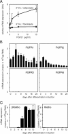
Fig. 1.
Fig. 1.
Gene Expression Differs in 3T3-L1 Fibroblasts and Adipocytes A, FGF21 increases GLUT1 mRNA expression in 3T3-L1 adipocytes but not in fibroblasts. Fourteen days after induction of differentiation, 3T3-L1 fibroblasts or 3T3-L1 adipocytes were treated with the indicated concentrations of FGF21 for 6 h, after which the copy number of the expressed GLUT1 mRNA was measured. Values represent means ± sd and are normalized by the control value (no treatment). Essentially the same results were obtained in three separate experiments. B, Changes in FGFR mRNA expression during adipocyte differentiation of 3T3-L1 cells. 3T3-L1 fibroblasts were seeded into 35-mm dishes and cultured in differentiation-inducing medium. Every other day, the cells in three dishes were separately harvested, total cellular RNAs were prepared, and the copy number of the expressed mRNA encoding each FGFR was measured. Values represent mean ± sd of triplicate samples and are expressed as copy numbers per microgram of total RNA. Essentially the same results were obtained in two separate experiments. C, Expression of βKlotho and Klotho mRNAs during adipocyte differentiation of 3T3-L1 cells. The copy numbers of the expressed mRNA for βKlotho and Klotho were measured as described in the legend to Fig. 2. Values represent mean ± sd of triplicate samples and are expressed as copy numbers per microgram of total RNA. Essentially the same results were obtained in two separate experiments.
Fig. 2.
Fig. 2.
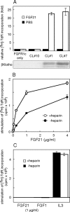
Coexpression of βKlotho Is Required for FGF21 to Stimulate DNA Synthesis in BaF3 Transfectants Expressing FGFR1c A, BaF3 transfectants expressing FGFR1c were further transfected with a βKlotho expression vector, and stable clones showing different levels of βKlotho expression (shown by immunoblotting in the lower panel) were isolated and examined. Each clone and the parental cells were treated with 1 μg/ml FGF21 or PBS (control) in the presence of 10 μg/ml heparin, and DNA synthesis was analyzed based on [3H]thymidine incorporation. The FGF21-induced increases in DNA synthesis were expressed as fold increases from control (PBS). Each columnrepresents the mean ± sd of triplicate samples. Essentially the same results were obtained in two separate experiments. CL, Clone. B, Clone no. 1, which expressed both FGFR1c and βKlotho, was stimulated with the indicated concentration of FGF21 in the presence or absence of 10 μg/ml heparin, and DNA synthesis was analyzed based on [3H]thymidine incorporation, which was calculated by subtracting the value of the untreated (PBS) sample from those of the FGF-treated ones. Each symbol represents the mean ± sd of triplicate samples. Essentially the same results were obtained in three separate experiments. C, BaF3 cells expressing only βKlotho were treated with FGF21 (1 μg/ml), FGF1 (1 μg/ml), or IL-3-containing WEHI-3-conditioned medium (5%) in the presence or absence of 10 μg/ml heparin, and the DNA synthesis was analyzed based on [3H]thymidine incorporation, which was calculated by subtracting the value of untreated (PBS) sample from those of the FGF- and IL-3-treated ones. Each column represents the mean ± sd of triplicate samples.
Fig. 3.
Fig. 3.
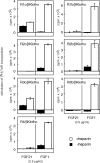
FGFR Specificity of FGF21-Induced DNA Synthesis in βKlotho-Expressing BaF3 Cells BaF3 cells coexpressing βKlotho and one of the FGFR subtypes were isolated as described in the text and stimulated with 0.5 μg/ml FGF21 in the presence or absence of 10 μg/ml heparin, after which DNA synthesis was analyzed based on [3H]thymidine incorporation, which was calculated by subtracting the value of the untreated (PBS) sample from those of the FGF-treated ones. As a positive control, some cells were stimulated with 0.5 μg/ml FGF1. Each column represents the mean ± sd of triplicate samples. Essentially the same results were obtained using at least two independent clones for each FGFR.
Fig. 4.
Fig. 4.
Dose-Dependent Stimulation of DNA Synthesis by FGF21 in FGFR1c/βKlotho/BaF3 and FGFR3c/βKlotho/BaF3 Cells BaF3 cells coexpressing βKlotho and one of the FGFR subtypes were treated with the indicated concentrations of FGF21, FGF19, or FGF1 in the presence of 10 μg/ml heparin. DNA synthesis was assessed based on [3H]thymidine incorporation. The incorporation by untreated cells was subtracted from that by FGF-treated cells to calculate stimulated [3H]thymidine incorporation. These values are presented as percentage of control, which was the stimulated [3H]thymidine incorporation in cells treated with 56 ng/ml FGF1. Each symbol represents the mean ± sd of quadruplicate samples. Essentially the same results were obtained with at least two independent clones for each transfectant.
Fig. 5.
Fig. 5.
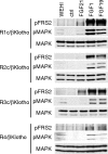
FGF21 Activates FGF Signaling Molecules FRS2α and MAPK in FGFR1c/βKlotho/BaF3 and FGFR3c/βKlotho/BaF3 Cells BaF3 cells coexpressing βKlotho and one of the FGFR subtypes were stimulated with FGF21 (0.56 μg/ml), FGF19 (0.56 μg/ml), or FGF1 (56 ng/ml) in the presence of 10 μg/ml heparin for 10 min. The negative control cells were untreated (ctrl), and the positive control cells were stimulated with 5% WEHI-3-conditioned medium (WEHI). The cells were then harvested, and activation of the signaling molecules was analyzed by Western blotting using phosphospecific antibodies against FRS2α and MAPK. Essentially the same results were obtained in two separate experiments.
Fig. 6.
Fig. 6.
FGF21 Stimulates DNA Synthesis in BaF3 Cells Coexpressing βKlotho and FGFR1c But Not Klotho and FGFR1c BaF3 transfectants coexpressing FGFR1c and βKlotho (R1c/βKlotho) or Klotho (R1c/Klotho) were stimulated with 1 μg/ml FGF21 or FGF23 in the presence or absence of 10 μg/ml heparin, after which DNA synthesis was analyzed based on [3H]thymidine incorporation, which was calculated by subtracting the value of the untreated (PBS) sample from those of the FGF-treated ones. Each column represents the mean ± sd of triplicate samples. Essentially the same results were obtained in three separate experiments.
Fig. 7.
Fig. 7.
FGF21 Activates the FGF Signaling Molecules FRS2α and MAPK in 3T3-L1 Fibroblasts Expressing βKlotho 3T3-L1 fibroblasts stably expressing βKlotho (βKlotho/3T3-L1 fibroblasts) were serum starved in OptiMEM-I medium for 18 h, incubated with or without 10 μm SU5402 for 3 h, and then stimulated with 1.1 or 4.5 μg/ml FGF21 for 15 min. The cells were then harvested, and activation of the signaling molecules was analyzed using phosphospecific antibodies against FRS2α and MAPK. FGF1 (18 ng/ml) and PBS served as positive and negative controls, respectively. Essentially the same results were obtained in two separate experiments.
Fig. 8.
Fig. 8.
FGF21 Increases Expression of GLUT1 mRNA in 3T3-L1 Fibroblasts Expressing βKlotho 3T3-L1 fibroblasts stably overexpressing βKlotho (βKlotho/3T3-L1 fibroblasts) were treated with the indicated concentrations of FGF21 for 6 h, after which the copy number of the expressed GLUT1 mRNA was measured by quantitative real-time PCR. Values represent the mean ± sd of quadruplicate samples and are normalized by the control value (no treatment). *, P < 0.05; **, P < 0.01 (Student’s t test). Essentially the same results were obtained in two separate experiments.

Similar articles

Cited by

References

    1. Ornitz DM, Itoh N 2001. Fibroblast growth factors. Genome Biol 2:REVIEWS3005 - PMC - PubMed
    1. Itoh N, Ornitz DM 2004. Evolution of the Fgf and Fgfr gene families. Trends Genet 20:563–569 - PubMed
    1. Yu X, White KE 2005. FGF23 and disorders of phosphate homeostasis. Cytokine Growth Factor Rev 16:221–232 - PubMed
    1. Inagaki T, Choi M, Moschetta A, Peng L, Cummins CL, McDonald JG, Luo G, Jones SA, Goodwin B, Richardson JA, Gerard RD, Repa JJ, Mangelsdorf DJ, Kliewer SA 2005. Fibroblast growth factor 15 functions as an enterohepatic signal to regulate bile acid homeostasis. Cell Metab 2:217–225 - PubMed
    1. Nishimura T, Nakatake Y, Konishi M, Itoh N 2000. Identification of a novel FGF, FGF-21, preferentially expressed in the liver. Biochim Biophys Acta 1492:203–206 - PubMed

Publication types

MeSH terms

Substances