. 2008 Jan 10;57(1):27-40.
doi: 10.1016/j.neuron.2007.11.025.

Suppression of neurodegeneration and increased neurotransmission caused by expanded full-length huntingtin accumulating in the cytoplasm

Affiliations

Suppression of neurodegeneration and increased neurotransmission caused by expanded full-length huntingtin accumulating in the cytoplasm

Eliana Romero et al. Neuron. .

Abstract

Huntington's disease (HD) is a dominantly inherited neurodegenerative disorder caused by expansion of a translated CAG repeat in the N terminus of the huntingtin (htt) protein. Here we describe the generation and characterization of a full-length HD Drosophila model to reveal a previously unknown disease mechanism that occurs early in the course of pathogenesis, before expanded htt is imported into the nucleus in detectable amounts. We find that expanded full-length htt (128Qhtt(FL)) leads to behavioral, neurodegenerative, and electrophysiological phenotypes. These phenotypes are caused by a Ca2+-dependent increase in neurotransmitter release efficiency in 128Qhtt(FL) animals. Partial loss of function in synaptic transmission (syntaxin, Snap, Rop) and voltage-gated Ca2+ channel genes suppresses both the electrophysiological and the neurodegenerative phenotypes. Thus, our data indicate that increased neurotransmission is at the root of neuronal degeneration caused by expanded full-length htt during early stages of pathogenesis.


Figures
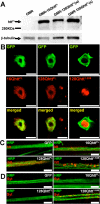
Figure 1
Figure 1. Human full-length huntingtin accumulates in the cytoplasm of Drosophila neurons and does not form visible axonal aggregates
(A) Western blot analysis of transgenic Drosophila heads using MAB2166 huntingtin-specific antibody reveals a (∼350KDa) band corresponding to full-length (unexpanded and expanded) human huntingtin. Based on densitometry analysis, the 16Q line expresses higher/similar protein levels than the 128Q(w) and 128Q(s) lines (∼2-fold and ∼1-fold, respectively). Genotypes: GMR (GMR-GAL4/UAS-GFP), GMR-16QhttFL (GMR-GAL4/UAS-16QhttFL[M28]), GMR-128QhttFL(w) (GMR-GAL4/+; UAS-128Qhtt(w)[F7]/+), GMR-128QhttFL(s) (GMR-GAL4/UAS-128Qhtt(s)[M36E2]) (B) Immunofluorescence confocal images of Drosophila apterous ventral nerve cord interneurons from 20-day-old flies expressing wild-type (left, 16QhttFL) or expanded (middle, 128QhttFL) full-length huntingtin. Right panels show similar images from animals expressing an expanded N-terminal huntingtin truncation (amino acids 1−208 excluding the polyglutamine tract, 128Qhtt1−208). Note co-localization of full-length huntingtin with the CD8-GFP cytoplasmic marker. The same results were obtained with 10-day-old and 30-day-old flies (data not shown). In contrast, the truncated version of huntingtin accumulates in the nucleus as early as the third-instar larval stage. MAB5374 was used at 1:100 for all stainings, which were done simultaneously. Flies raised at 27°C. Scale bar= 5μm. Genotypes: 16QhttFL (UAS-16QhttFL[M28]/UAS-CD8-GFP; apVNC-GAL4/+), 128QhttFL (UAS-128QhttFL(s)[M36E2]/UAS-CD8-GFP; apVNC-GAL4/+), 128Qhtt1−208 (UAS-CD8-GFP/+; UAS-128Qhtt1−208[M64]/apVNC-GAL4). (C-D) Immunolabeling of huntingtin (htt) (C) or endogenous synaptotagmin I (Syt) (D) proteins in motor neuron axon bundles from third instar larvae of the genotype indicated in each panel. Note diffuse pattern and absence of aggregates in wild-type (16QhttFL) and expanded full-length huntingtin (128QhttFL) axons. In contrast, a long N-terminal huntingtin truncation (amino acids 1−548, 128Qhtt1−548) causes large huntingtin and synaptotagmin I axonal aggregates. MAB5374 1:100. Flies were at 29°C. Scale bar= 5μm. Genotypes: GFP (Elav-GAL4/+; UAS-GFP/+), 16QhttFL (Elav-GAL4/+; UAS-16QhttFL[M28]/+), 128QhttFL (Elav-GAL4/+; UAS-128QhttFL(s)[M36E2]/+), 128Qhtt1−548 (Elav-GAL4/+; UAS-128QHtt1−548/+).
Figure 2
Figure 2. Progressive neurodegenerative eye phenotype produced by overexpression of expanded (128Q), but not unexpanded (16Q) full-length huntingtin
(A-D) Light microscope (left), and SEM (center) images of transgenic flies expressing (A) non-toxic LacZ control protein, (B) wild-type huntingtin, (C) low levels of expanded huntingtin, and (D) high levels of expanded huntingtin. Insets in SEM images show magnification (600×) of the ommatidia field. Note disorganization of ommatidia in D. Right panels show phalloidin staining of the corresponding eyes from 20-day-old flies showing arrangement of rhabdomeres. Flies for light microscope and SEM images were raised at 25°C. Flies for phalloidin staining were raised at 27°C. Genotypes for light microscope and SEM: GMR-lacZ (GMR-GAL4(s)/UAS-lacZ), GMR-16QhttFL (GMR-GAL4(s)/UAS-16QhttFL[M28]), GMR-128QhttFL(w) (GMR-GAL4(s)/+; UAS-128QhttFL(w)[F7]/+), GMR-128QhttFL (GMR-GAL4(s)/UAS-128QhttFL(s)[M36E2]). Genotypes for phalloidin staining: GMR-lacZ (GMR-GAL4/UAS-lacZ), GMR-16QhttFL (GMR-GAL4/UAS-16QhttFL[M28]), GMR-128QhttFL (GMR-GAL4/+; UAS-128QhttFL(w)[F7]), GMR-128QhttFL (GMR-GAL4/UAS-128QhttFL[M36E2]). (E-F) Quantification of the number of rhabdomeres per ommatidium in 1-day-old (E) or 20-day-old (F) flies expressing the following proteins. Green: non-toxic LacZ control, yellow: wild-type huntingtin, red: expanded huntingtin at relatively low levels, and black: expanded huntingtin at relatively high levels. Flies raised at 27°C. n=100 ommatidia per genotype. The distribution of the rhabdomeres at day 20 for 128QhttFL(w) and 128QhttFL(s) is significantly different from LacZ and 16QhttFL controls (p<0.001, Mann-Whitney test). Genotypes: GMR-lacZ (GMR-GAL4/UAS-lacZ), GMR-16QhttFL (GMR-GAL4/UAS-16QhttFL[M28]), GMR-128QhttFL(w) (GMR-GAL4/+; UAS-128QhttFL(w)[F7]), GMR-128QhttFL(s) (GMR-GAL4/UAS-128QhttFL(s)[M36E2]).
Figure 3
Figure 3. Reduced survival, impaired motor performance, and neuronal degeneration in Drosophila expressing expanded full-length huntingtin in the CNS
(A) Average survival of flies expressing GFP (dark bars) or 128QhttFL (light bars) in the CNS (C164-GAL4) at days 25, 30 and 35. The survival rate of flies expressing 128QhttFL is significantly lower than the survival rate of control flies expressing a non-toxic GFP protein (p<0.05, 0.01 and 0.01, respectively, Mann-Whitney test). Dots denote single data points for individual populations. Error bars= SEM. n= 7 and 10 populations for GFP and 128QhttFL respectively. (B) Climbing performance as a function of age in control and 128QhttFL-expressing flies. Normal decline in climbing performance is observed after day 25 in flies expressing the non-toxic GFP protein (silver line). In contrast, flies expressing 128QhttFL (black lines) perform poorly after day 20. All flies raised at 27°C. Two independent experiments are shown for flies expressing 128QhttFL. Error bars= SEM of 10 trials per time point. (C) Flying ability in 25-day-old control and huntingtin-expressing flies. Flying ability is represented in arbitrary units with 12 being a perfect ability and 0 being no ability (see Experimental Procedures). Note that most control flies that carry only the motor neuron driver (silver line) or express wild-type huntingtin (16QhttFL, grey dotted lines) perform well in this assay with no significant difference between them (p>0.1, Mann-Whitney test). Most flies expressing expanded huntingtin (128QhttFL, black lines), on the other hand, show impaired flying ability when compared to either control (p<0.05, Mann-Whitney test). (D) Stacks of confocal images of neurons projecting into indirect flight muscles (IFM) 3 and 4 of 10-day (upper panels) and 25-day (lower panels) old flies expressing wild-type (left) or expanded (right) huntingtin. Note the loss of neuronal projections and NMJs in flies expressing expanded huntingtin. Scale bar=10μm. (E) Quantification of the number of neuronal projections in a specified 100μm × 100μm area of IFM 3/ 4 shown in D. 20-day and 25-day, but not 10-day old flies expressing 128QhttFL have significantly fewer neuronal projections than control flies of the same age expressing wild-type 16QhttFL protein (ns: p>0,05; *: p<0.05, Student-t test; n=5). All flies raised at 27°C. Genotypes: C164 (C164-GAL4/+), C164-GFP (C164-GAL4/UAS-GFP), C164−16QhttFL (C164-GAL4/UAS-16QhttFL[M28]), C164−128QhttFL (C164-GAL4/UAS-128QhttFL(s)[M36E2]).
Figure 4
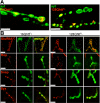
Figure 4. Uneven distribution of full-length huntingtin across boutons does not affect distribution of key synaptic proteins
(A) Immunofluorescence confocal images of neuromuscular junctions from third-instar larvae expressing wild-type (left) or expanded (right) huntingtin reveal its uneven distribution from one bouton to another within individual axons. Boutons are visualized with anti-synaptotagmin antibody (green) and huntingtin is labeled in red. Scale bar= 10μm. (B) Immunofluorescence confocal images of neuromuscular junctions from third-instar larvae expressing wild-type (left) or expanded (right) huntingtin show morphologically normal boutons with no abnormal distribution of proteins involved in neurotransmitter secretion. Boutons are visualized with anti-dlg (green) and stained for (from top to bottom) GluRIIA, Rop, Snap and Syx (red). No differences are detected in the patterns of accumulation of any of these proteins in boutons from larvae expressing expanded huntingtin or boutons from control larvae expressing wild-type huntingtin (p>0.5, 0.1, 0.1 and 0.1, respectively). Scale bar= 5μm. All larvae raised at 29°C. Genotypes: 16QhttFL (Elav-GAL4/+; UAS-16QhttFL(s)[M28]/+), 128QhttFL (Elav-GAL4/+; UAS-128QhttFL(s)[M36E2]/+).
Figure 5
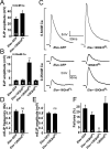
Figure 5. Neurotransmitter release probability is increased upon expression of full-length expanded huntingtin
(A-B) Quantification of EJP amplitudes recorded at (A) 1Hz in 0.6mM Ca2+ or (B) 0.25mM Ca2+ in Drosophila larvae expressing a non-toxic control protein (GFP), wild-type or expanded full-length huntingtin or controls carrying the huntingtin transgene without a GAL4 driver. ns: p>0.05; *p<0.05; **p<0.01 throughout the figure. (C) Sample EJP traces recorded from Drosophila larvae expressing GFP control protein and expanded full-length huntingtin in HL3 buffer at 0.6mM Ca2+ (top) or 0.25mM Ca2+ (bottom). For 0.25mM Ca2+, sample traces are also shown for transgenic larvae expressing wild-type huntingtin and for controls carrying the expanded huntingtin transgene without a GAL4 driver. Arrows indicate blanked-out stimulus artifact. (D-E) Average (D) frequency and (E) amplitude of mEJPs recorded from abdominal muscle 6 or 7 in GFP control and larvae expressing expanded huntingtin. Error bars: SEM. Number inside bars is the number of animals studied. (F) Percent failures measured at 1Hz in 0.25mM Ca2+ following stimulation at 2−3× threshold of motor neurons in larvae expressing a non-toxic GFP protein, wild-type huntingtin, expanded huntingtin or controls carrying the huntingtin transgene without a GAL4 driver. All experiments done at 29°C. Genotypes: Elav-GFP (Elav-GAL4/+; UAS-GFP/+), Elav-16QhttFL (Elav-GAL4/+; UAS-16QhttFL[M28]/+), 128QhttFL (UAS-128QhttFL(s)[M36E2]/+), Elav-128QhttFL (Elav-GAL4/+; UAS-128QhttFL(s)[M36E2]/+).
Figure 6
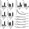
Figure 6. Heterozygous mutations in genes affecting secretion suppress the increased release probability caused by full-length expanded huntingtin
(A-H) Quantification of EJP amplitudes (A, C, E and G) and percent failures (B, D, F and H) in larvae expressing a non-toxic control protein GFP, heterozygous mutant for (A,B) Snap, (C,D) Rop, (E,F) Syntaxin 1A, or (G, H) Vha100kDa, expressing expanded huntingtin or expressing expanded huntingtin as well as heterozygous mutant for either (A,B) Snap, (C,D) Rop, (E,F) Syntaxin 1A, or (G,H) Vha100−1 at 29°C. Error bars: SEM. Number inside bars is the number of animals studied. ns, p>0.05; *, p<0.05; **, p<0.01. Experiments done at 29°C. (I-N) Sample EJP traces (5 consecutive traces recorded at 1Hz interval) recorded from (I) larvae expressing expanded huntingtin, (J) the control protein, GFP, (K) heterozygous mutant for Snap, (L) expressing expanded huntingtin and heterozygous mutant for Snap, (M) heterozygous mutant for Rop and (N) expressing expanded huntingtin and heterozygous mutant for Rop. Arrows indicate blanked-out stimulus artifact. All experiments done at 29°C. All recordings at 1Hz in HL3 buffer at 0.25mM Ca2+. Genotypes: GFP (Elav-GAL4/+; UAS-GFP/+), 128QhttFL (Elav-GAL4/+; UAS-128QhttFL(s)[M36E2]/+), SnapM4/+ (Elav-GAL4/+; SnapM4/+), 128QhttFL/SnapM4 (Elav-GAL4/+; UAS-128QhttFL(s)[M36E2]/+; SnapM4/+), Rop27/+ (Elav-GAL4/+; RopG27/+), 128QhttFL/RopG27 (Elav-Gal4/+; UAS-128QhttFL(s)[M36E2]/+; RopG27/+), Syx229/+ (Elav-GAL4/+; Syx229/+), 128QhttFL/Syx229 (Elav-Gal4/+; UAS-128QhttFL(s)[M36E2]/+; Syx229/+), vha1/+ (Elav-GAL4/+; vha1/+), 128QhttFL/vha1 (Elav-GAL4/+; UAS-128QhttFL(s)[M36E2]/+; vha1/+).
Figure 7
Figure 7. Heterozygous mutations in genes affecting neurotransmitter secretion also suppress neurodegeneration and motor impairments in animals expressing expanded full-length huntingtin
(A) Number of rhabdomeres per ommatidium in 20-day-old flies of the indicated genotypes. The distribution of the rhabdomeres is significantly different (p<0.001, Mann-Whitney test) between 128QhttFL and 128QhttFL animals also carrying one mutant copy of SNAP, Syx, Rop, or Dmca1D. Flies grown at 27°C. Genotypes: 128QhttFL (GMR-GAL4/+; UAS-128QhttFL(w)[F7]), 128QhttFL/SnapM4 (GMR-GAL4/+; UAS-128QhttFL(w)[F7]/SnapM4), 128QhttFL/Syx229 (GMR-GAL4/+; UAS-128QhttFL(w)[F7]/Syx229), 128QhttFL/RopG27 (GMR-GAL4/+; UAS-128QhttFL(w)[F7]/RopG27), 128QhttFL/Dmca1DX10 (GMR-GAL4/Dmca1DX10; UAS-128QhttFL(w)[F7]/+), 128QhttFL/vha1 (GMR-GAL4/+; UAS-128QhttFL(w)[F7]/vha1), GFP (GMR-GAL4/ UAS-GFP). (B-H) Phalloidin staining of dissected retinas from 20-day-old flies of the genotypes indicated in each panel. Scale bar= 5μM. Flies grown at 27°C Genotypes in order: (B)128QhttFL (GMR-GAL4/+; UAS-128QhttFL(w)[F7]/+), (C)128QhttFL/SnapM4 (GMR-GAL4/+; UAS-128QhttFL(w)[F7]/SnapM4), (D) 128QhttFL/Syx229 (GMR-GAL4/+; UAS-128QhttFL(w)[F7]/Syx229), (E) 128QhttFL/RopG27 (GMR-GAL4/+; UAS-128QhttFL(w)[F7]/RopG27), (F) 128QhttFL/Dmca1DX10 (GMR-GAL4/Dmca1DX10; UAS-128QhttFL(w)[F7]/+),.(G) 128QhttFL/Vha1 (GMR-GAL4/+; UAS-128QhttFL(w)[F7]/Vha1), (H) GFP (GMR-GAL4/UAS-GFP). (I) Suppression of climbing performance phenotype in flies expressing 128QhttFL that also carry a heterozygous loss-of-function mutation in Syx. Flies expressing the non-toxic GFP protein or 16QhttFL show normal decline in climbing performance with age. Climbing performance impairment occurs prematurely in flies expressing 128QhttFL, but it is restored to almost normal levels in a background heterozygous mutant for Syx. Error bars= SEM of 10 trials per time point. Flies grown at 25°C. Genotypes: GFP (C164-GAL4/UAS-GFP), 16QhttFL (C164-GAL4/UAS-16QhttFL[M28]), 128QhttFL (C164-GAL4/UAS-128QhttFL(s)[M36E2]), 128QhttFL/Syx229 (C164-GAL4/UAS-128QhttFL(s)[M36E2]; Syx229/+). (J-L) SEM eye images of flies expressing (J) the non-toxic control protein GFP, (K) 128QhttFL (L) 128QhttFL and heterozygous mutant for Syx. Note partial suppression of the disorganized ommatidia phenotype in Syx heterozygous mutant animals. Genotypes: lacZ (GMR-GAL4(s)/UAS-GFP), 128QhttFL (GMR-GAL4(s)/UAS-128QhttFL(s)[M36E2]), 128QhttFL(w)/Syx229 (GMR-GAL4(s)/UAS-128QhttFL(s)[M36E2]/+; Syx229/+). Flies grown at 25°C.
Figure 8
Figure 8. Increased Ca2+ levels caused by expanded full-length huntingtin can be suppressed genetically
(A) Fluorescence ratios (F340/F380) measured from fura dextran-filled larval NMJs. Neuronal expression of 128QhttFL and 16QhttFL leads to increased fluorescence ratios compared to controls (GFP), indicating elevated resting Ca2+ levels at these synapses (* denotes p< 0.05, t-test). These elevated fluorescence ratios are restored to control levels in 128QhttFL animals that are also heterozygous for either Syntaxin or Dmca1D mutations (* denotes p< 0.05, t-test). Genotypes: Elav-Gal4/+; UAS-128QhttFL(s)[M36E2]/+; Syx229/+ and Elav-GAL4/+; UAS-128QhttFL(s)[M36E2]/Dmca1DX10. Note that animals carrying Syx or Dmca1D heterozygous mutations but no huntingtin transgene show no effect on resting Ca2+ levels compared to GFP controls. Genotypes: Elav-GAL4/+; Syx229/+ and Elav-GAL4/+; Dmca1DX10/+. Boutons were measured from 2−3 synapses in 6 larvae per genotype. Dots denote single data points from individual boutons. (B) Fluorescence from boutons forward-filled with fura dextran and excited at both 340nm and 380nm. Ca2+ -free dye absorbs optimally at 380nm while Ca2+-bound dye is excited primarily at 340nm. NMJ synapses loaded with fura dextran were clearly visualized (top panel) and individual boutons could be spatially resolved (dashed box). In contrast to controls (Elav-GAL4/+; UAS-GFP/+), NMJ boutons from larvae expressing expanded huntingtin (Elav-GAL4/+; UAS-128QhttFL(s)[M36E2]/+) emit more intensely when excited at 340nm compared to 380nm. However, Syx heterozygosity (Elav-Gal4/+; UAS-128QhttFL(s)[M36E2]/+; Syx229/+) alleviates this phenotype. (C-D) Quantification of EJP amplitudes and percent failures recorded at 1Hz in 0.25mM Ca2+ in larvae expressing a non-toxic GFP control protein, expressing 128QhttFL, heterozygous mutant for Dmca1D (C), Dmca1A (D), or expressing 128QhttFL as well as heterozygous mutant for either Dmca1D (C) or Dmca1A (D). All experiments at done at 29°C. Error bars: SEM. The number of recordings from at least 3 animals is indicated inside the bars. ns, p>0.05; *, p<0.05; **, p<0.01 throughout the figure. Genotypes: GFP (Elav-GAL4/+; UAS-GFP/+), 16QhttFL (Elav-GAL4/+; UAS-16QhttFL(s)[M28]/+), 128QhttFL (Elav-GAL4/+; UAS-128QhttFL(s)[M36E2]/+), Syx229/+ (Elav-GAL4/+; Syx229/+), 128QhttFL/Syx229 (Elav-Gal4/+; UAS-128QhttFL(s)[M36E2]/+; Syx229/+), Dmca1DX10/+ (Elav-GAL4/+; Dmca1DX10/+), 128QhttFL/Dmca1DX10 (Elav-GAL4/+; UAS-128QhttFL(s)[M36E2]/Dmca1DX10), Dmca1AHC129/+ (Elav-GAL4/Dmca1AHC129), 128QhttFL/Dmca1AHC129 (Elav-GAL4/Dmca1AHC129; UAS-128QhttFL(s)[M36E2]/+).

Similar articles

Cited by

References

    1. The Huntington's Disease Collaborative Research Group A novel gene containing a trinucleotide repeat that is expanded and unstable on Huntington's disease chromosomes. Cell. 1993;72:971–983. - PubMed
    1. Auluck PK, Chan HY, Trojanowski JQ, Lee VM, Bonini NM. Chaperone suppression of alpha-synuclein toxicity in a Drosophila model for Parkinson's disease. Science. 2002;295:865–868. - PubMed
    1. Bezprozvanny I, Hayden MR. Deranged neuronal calcium signaling and Huntington disease. Biochem Biophys Res Commun. 2004;322:1310–1317. - PubMed
    1. Bilen J, Bonini NM. Drosophila as a model for human neurodegenerative disease. Annu Rev Genet. 2005;39:153–171. - PubMed
    1. Brand AH, Perrimon N. Targeted gene expression as a means of altering cell fates and generating dominant phenotypes. Development. 1993;118:401–415. - PubMed

Publication types

MeSH terms

LinkOut - more resources