. 2008 Jan 9;3(1):e1409.
doi: 10.1371/journal.pone.0001409.

A potential new pathway for Staphylococcus aureus dissemination: the silent survival of S. aureus phagocytosed by human monocyte-derived macrophages

Affiliations

A potential new pathway for Staphylococcus aureus dissemination: the silent survival of S. aureus phagocytosed by human monocyte-derived macrophages

Malgorzata Kubica et al. PLoS One. .

Abstract

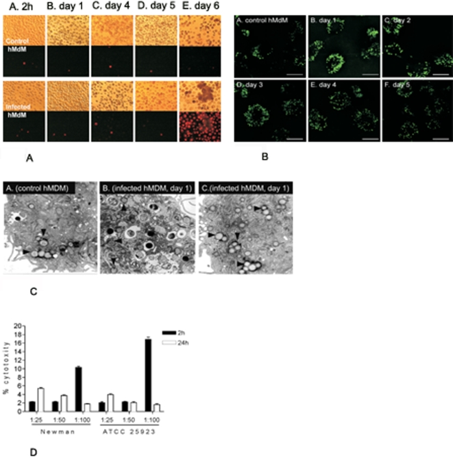
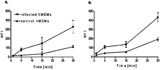
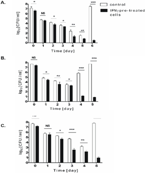
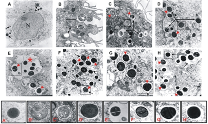
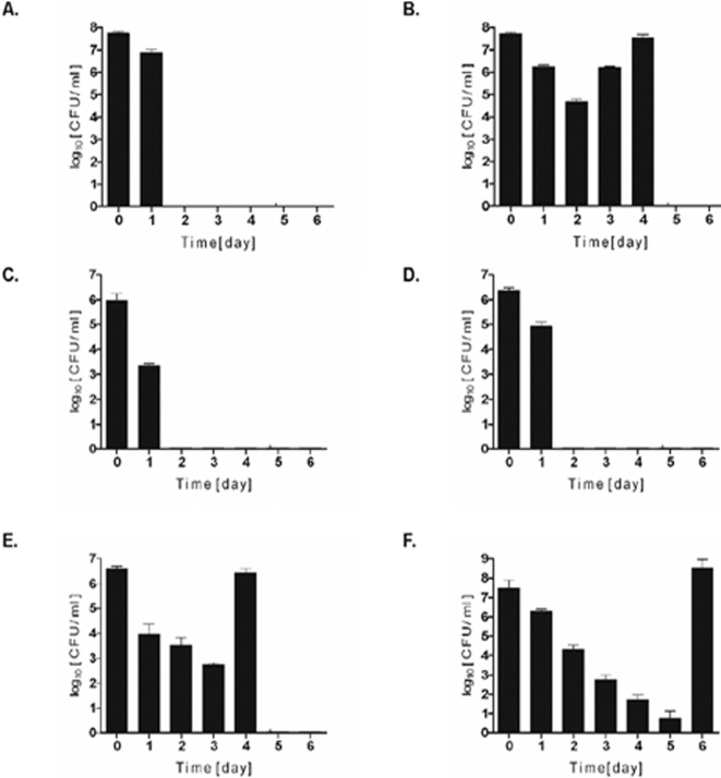
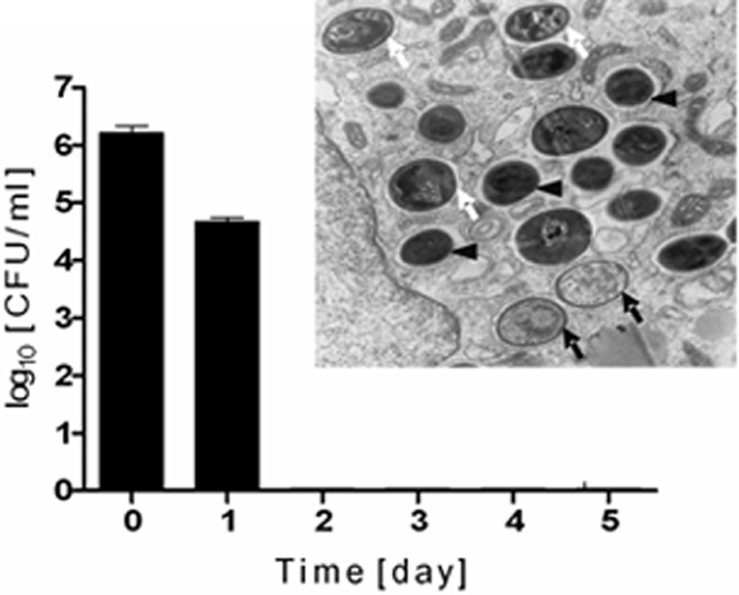
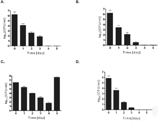
Although considered to be an extracellular pathogen, Staphylococcus aureus is able to invade a variety of mammalian, non-professional phagocytes and can also survive engulfment by professional phagocytes such as neutrophils and monocytes. In both of these cell types S. aureus promptly escapes from the endosomes/phagosomes and proliferates within the cytoplasm, which quickly leads to host cell death. In this report we show that S. aureus interacted with human monocyte-derived macrophages in a very different way to those of other mammalian cells. Upon phagocytosis by macrophages, S. aureus persisted intracellularly in vacuoles for 3-4 days before escaping into the cytoplasm and causing host cell lysis. Until the point of host cell lysis the infected macrophages showed no signs of apoptosis or necrosis and were functional. They were able to eliminate intracellular staphylococci if prestimulated with interferon-gamma at concentrations equivalent to human therapeutic doses. S. aureus survival was dependent on the alternative sigma factor B as well as the global regulator agr, but not SarA. Furthermore, isogenic mutants deficient in alpha-toxin, the metalloprotease aureolysin, protein A, and sortase A were efficiently killed by macrophages upon phagocytosis, although with different kinetics. In particular alpha-toxin was a key effector molecule that was essential for S. aureus intracellular survival in macrophages. Together, our data indicate that the ability of S. aureus to survive phagocytosis by macrophages is determined by multiple virulence factors in a way that differs considerably from its interactions with other cell types. S. aureus persists inside macrophages for several days without affecting the viability of these mobile cells which may serve as vehicles for the dissemination of infection.


Conflict of interest statement

Competing Interests: The authors have declared that no competing interests exist.

Figures

Figure 1
Figure 1. Internalization of S. aureus strain Newman does not affect hMDM viability until the plasma membrane is permeabilized.
A. Transmission light (upper two rows) and fluorescence (lower two rows) micrographs (x 40) of control (upper panel) and S. aureus-infected cells (bottom panel) maintained in culture for 2h (A) and 1 day (B), 4- (C), 5- (D), and 6-days (E). Propidium iodide-positive cells represent infected host cells with leaky plasma membranes. B. BODIPY495/503 staining of lipid droplets of control and S. aureus infected hMDM cultures on five consecutive days post-phagocytosis. Cells were permeabilized with 0.2 % Triton X-100 and stained as described in the Materials and Methods section. All scale bars = 10 µm. C. Transmission electron micrographs of control (panel A) and infected cells (panels B and C) one day post-phagocytosis. Black arrowheads point to lipid droplets. Magnification: x10,000 (A and C) and ×7,500 (B). The photographs presented are representative of a minimum of 20 fields observed. D. Cytotoxicity (%) of S. aureus infection. Plasma membrane permeabilization or cell lysis induced in macrophage cultures by S. aureus infection at different MOI was determined as LDH activity levels. Cytotoxicity was calculated according to the formula: % cytotoxicity = [(experimental value–low control)/(high control–low control)] × 100, where a low control is the LDH activity in the conditioned medium of the control non-infected culture, while the LDH activity in the whole cell culture with cells lysed with detergent (2% Triton X-100) constitutes a high control. An experimental value was the activity in the conditioned medium from the culture infected with S. aureus. According to this calculation the control non-infected culture was assumed to show 0% cytotoxicity. All assays were performed in triplicate.
Figure 2
Figure 2. S. aureus infected macrophages retained their bactericidal functions.
Control hMDMs, and cells 4 days post infection with S. aureus strain Newman, were stimulated to phagocytose either live bacteria at a MOI of 1∶25 (1.25×10⁷ CFU) (panel A) or latex beads (panel B). Generation of reactive oxygen species (ROS) determined as the level of the mean fluorescence intensity (MFI) was measured at various time intervals. The data shown is representative of at least three separate experiments, performed in triplicate, using hMDMs derived from different donors.
Figure 3
Figure 3. After being phagocytosed by hMDM S. aureus strain Newman decreasingly persists intracellularly for several days until a burst of growth on day 6.
A. CFU of S. aureus in cell lysates and culture medium on six consecutive days post-infection. Macrophages were allowed to engulf S. aureus at a MOI of 25 for 2 h, washed, and extracellular bacteria killed by gentamycin. Macrophages were cultured in media without antibiotics. At consecutive days post-phagocytosis media was aspired and hMDMs were lysed. Both conditioned media and cell lysates were plated onto TSA for CFU enumeration. The data shown details of one representative experiment (means±SD) of the 76 we performed in triplicate. B. CFU of S. aureus and LDH activity levels (A490 nm) in the conditioned culture medium on six consecutive days post infection (left panel); and transmission (upper row) and fluorescent (propidium iodide staining, lower row) light micrographs (x40) of S. aureus-infected cells maintained for the indicated time interval post infection. C. Confocal fluorescence microscopy images of hMDMs after 1, 3, and 5 days post-infection at MOI of 25. Cells were fixed, treated with RNase and stained with acridine orange (see Experimental Procedures section). Internalized bacteria are observed as green spots. All scale bars = 10 µm. D. Confocal fluorescence microscopy images of viable bacteria in hMDMs on the 5th day post-phagocytosis. Infected hMDMs were collected, permeabilized with 0.2 % Triton X-100 and double- labeled with propidium iodide and Syto9 (LIVE/DEAD BacLight Kit). Viable S. aureus cells are stained green while red signals represent dead bacteria and the host cell's nuclear DNA stained mainly with propidium. Scale bar = 7.5 µm. E. Specific PCR amplification of the S. aureus 16S rRNA gene (left panel) from cell lysates and the MLVF pattern (right panel) of bacteria infecting cells on consecutive days post-phagocytosis (numbers above lanes). Lane 0, sample collected immediately upon completion of phagocytosis (2h). Lane N, the MLVF pattern of S. aureus Newman before infection.
Figure 4
Figure 4. Macrophages stimulated with interferon-γ kill engulfed S. aureus strains: Newman (A), ATCC 25923 (B), and COL (C) more efficiently than non-stimulated cells.
Cells were stimulated with recombinant human IFNγ overnight at concentrations equivalent to human therapeutic doses (100 U ml−1), and then allowed to phagocytose three different strains of S. aureus for 2 h. Infected cultures were processed as described in the legend for Fig. 3A and live bacteria in the whole cultures (CFU) were enumerated up to 14 days postphagocytosis. Since at the longer timepoints no bacterial growth was detected (CFU = 0) these points were not presented on the graph. The data shown is representative of at least three separate experiments, performed in triplicate, using hMDM derived from different donors. Bars represent mean CFU value ±SD. *, p<0.05; **, p<0.01; ***, p<0.001. NS-not significant.
Figure 5
Figure 5. Apparently structurally normal, intact dividing and viable S. aureus Newman cells persist in the intracellular vacuolar compartment on four consecutive days post-phagocytosis.
The photographs presented are representative of a minimum of 20 fields observed. hMDMs were allowed to engulf bacteria for 2h at a MOI of 25, and infected macrophages were fixed with Karnovsky's fixative either immediately following phagocytosis (2 h), or on consecutive days post-phagocytosis, before being processed by standard electron microscopic techniques. At any given point post-phagocytosis dividing bacterial cells were visible (arrowheads). Magnified views of selected bacteria framed on main micrographs are shown in the bottom row labeled from A' to H', respectively. A. Control, non-infected hMDM has a morphological appearance typical for that of professional phagocyte, with vesicles localized around the nucleus (N) and only few lipid droplets (arrows). Magnification: x14,000. B. Immediately post-phagocytosis (2h), the bacteria were located predominantly in very tight membranous compartments (arrows). Magnification: x50,000. C. Already 1 day post-phagocytosis the majority of S. aureus cells were found in clearly defined membrane-enclosed vacuoles (arrows). A partially degraded bacterium in the vacuole is framed. Magnification: x20,000. D and E. At day 2 the cells of S. aureus can only be found in well defined vacuoles (arrows), occasionally spacious vacuoles (asterisk) containing several bacteria. Magnification: x32,500. F. On day 3 S. aureus persists in vacuoles which often reveal signs of partial membrane discontinuity (arrows). Magnification: x32,500. G and H. On day 4, the majority of bacteria were found in vacuoles with profoundly damaged or fully disintegrated membranes (arrow). Magnification: x54,000 (G) and x32,000 (H).
Figure 6
Figure 6. Functional sigB and agr operons, but not sarA, are indispensable for S. aureus to survive phagocytosis by hMDMs.
Macrophages were allowed to engulf defined strains of S. aureus for 2h at a MOI of 25, and the intracellular survival of bacteria on consecutive days post-phagocytosis was monitored by enumeration of the CFU of cell lysates (see Fig. 3A legend). The data shown is representative of at least three separate experiments, performed in triplicate, using hMDMs derived from different donors. Bars represent mean CFU value ±SD. A. 8325-4, a natural rsbU defective strain lacking a functional SigB. B. SH1000, a derivative of strain 8325-4 with a restored rsbU gene and SigB activity. C. Newman sigB mutant D. Newman agr mutant E. Newman sarA mutant F. Newman wild-type
Figure 7
Figure 7. An α-hemolysin mutant of S. aureus strain Newman is efficiently killed in phago(lyso)somes within two days of phagocytosis.
Intracellular persistence of S. aureus was determined as described in the legend of Fig. 3A. Bars represent mean CFU values ±SD and are representative of at least three separate experiments, performed in triplicate, using hMDMs derived from different donors. An electron microphotograph of infected hMDMs was obtained 1 day post-phagocytosis and shows partially (white arrows) and totally degraded (black arrows) bacterial cells inside tight vacuoles. At this time point some apparently intact bacterial cells are also present (black arrowheads). Magnification: x32,500. The photograph presented is a representative of a minimum of 20 fields observed.
Figure 8
Figure 8. Metalloprotease (Aur), protein A, and MSCRAMs, but not the V8 protease (SspA) and staphopain B (SspB), contribute to S. aureus intracellular persistence in infected hMDMs.
Intracellular persistence of S. aureus was determined as described in the legend of Fig. 3A. Bars represent mean CFU value ±SD and are representative of at least three separate experiments, performed in triplicate, using hMDMs derived from different donors. A. Newman aur mutant B. Newman srtA mutant C. Newman ssp operon mutant D. Newman spA mutant

Similar articles

Cited by

References

    1. Archer GL. Staphylococcus aureus: A well-armed pathogen. Clin Infect Dis. 1998;26:1179–1181. - PubMed
    1. Lowy FD. Staphylococcus aureus infections. New Engl J Med. 1998;339:520–532. - PubMed
    1. Zetola N, Francis JS, Nuermberger EL, Bishai WR. Community-acquired meticillin-resistant: an emerging threat. Lancet Infect Dis. 2005;5:275–286. - PubMed
    1. Foster TJ, Hook M. Surface protein adhesins of Staphylococcus aureus. Trends Microbiol. 1998;6:484–488. - PubMed
    1. Foster TJ. Immune evasion by staphylococci. Nat Rev Microbiol. 2005;3:948–958. - PubMed

Publication types