Tuning lipid mixtures to induce or suppress domain formation across leaflets of unsupported asymmetric bilayers
- PMID: 18172219
- PMCID: PMC2224171
- DOI: 10.1073/pnas.0702970105
Tuning lipid mixtures to induce or suppress domain formation across leaflets of unsupported asymmetric bilayers
Abstract
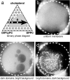
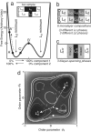
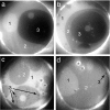
Plasma membranes of cells are asymmetric in both lipid and protein composition. The mechanism by which proteins on both sides of the membrane colocalize during signaling events is unknown but may be due to the induction of inner leaflet domains by the outer leaflet. Here we show that liquid domains form in asymmetric Montal-Mueller planar bilayers in which one leaflet's composition would phase-separate in a symmetric bilayer and the other's would not. Equally important, by tuning the lipid composition of the second leaflet, we are able to suppress domains in the first leaflet. When domains are present in asymmetric membranes, each leaflet contains regions of three distinct lipid compositions, implying strong interleaflet interactions. Our results show that mechanisms of domain induction between the outer and inner leaflets of cell plasma membranes do not necessarily require the participation of membrane proteins. Based on these findings, we suggest mechanisms by which cells could actively regulate protein function by modulating local lipid composition or interleaflet interactions.
Conflict of interest statement
The authors declare no conflict of interest.
Figures


Similar articles
-
Interleaflet interaction and asymmetry in phase separated lipid bilayers: molecular dynamics simulations.J Am Chem Soc. 2011 May 4;133(17):6563-77. doi: 10.1021/ja106626r. Epub 2011 Apr 7. J Am Chem Soc. 2011. PMID: 21473645
-
Transbilayer effects of raft-like lipid domains in asymmetric planar bilayers measured by single molecule tracking.Biophys J. 2006 Nov 1;91(9):3313-26. doi: 10.1529/biophysj.106.091421. Epub 2006 Aug 11. Biophys J. 2006. PMID: 16905614 Free PMC article.
-
Behavior of Bilayer Leaflets in Asymmetric Model Membranes: Atomistic Simulation Studies.J Phys Chem B. 2016 Aug 25;120(33):8438-48. doi: 10.1021/acs.jpcb.6b02148. Epub 2016 May 10. J Phys Chem B. 2016. PMID: 27121138
-
Domain coupling in asymmetric lipid bilayers.Biochim Biophys Acta. 2009 Jan;1788(1):64-71. doi: 10.1016/j.bbamem.2008.09.003. Epub 2008 Sep 20. Biochim Biophys Acta. 2009. PMID: 18848518 Free PMC article. Review.
-
Model systems, lipid rafts, and cell membranes.Annu Rev Biophys Biomol Struct. 2004;33:269-95. doi: 10.1146/annurev.biophys.32.110601.141803. Annu Rev Biophys Biomol Struct. 2004. PMID: 15139814 Review.
Cited by
-
Biomimetic Lipid Raft: Domain Stability and Interaction with Physiologically Active Molecules.Adv Exp Med Biol. 2024;1461:15-32. doi: 10.1007/978-981-97-4584-5_2. Adv Exp Med Biol. 2024. PMID: 39289271 Review.
-
Disruption of liquid/liquid phase separation in asymmetric GUVs prepared by hemifusion.bioRxiv [Preprint]. 2024 Jun 25:2024.06.21.600037. doi: 10.1101/2024.06.21.600037. bioRxiv. 2024. PMID: 38979299 Free PMC article. Preprint.
-
Methyl-β-cyclodextrin asymmetrically extracts phospholipid from bilayers, granting tunable control over differential stress in lipid vesicles.Soft Matter. 2024 May 29;20(21):4291-4307. doi: 10.1039/d3sm01772a. Soft Matter. 2024. PMID: 38758097 Free PMC article.
-
Multispherical shapes of vesicles with intramembrane domains.Eur Phys J E Soft Matter. 2024 Jan 11;47(1):4. doi: 10.1140/epje/s10189-023-00399-z. Eur Phys J E Soft Matter. 2024. PMID: 38206459 Free PMC article.
-
Coupling liquid phases in 3D condensates and 2D membranes: Successes, challenges, and tools.Biophys J. 2024 Jun 4;123(11):1329-1341. doi: 10.1016/j.bpj.2023.12.023. Epub 2023 Dec 29. Biophys J. 2024. PMID: 38160256 Review.
References
Publication types
MeSH terms
Substances
LinkOut - more resources
Full Text Sources

