. 2008 Jan 2;3(1):e1376.
doi: 10.1371/journal.pone.0001376.

Nitrated alpha-synuclein immunity accelerates degeneration of nigral dopaminergic neurons

Affiliations

Nitrated alpha-synuclein immunity accelerates degeneration of nigral dopaminergic neurons

Eric J Benner et al. PLoS One. .

Abstract

Background: The neuropathology of Parkinson's disease (PD) includes loss of dopaminergic neurons in the substantia nigra, nitrated alpha-synuclein (N-alpha-Syn) enriched intraneuronal inclusions or Lewy bodies and neuroinflammation. While the contribution of innate microglial inflammatory activities to disease are known, evidence for how adaptive immune mechanisms may affect the course of PD remains obscure. We reasoned that PD-associated oxidative protein modifications create novel antigenic epitopes capable of peripheral adaptive T cell responses that could affect nigrostriatal degeneration.

Methods and findings: Nitrotyrosine (NT)-modified alpha-Syn was detected readily in cervical lymph nodes (CLN) from 1-methyl-4-phenyl-1,2,3,6-tetrahydropyridine (MPTP) intoxicated mice. Antigen-presenting cells within the CLN showed increased surface expression of major histocompatibility complex class II, initiating the molecular machinery necessary for efficient antigen presentation. MPTP-treated mice produced antibodies to native and nitrated alpha-Syn. Mice immunized with the NT-modified C-terminal tail fragment of alpha-Syn, but not native protein, generated robust T cell proliferative and pro-inflammatory secretory responses specific only for the modified antigen. T cells generated against the nitrated epitope do not respond to the unmodified protein. Mice deficient in T and B lymphocytes were resistant to MPTP-induced neurodegeneration. Transfer of T cells from mice immunized with N-alpha-Syn led to a robust neuroinflammatory response with accelerated dopaminergic cell loss.

Conclusions: These data show that NT modifications within alpha-Syn, can bypass or break immunological tolerance and activate peripheral leukocytes in draining lymphoid tissue. A novel mechanism for disease is made in that NT modifications in alpha-Syn induce adaptive immune responses that exacerbate PD pathobiology. These results have implications for both the pathogenesis and treatment of this disabling neurodegenerative disease.


Conflict of interest statement

Competing Interests: The authors have declared that no competing interests exist.

Figures

Figure 1
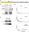
Figure 1. Drainage of N-α-Syn and MBP to CLN with macrophage activation and production of α-Syn serum antibodies after MPTP intoxication.
(A) Western blot of tissue homogenates from VMB and CLN of mice 20 hrs following treatment with PBS or MPTP, were probed with antibodies to α-Syn. (B) N-α/β Syn IP with (clone nSyn12 antibodies) against CLN homogenates from PBS or MPTP-treated mice. Immunoprecipitates were fractionated on a 16% polyacrylamide gel and the gel stained with SYPRO Red or blotted. The Western blot was probed with anti-α-Syn. (C) Proteins recovered from in-gel digestion of 12–18 kD fragments from anti-N-α/β Syn of CLN immunoprecipitates were were identified by LC-MS/MS. The sequence coverage by peptides identified by LC-MS/MS from the CLN of MPTP-treated mice is highlighted in yellow within the primary aa sequence of full-length mouse α-Syn. (D) Western blots of lymph node homogenates (Cervical, Axillary, and Inquinal) from mice treated with PBS or MPTP. Blots were probed with antibodies to nitrotyrosine (NT) or anti-myelin basic protein (MBP). (E) Flow cytometric analysis of CD11b and I-Ab expression in cells from CLN, show an increased number of CD11b+I-A+ cells 24 hrs after MPTP treatment compared to PBS administered animals (n = 3 mice/group). (F) Antibodies against α-Syn and N-α-Syn in sera of B6 WT mice on day 21 following MPTP intoxication (n = 8) or PBS control treatment (n = 5) as determined by anti-α-Syn specific ELISA. Sera from MPTP treated group contained significantly higher IgG antibodies directed against 4YSyn (p = 0.021) and N-4YSyn (p = 0.016) compared to PBS treated control sera. Comparisons of mean IgG concentrations ± SEM was by Student's t test.
Figure 2
Figure 2. Nigral degeneration following MPTP-intoxication in B6 SCID mice before and after lymphoid cell reconstitution.
(A) Photomicrographs of TH-immunostained SN (left panels) and CD3-immunostained spleen sections (right panels) from B6 (WT), SCID, and reconstituted SCID (RCS-SCID) mice treated with PBS or MPTP and obtained on day 21 post-MPTP intoxication. Immunostaining for expression of CD3 in spleens show normal distributions of CD3+ T cells in B6 WT and RCS-SCID mice treated with PBS or MPTP. Note the absence of CD3+ T cells in spleens from SCID MPTP mice. (B) Quantification of TH+ neurons in the SN of B6 WT, SCID, or reconstituted (RCS) SCID mice treated with PBS or MPTP. Values represent mean number of TH+ neurons ± SEM for 5-9 mice per group. abcdefgPair-wise comparisons by Bonferroni post-hoc test: acd p<0.0001, bef p<0.001, g p<0.05. (C) Coronal VMB sections of MPTP intoxicated B6 mice reacted with antibodies against CD3, CD4 and CD8 show positive immunostaining of cells with small, round lymphocytic morphology (magnification = 400X).
Figure 3
Figure 3. Characterization of purified and nitrated recombinant 4YSyn.
(A) Primary aa sequence of His-tagged 4YSyn peptide. The His-Tag sequence is highlighted in yellow. The sequence of 4YSyn (Syn100–140) is shown underlined with 4 Tyr residues (magenta) as potent sites for nitration. Trypsin cleavage sites at Arg (arrowhead) and Lys (arrow) are shown. (B) Purified 4YSyn (lane 1) and N-4YSyn following nitration with peroxynitrite (lane 2) fractionated on a 10–20% polyacrylamide gel and visualized using silver stain. Covalently cross-linked oligomers are indicated by arrowheads. (C) Western blot confirmation of purified 4YSyn and its associated NT modifications following peroxynitrite treatment. (D) MALDI-TOF spectra of purified 4YSyn (top panel), N-4YSyn (middle panel), and 4YSyn after tryptic digest (lower panel).
Figure 4
Figure 4. Experimental protocol for adoptive transfer and lymphocyte proliferation assessment of donor SPC in B10.BR mice.
(A) B10.BR (H-2K) mice were immunized with PBS, 50 µg 4YSyn, or 50 µg N-4YSyn emulsified in CFA. Mice were boosted 14 days later with PBS or their respective antigens in IFA. After 5 days, donor mice were sacrificed and single cell suspensions were prepared from the draining inguinal lymph nodes and spleen, and T cells were enriched by negative selection. Twelve hours after the final MPTP injection, 5×10⁷ donor immune SPC or 2.5×10⁷ T cells were adoptively transferred to MPTP-treated recipient mice. SPC were evaluated for antigen specificity prior to adoptive transfer by lymphocyte proliferation assays. SN of recipients were evaluated after 28 days of MPTP treatment for migration of T cells, survival of dopaminergic neurons, and reactive microglia. (B). SPC were tested for antigen specific proliferation by culturing in the presence of media alone or media containing 3 µg/ml of immunizing antigens for 5 days and using standard 3H-thymidine incorporation assays.
Figure 5
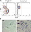
Figure 5. Adoptive transfer of SPC and purified T cells from N-4YSyn vaccinated B10.BR donors leads to infiltration of T cells in the SNpc of MPTP mice on day 2.
(A) Frequency of CD3+ T cells and CD19+ B cells before and after enrichment of T cells. Population of enriched T-cells was 94% CD3+ prior to adoptive transfer to B10.BR mice. (B) Sections throughout the SNpc were immunostained for CD3 and counterstained with thionin. Clusters of CD3+ cells are observed within the SNpc (arrowheads) as seen at 100× magnification (left). Magnification (600X) of boxed area (left panel) is shown (right panel). CD3+ cells are small and round exhibiting lymphocyte morphology.
Figure 6
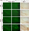
Figure 6. SPC from N-4YSyn immunized B10.BR mice exacerbate MPTP-induced dopaminergic neurodegeneration and induce microglial responses in the SNpc.
Photomicrographs from VMB sections stained with Fluoro-Jade C (left and middle panels) and Mac-1 antibody (right panels). PBS controls (PBS/none) exhibit an absence of Fluoro-Jade C stained dead neurons on days 2 and 7, and only faint Mac-1 immunoreactivity on day 4 post-treatment. In MPTP-treated mice that received SPC from PBS/adjuvant treated donors (MPTP/PBS), Fluoro-Jade C stained neurons are evident at day 2, but not detectable by day 7. MPTP-treated mice that received SPC from 4YSyn immunized donors (MPTP/4YSyn), also exhibits dead fluorescent neurons by day 2 comparable to the MPTP/PBS control group, and only rare degenerating neurons are visible by day 7. Mac-1 immunoreactivity in those mice is comparably resolved to levels seen in MPTP/PBS control group. SPC transfers from N-4YSyn immunized donors to MPTP-treated mice (MPTP/N-4YSyn) induced a robust and prolonged microglial response, conspicuously enhanced when compared to MPTP/PBS-treated controls, with concomitant neuronal death still evident by Fluoro-Jade C staining at day 7.
Figure 7
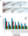
Figure 7. N-4YSyn immunization with adjuvant exacerbates dopaminergic neuronal cell loss in B10.BR mice.
(A) All panels show TH-positive neurons in the SN from mice treated with: [top row, L to R] PBS or MPTP alone, MPTP and SPC from N-4YSyn immunized donors (MPTP+N-4YSyn), [bottom row, L to R] PBS and SPC from N-4YSyn immunized donors (PBS+N-4YSyn), MPTP and SPC from 4YSyn immunized donors (MPTP+4YSyn), and MPTP and T cells from N-4YSyn immunized donors (MPTP+N-4YSyn T Cells). Tissues collected 28 days post-MPTP treatment (B) Counts of nigral Nissl+ (blue bars), TH+ (red bars), and TH- (green bars) neurons on day 28 after MPTP treatment as determined by stereological analysis. Control groups included mice treated with PBS alone (n = 4), MPTP alone (n = 8), and PBS animals that received immune effector SPC from N-4YSyn immunized donor mice PBS/N-4YSyn/SPC (n = 6). Experimental groups included MPTP/PBS/SPC (n = 6), MPTP/4YSyn/SPC (n = 8), MPTP/N-4YSyn/SPC (n = 9), and MPTP mice which received purified T cells from N-4YSyn vaccinated donors. Values are means ± SEM. p<0.01 compared to the following treatment groups: aPBS, bMPTP, cMPTP/4YSyn/SPC.
Figure 8
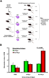
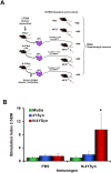
Figure 8. Scheme for immunization, lymphocyte proliferation assessment, and adoptive transfer of donor SPC in B6 mice.
(A) B6 (H-2b) mice were immunized with 10 µg 4YSyn in PBS, 50 µg N-4YSyn in CFA, 10 µg N-4YSyn in PBS or PBS in CFA. Mice were boosted 14 days later with their respective antigens as formulated previously with or without IFA. After 5 days, single lymphoid cell suspensions were prepared and assessed for antigen-specific responses in standard lymphocyte proliferation assays. Single cell suspensions were pooled and adoptively transferred to MPTP-treated syngeneic recipients 12 hrs after the final MPTP injection. 5×10⁷ donor immune SPC were adoptively transferred to MPTP-treated recipient mice. Survival of dopaminergic neurons in the SN of recipient mice were evaluated after 7 days. (B) Antigen specific proliferation of SPC from B6 (H-2b) mice (n = 5/group) immunized with PBS/CFA or N-4YSyn/CFA, and cultured for 5 days in media alone (green bars) or in the presence 1 µg/ml of 4YSyn (blue bars) or N-4YSyn (red bars). Cultures were pulsed for 18 hrs, cells harvested and 3H-thymidine incorporation counted by β-scintillation spectrometry. Values represent mean stimulation indices±SEM and analyzed by ANOVA and Bonferroni post-hoc tests. ap = 0.0478.
Figure 9
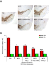
Figure 9. Lymphocytes from N-4YSyn immunization exacerbate nigral dopaminergic neuronal loss in B6 mice.
(A) All panels show TH+ neurons in the SN from mice treated with PBS or MPTP alone, MPTP and SPC from 4YSyn immunized donors (MPTP+4YSyn), MPTP and SPC from N-4YSyn immunized donors (MPTP+N-4YSyn) and lastly, MPTP and SPC from N-4YSyn+CFA immunized donors (MPTP+N-4YSyn CFA). (B) Counts of nigral TH+ and TH neurons on day 7 after MPTP treatment. Experimental groups included mice treated with PBS alone (n = 7), MPTP alone (n = 7), MPTP/4YSyn (n = 7), MPTP/N-4YSyn/CFA (n = 6) and MPTP/N-4YSyn (n = 6). Values are means ± SEM. Analysis by ANOVA with Bonferroni post-hoc tests indicated ap<0.0001 compared to PBS control: bp<0.001 compared to MPTP group; cp<0.001 compared to MPTP/4YSyn group; and dp<0.03 compared to MPTP/4YSynCFA SPC.
Figure 10
Figure 10. N-4YSyn-mediated inhibition of T cell proliferation.
Proliferative responses of anti-CD3 stimulated T cells from naïve B6 mice in presence of graded concentrations of 4YSyn or N-4YSyn (1, 3, 10, 30 µg/ml) or in media alone (0 µg/ml). T cells were cultured for 72 hrs and pulsed with 3H-thymidine for the final 18 hrs of culture. Harvested cells were counted for 3H-thymidine uptake by β-scintillation spectrometry and proliferation was expressed as mean counts per min (CPM)±SEM for quadruplicate samples and evaluated by ANOVA with Bonferroni post-hoc tests. ap<0.01 compared with T cells stimulated with anti-CD3 and cultured in media alone.
Figure 11
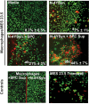
Figure 11. N-4YSyn activated immune SPC induces macrophage-mediated dopaminergic cell death.
Representative fluorescence photomicrographs are shown of live (green) and dead (red) cells from 24 hr macrophage/MES 23.5 co-cultures in the presence of media alone, N-4YSyn, N-4YSyn and antigen-stimulated SPC of N-4YSyn-immunized mice (N-4YSyn+SPC), or N-4YSyn and the supernatants from antigen-stimulated SPC of N-4YSyn-immunized mice (N-4YSyn+SPC Sup). Antigen-stimulated SPC from N-4YSyn immune mice were induced in vitro with N-4YSyn, and cells and supernatants for use in the assay were harvested after 5 days of culture. Controls included macrophages cultured in the presence of SPC supernatants from antigen-stimulated SPC of N-4YSyn-immunized mice (Macrophages+SPC Sup); macrophages cultured in the presence of N-4YSyn alone (Macrophages+N4YSyn); and transwell cultures of plated MES 23.5 cells and macrophages in the transwell stimulated with N-4YSyn. Frequencies (±SEM) of dead (red) cells for 4-8 fields/assay are provided in the lower right corner of each panel. Differences in the mean frequencies of dead cells were evaluated by ANOVA and Bonferroni post-hoc tests. p<0.01 compared to cultures treated with aMedia, bN-4YSyn, or cN-4YSyn+SPC.

Similar articles

Cited by

References

    1. Dauer W, Przedborski S. Parkinson's disease: mechanisms and models. Neuron. 2003;39:889–909. - PubMed
    1. Klockgether T. Parkinson's disease: clinical aspects. Cell Tissue Res. 2004;318:115–120. - PubMed
    1. Spillantini MG, Schmidt ML, Lee VM, Trojanowski JQ, Jakes R, et al. Alpha-synuclein in Lewy bodies. Nature. 1997;388:839–840. - PubMed
    1. Sidhu A, Wersinger C, Vernier P. Does alpha-synuclein modulate dopaminergic synaptic content and tone at the synapse? FASEB J. 2004;18:637–647. - PubMed
    1. Paxinou E, Chen Q, Weisse M, Giasson BI, Norris EH, et al. Induction of alpha-synuclein aggregation by intracellular nitrative insult. J Neurosci. 2001;21:8053–8061. - PMC - PubMed

Publication types