Fractalkine deficiency markedly reduces macrophage accumulation and atherosclerotic lesion formation in CCR2-/- mice: evidence for independent chemokine functions in atherogenesis
- PMID: 18165355
- PMCID: PMC3589525
- DOI: 10.1161/CIRCULATIONAHA.107.743872
Fractalkine deficiency markedly reduces macrophage accumulation and atherosclerotic lesion formation in CCR2-/- mice: evidence for independent chemokine functions in atherogenesis
Abstract
Background: Monocyte-derived foam cells are the hallmark of early atherosclerosis, and recent evidence indicates that chemokines play important roles in directing monocyte migration from the blood to the vessel wall. Genetic deletions of monocyte chemoattractant protein-1 (MCP-1, CCL2), fractalkine (CX3CL1), or their cognate receptors, CCR2 and CX3CR1, markedly reduce atherosclerotic lesion size in murine models of atherosclerosis. The aim of this study was to determine whether these 2 chemokines act independently or redundantly in promoting atherogenesis.
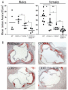
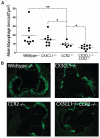
Methods and results: We crossed CX3CL1(-/-)ApoE(-/-) and CCR2(-/-)ApoE(-/-) mice to create CX3CL1(-/-)CCR2(-/-)ApoE(-/-) triple knockouts and performed a 4-arm atherosclerosis study. Here, we report that deletion of CX3CL1 in CCR2(-/-) mice dramatically reduced macrophage accumulation in the artery wall and the subsequent development of atherosclerosis. Deletion of CX3CL1 did not reduce the number of circulating monocytes in either "wild-type" ApoE(-/-) mice or CCR2(-/-)ApoE(-/-) mice, which suggests a role for CX3CL1 in the direct recruitment and/or capture of CCR2-deficient monocytes.
Conclusions: These data provide the first in vivo evidence for independent roles for CCR2 and CX3CL1 in macrophage accumulation and atherosclerotic lesion formation and suggest that successful therapeutic strategies may need to target multiple chemokines or chemokine receptors.
Figures


Similar articles
-
Roles for the CX3CL1/CX3CR1 and CCL2/CCR2 Chemokine Systems in Hypoxic Pulmonary Hypertension.Am J Respir Cell Mol Biol. 2017 May;56(5):597-608. doi: 10.1165/rcmb.2016-0201OC. Am J Respir Cell Mol Biol. 2017. PMID: 28125278
-
Retinal Phenotype following Combined Deletion of the Chemokine Receptor CCR2 and the Chemokine CX3CL1 in Mice.Ophthalmic Res. 2016;55(3):126-34. doi: 10.1159/000441794. Epub 2015 Dec 16. Ophthalmic Res. 2016. PMID: 26670885
-
Decreased atherosclerosis in CX3CR1-/- mice reveals a role for fractalkine in atherogenesis.J Clin Invest. 2003 Feb;111(3):333-40. doi: 10.1172/JCI15555. J Clin Invest. 2003. PMID: 12569158 Free PMC article.
-
The role of chemokines in atherosclerosis: recent evidence from experimental models and population genetics.Curr Opin Lipidol. 2004 Apr;15(2):145-9. doi: 10.1097/00041433-200404000-00007. Curr Opin Lipidol. 2004. PMID: 15017357 Review.
-
Involvement of chemokine receptor 2 and its ligand, monocyte chemoattractant protein-1, in the development of atherosclerosis: lessons from knockout mice.Curr Opin Lipidol. 2001 Apr;12(2):175-80. doi: 10.1097/00041433-200104000-00011. Curr Opin Lipidol. 2001. PMID: 11264989 Review.
Cited by
-
wRAPping up early monocyte and neutrophil recruitment in atherogenesis via Annexin A1/FPR2 signaling.Circ Res. 2015 Feb 27;116(5):774-7. doi: 10.1161/CIRCRESAHA.115.305920. Circ Res. 2015. PMID: 25722438 Free PMC article. No abstract available.
-
Having an Old Friend for Dinner: The Interplay between Apoptotic Cells and Efferocytes.Cells. 2021 May 20;10(5):1265. doi: 10.3390/cells10051265. Cells. 2021. PMID: 34065321 Free PMC article. Review.
-
Statins promote the regression of atherosclerosis via activation of the CCR7-dependent emigration pathway in macrophages.PLoS One. 2011;6(12):e28534. doi: 10.1371/journal.pone.0028534. Epub 2011 Dec 6. PLoS One. 2011. PMID: 22163030 Free PMC article.
-
Trafficking of Mononuclear Phagocytes in Healthy Arteries and Atherosclerosis.Front Immunol. 2021 Oct 25;12:718432. doi: 10.3389/fimmu.2021.718432. eCollection 2021. Front Immunol. 2021. PMID: 34759917 Free PMC article. Review.
-
Monocyte-to-High Density Lipoprotein Cholesterol Ratio Positively Predicts Coronary Artery Disease and Multi-Vessel Lesions in Acute Coronary Syndrome.Int J Gen Med. 2023 Aug 28;16:3857-3868. doi: 10.2147/IJGM.S419579. eCollection 2023. Int J Gen Med. 2023. PMID: 37662500 Free PMC article.
References
-
- Ross R. Cell biology of atherosclerosis. Annu Rev Physiol. 1995;57:791–804. - PubMed
-
- Hansson GK, Libby P. The immune response in atherosclerosis: a double-edged sword. Nat Rev Immunol. 2006;6:508–519. - PubMed
-
- Charo IF, Ransohoff RM. The many roles of chemokines and chemokine receptors in inflammation. N Engl J Med. 2006;354:610–621. - PubMed
-
- Gordon S, Taylor PR. Monocyte and macrophage heterogeneity. Nat Rev Immunol. 2005;5:953–964. - PubMed
Publication types
MeSH terms
Substances
Grants and funding
LinkOut - more resources
Full Text Sources
Other Literature Sources
Molecular Biology Databases
Research Materials
Miscellaneous

