4E10-resistant variants in a human immunodeficiency virus type 1 subtype C-infected individual with an anti-membrane-proximal external region-neutralizing antibody response
- PMID: 18094155
- PMCID: PMC2258954
- DOI: 10.1128/JVI.02161-07
4E10-resistant variants in a human immunodeficiency virus type 1 subtype C-infected individual with an anti-membrane-proximal external region-neutralizing antibody response
Abstract
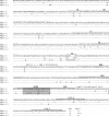
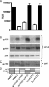
The broadly neutralizing monoclonal antibody (MAb) 4E10 recognizes a linear epitope in the C terminus of the membrane-proximal external region (MPER) of gp41. This epitope is particularly attractive for vaccine design because it is highly conserved among human immunodeficiency virus type 1 (HIV-1) strains and neutralization escape in vivo has not been observed. Multiple env genes were cloned from an HIV-1 subtype C virus isolated from a 7-year-old perinatally infected child who had anti-MPER neutralizing antibodies. One clone (TM20.13) was resistant to 4E10 neutralization as a result of an F673L substitution in the MPER. Frequency analysis showed that F673L was present in 33% of the viral variants and in all cases was linked to the presence of an intact 2F5 epitope. Two other envelope clones were sensitive to 4E10 neutralization, but TM20.5 was 10-fold less sensitive than TM20.6. Substitutions at positions 674 and 677 within the MPER rendered TM20.5 more sensitive to 4E10 but had no effect on TM20.6. Using chimeric and mutant constructs of these two variants, we further demonstrated that the lentivirus lytic peptide-2 domain in the cytoplasmic tail affected the accessibility of the 4E10 epitope, as well as virus infectivity. Collectively, these genetic changes in the face of a neutralizing antibody response to the MPER strongly suggested immune escape from antibody responses targeting this region.
Figures


Similar articles
-
Anti-human immunodeficiency virus type 1 (HIV-1) antibodies 2F5 and 4E10 require surprisingly few crucial residues in the membrane-proximal external region of glycoprotein gp41 to neutralize HIV-1.J Virol. 2005 Jan;79(2):1252-61. doi: 10.1128/JVI.79.2.1252-1261.2005. J Virol. 2005. PMID: 15613352 Free PMC article.
-
Evolution of antibody landscape and viral envelope escape in an HIV-1 CRF02_AG infected patient with 4E10-like antibodies.Retrovirology. 2009 Dec 14;6:113. doi: 10.1186/1742-4690-6-113. Retrovirology. 2009. PMID: 20003438 Free PMC article.
-
An affinity-enhanced neutralizing antibody against the membrane-proximal external region of human immunodeficiency virus type 1 gp41 recognizes an epitope between those of 2F5 and 4E10.J Virol. 2007 Apr;81(8):4033-43. doi: 10.1128/JVI.02588-06. Epub 2007 Feb 7. J Virol. 2007. PMID: 17287272 Free PMC article.
-
The membrane-proximal external region of the human immunodeficiency virus type 1 envelope: dominant site of antibody neutralization and target for vaccine design.Microbiol Mol Biol Rev. 2008 Mar;72(1):54-84, table of contents. doi: 10.1128/MMBR.00020-07. Microbiol Mol Biol Rev. 2008. PMID: 18322034 Free PMC article. Review.
-
Antigp41 membrane proximal external region antibodies and the art of using the membrane for neutralization.Curr Opin HIV AIDS. 2017 May;12(3):250-256. doi: 10.1097/COH.0000000000000364. Curr Opin HIV AIDS. 2017. PMID: 28422789 Review.
Cited by
-
An MPER antibody neutralizes HIV-1 using germline features shared among donors.Nat Commun. 2019 Nov 26;10(1):5389. doi: 10.1038/s41467-019-12973-1. Nat Commun. 2019. PMID: 31772165 Free PMC article.
-
Direct antibody access to the HIV-1 membrane-proximal external region positively correlates with neutralization sensitivity.J Virol. 2011 Aug;85(16):8217-26. doi: 10.1128/JVI.00756-11. Epub 2011 Jun 8. J Virol. 2011. PMID: 21653673 Free PMC article.
-
Genotypic and functional properties of early infant HIV-1 envelopes.Retrovirology. 2011 Aug 15;8:67. doi: 10.1186/1742-4690-8-67. Retrovirology. 2011. PMID: 21843318 Free PMC article.
-
4E10-resistant HIV-1 isolated from four subjects with rare membrane-proximal external region polymorphisms.PLoS One. 2010 Mar 23;5(3):e9786. doi: 10.1371/journal.pone.0009786. PLoS One. 2010. PMID: 20352106 Free PMC article.
-
Variations in autologous neutralization and CD4 dependence of b12 resistant HIV-1 clade C env clones obtained at different time points from antiretroviral naïve Indian patients with recent infection.Retrovirology. 2010 Sep 22;7:76. doi: 10.1186/1742-4690-7-76. Retrovirology. 2010. PMID: 20860805 Free PMC article.
References
-
- Binley, J. M., T. Wrin, B. Korber, M. B. Zwick, M. Wang, C. Chappey, G. Stiegler, R. Kunert, S. Zolla-Pazner, H. Katinger, C. J. Petropoulos, and D. R. Burton. 2004. Comprehensive cross-clade neutralization analysis of a panel of anti-human immunodeficiency virus type 1 monoclonal antibodies. J. Virol. 7813232-13252. - PMC - PubMed
Publication types
MeSH terms
Substances
LinkOut - more resources
Full Text Sources
Other Literature Sources
Medical
Molecular Biology Databases

