. 2008 Mar;36(4):1057-71.
doi: 10.1093/nar/gkm1036. Epub 2007 Dec 17.

The cucumovirus 2b gene drives selection of inter-viral recombinants affecting the crossover site, the acceptor RNA and the rate of selection

Affiliations

The cucumovirus 2b gene drives selection of inter-viral recombinants affecting the crossover site, the acceptor RNA and the rate of selection

Bu-Jun Shi et al. Nucleic Acids Res. 2008 Mar.

Abstract

RNA-RNA recombination is an important pathway in virus evolution and has been described for many viruses. However, the factors driving recombination or promoting the selection of recombinants are still unclear. Here, we show that the small movement protein (2b) was able to promote selection of RNA 1/2-RNA 3 recombinants within a chimeric virus having RNAs 1 and 2 from cucumber mosaic virus, and RNA 3 from the related tomato aspermy virus, along with heterologous 2b genes. The source of the 2b also determined the selection of the acceptor RNA and the crossover site, as well as affecting the rate of selection of the recombinant RNAs. The nature of the RNA 3 also influenced the selection of the recombinant RNAs. A 163-nt tandem repeat in RNA 3 significantly affected the rate of selection of the recombinant RNA, while a single nucleotide within the repeat affected the crossover site. The recombination occurred in a non-random manner, involved no intermediates and probably was generated via a copy-choice mechanism during (+) strand RNA synthesis.


Figures
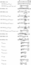
Figure 1.
Figure 1.
Schematic representation of constructs used in this article. The constructs regulated from the 35S promoter and 35S terminator are indicated. The open reading frames encoding various cucumoviral proteins on the constructs are shown. The 23 nt of sequence identity between all genomic RNAs is indicated by a short thick bar. The tandem repeat of 163 nt is indicated by an arrow. The crosshatch lines represent the amino acid sequences different from the native sequence for either 2a or 2b.
Figure 2.
Figure 2.
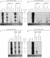
Northern hybridization of accumulated viral RNAs in N. glutinosa. Total RNAs extracted from inoculated and passage N. glutinosa plants were fractionated in 1.2% of agarose gel containing 1.1% formaldehyde, and then blotted to membranes. The hybridization of the membranes was done with probes specific for the 3′ NTRs of the Q-CMV or TAV RNAs. The positions of RNAs 1, 2, 3, 4, 4A, 3B and 5 are indicated. CMV RNA 5 is 307–310 nt and represents the 3′ NTR of the genomic RNAs 1–3. TAV RNA 5 is 323 nt, while TAV RNA 3B is 486 nt; both are derived from the 3′ NTR of TAV RNA 3.
Figure 3.
Figure 3.
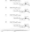
Genome organization of C1C2T2BT3, C1C2W2BT3 and their derived recombinant viruses. The genomic RNAs or 2b genes of each virus are indicated by different pattern fills. The short black bars on the RNAs represent the 23 nt of sequence identity. The arrows on RNA 3 represent the tandem repeats (first repeat, A, or second repeat, G). The junction site of each RNA 3 recombinant is indicated. The nucleotide sequence in the junction site of the RNA 3 recombinant derived from C1C2W2BT3 is indicated. The sequence in italics is from TAV RNA 3. The sequences underlined represent the imperfect 19-nt repeats. The single nucleotide difference in the repeats is indicated by an asterisk.
Figure 4.
Figure 4.
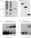
TAV 2b protein expression, purification and RNA 3 binding. (A) The TAV 2b protein fused with GST and transformed into the bacteria strain BL21 (DE3) was analysed by SDS–PAGE. Total bacterial lysate after the induction of protein expression (lane 2; tp) was passaged through glutathione beads to purify the GST-2b fusion protein (lane 3; pp). Molecular mass markers (M) are shown in lane 1. (B) Purified GST-2b fusion protein (lane 1) was cleaved using thrombin and the resulting products, GST (lane 2) and 2b proteins (lane 3), were gel-purified. GST protein was treated with thrombin (lane 2). The gels in A and B were stained with Coomassie Blue. (C and D) Autoradiograms of gel retardation electrophoresis assays of the 2b protein binding to RNA 3. Increasing amounts of the 2b protein were incubated in the binding buffer as with 4 ng of either 32P-labelled TAV RNA 3 (+) transcript (C) or 32P-labelled TAV RNA 3(−) transcript (D). The samples were electrophoresed in 4% non-denaturing polyacrylamide gels. The amounts of the 2b protein, the position of free RNA 3 (f) and the position of retarded RNA 3 (r) are indicated.
Figure 5.
Figure 5.
Genome organization of C1C2T2BT3Δ163(A), C1C2T2BT3Δ163(G), C1C2W2BT3Δ163(A), C1C2W2BT3Δ163(G) and their derived recombinant viruses. The origin of the 3′ NTR of each RNA 3 is differentiated by fill patterns (stippled for RNA 1 and crosshatched for RNA 2). The short black bars on the RNAs represent the 23 nt of sequence identity. The arrows on RNA 3 represent one of the tandem repeats (A or G). The junction site of each RNA 3 recombinant is indicated. The nucleotide sequence in the junction site of the RNA 3 recombinant derived from C1C2W2BT3Δ163(G) is indicated. The sequence in italics is from TAV RNA 3. The sequences underlined represent the imperfect 19-nt repeats. The single nucleotide difference in the repeats is indicated by an asterisk.
Figure 6.
Figure 6.
Agarose gel and Southern hybridization of RT-PCR products from C1C2T2BT3Δ163(G). RT-PCR was done using primer pair C3′/T3-1187 and total RNAs extracted from the plants at day 22–26 after inoculation with C1C2T2BT3Δ163(G). Top row: The RT-PCR products were analysed on a 1% agarose gel and stained with ethidium bromide (EB). The sizes of the products are indicated. M in the left lane indicates the DNA ladder, while CMV in the right lane is used as a negative control showing no RT-PCR product is obtained from the plants infected with Q-CMV alone. Middle row: The RT-PCR products on the gel were blotted to membranes and hybridized with the Q-CMV-specific probe. Bottom row: The same RT-PCR products were blotted and hybridized with the TAV-specific probe.
Figure 7.
Figure 7.
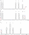
Proposed secondary structure of RNA 3 sequences complementary to the 3′ NTR of TAV RNA 3. The residues are numbered according to the corresponding (+) RNA 3, with nucleotide 1902 (underlined) as the position complementary to the 5′ first nucleotide of TAV RNA 3B. Stem-loops are labelled starting from the 5′ end of the (−) TAV RNA 3, as complementary to those stem-loops of the 3′ end of (+) TAV RNA 3 (A–H) described previously (64). Those stem-loops present in the first 163-nt repeat (L2-H2) are shown in (A), while those present in the second 163-nt repeat (L1-H1) and loop G are shown in (B), with the remaining 5′ proximal stem-loops and non-base-paired sequences of (−) TAV RNA 3 shown in (C). The sites of recombination observed here are indicated by an asterisk (nucleotide 2060), a dagger (nucleotide 2064) or an arrow (nucleotide 2245) in (B and C). The site of recombination described in Ref. (30) is at position 1973 and is indicated by an arrow in (A). The single nucleotide difference (nucleotides 1966 and 2129) between the first and second 163-nt repeat is shown in blue and is underlined. A possible pseudoknot can be formed between the three As at the top of stem-loop G and the three Us (shown in blue) in the non-base-paired region between stem-loops K2 and J2 in the first 163-nt repeat, but not with the corresponding CUU sequence shown in the second 163-nt repeat. [Note: Neither stem-loops B nor F are indicated, since loop B did not contain a stem in (+) TAV RNA 3 (64) and small stem-loop F was composed of several non-canonical G–U base pairs in (+) TAV RNA 3 (54) and is not present in (−) TAV RNA 3]. The ΔG values (kcal/mol) for the various stem-loops are A = −11.5, C = −7.8, D = −8.4, E = −6.8, G = −1.3, H = −2.9, I = −4.8, J = −3.8, K = −2.7, L1 = −6.9 and L2 = −5.7, as calculated from Ref. (65).

Similar articles

Cited by

References

    1. Hirst GK. Genetic recombination with Newcastle disease virus, polioviruses, and influenza. Cold Spring Harb. Symp. Quant. Biol. 1962;27:303–309. - PubMed
    1. Ledinko N. Genetic recombination with poliovirus type 1. Studies of crosses between a normal horse serum-resistant mutant and several guanidine-resistant mutants of the same strain. Virology. 1963;20:107–119. - PubMed
    1. King AM, McCahon D, Saunders K, Newman JW, Slade WR. Multiple sites of recombination within the RNA genome of foot-and-mouth disease virus. Virus Res. 1985;3:373–384. - PMC - PubMed
    1. McCahon D. The genetics of aphthovirus. Brief review. Arch. Virol. 1981;69:1–23. - PubMed
    1. Luytjes W, Bredenbeek PJ, Noten AF, Horzinek MC, Spaan WJ. Sequence of mouse hepatitis virus A59 mRNA 2: indications for RNA recombination between coronaviruses and influenza C virus. Virology. 1988;166:415–422. - PMC - PubMed

Publication types