Comparative Study
. 2008 Feb;14(2):297-309.
doi: 10.1261/rna.698108. Epub 2007 Dec 14.

Analysis of the human polynucleotide phosphorylase (PNPase) reveals differences in RNA binding and response to phosphate compared to its bacterial and chloroplast counterparts

Affiliations
Comparative Study

Analysis of the human polynucleotide phosphorylase (PNPase) reveals differences in RNA binding and response to phosphate compared to its bacterial and chloroplast counterparts

Victoria Portnoy et al. RNA. 2008 Feb.

Abstract

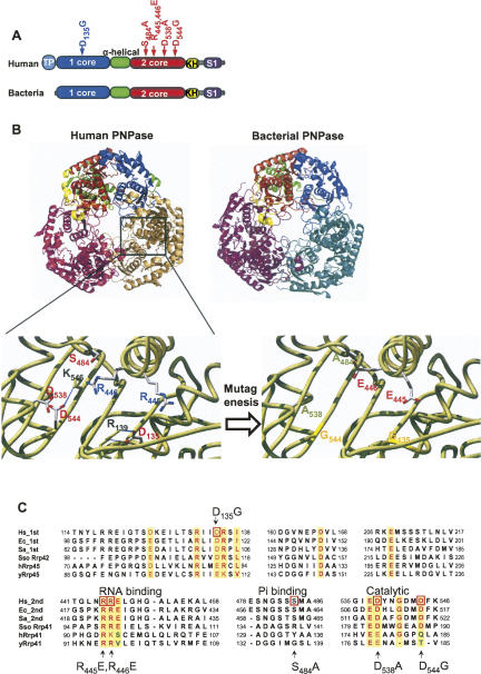
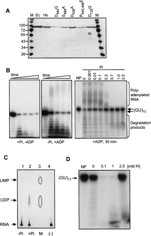
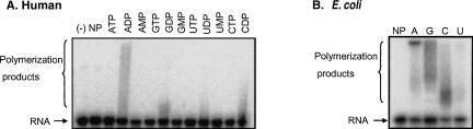
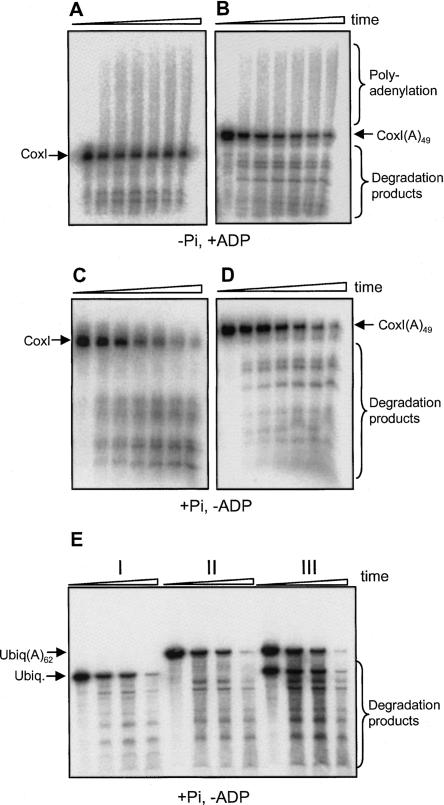
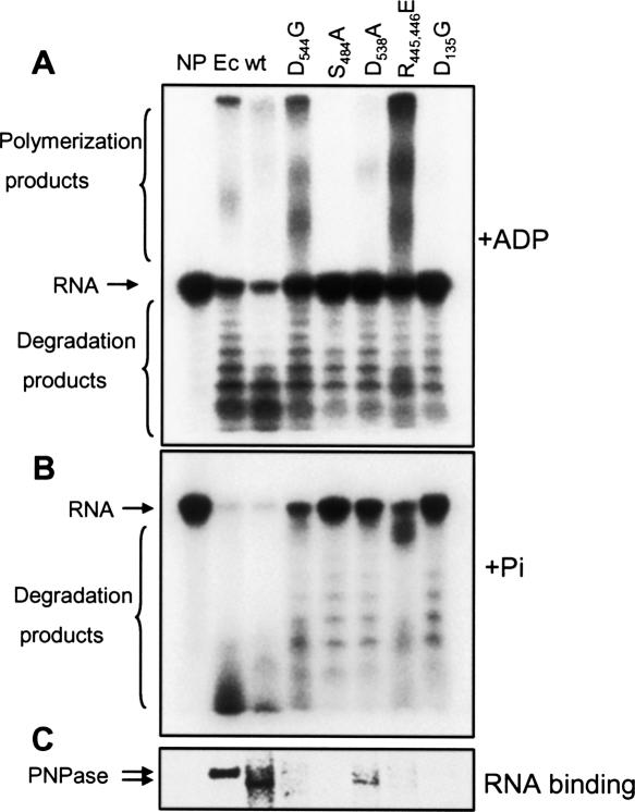
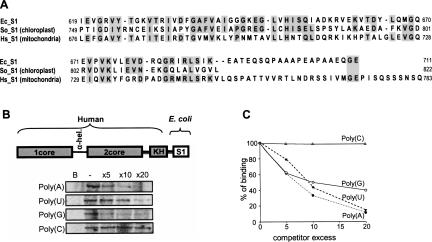
PNPase is a major exoribonuclease that plays an important role in the degradation, processing, and polyadenylation of RNA in prokaryotes and organelles. This phosphorolytic processive enzyme uses inorganic phosphate and nucleotide diphosphate for degradation and polymerization activities, respectively. Its structure and activities are similar to the archaeal exosome complex. The human PNPase was recently localized to the intermembrane space (IMS) of the mitochondria, and is, therefore, most likely not directly involved in RNA metabolism, unlike in bacteria and other organelles. In this work, the degradation, polymerization, and RNA-binding properties of the human PNPase were analyzed and compared to its bacterial and organellar counterparts. Phosphorolytic activity was displayed at lower optimum concentrations of inorganic phosphate. Also, the RNA-binding properties to ribohomopolymers varied significantly from those of its bacterial and organellar enzymes. The purified enzyme did not preferentially bind RNA harboring a poly(A) tail at the 3' end, compared to a molecule lacking this tail. Several site-directed mutations at conserved amino acid positions either eliminated or modified degradation/polymerization activity in different manners than observed for the Escherichia coli PNPase and the archaeal and human exosomes. In light of these results, a possible function of the human PNPase in the mitochondrial IMS is discussed.


Figures

FIGURE 1.
FIGURE 1.
Human and bacterial PNPases and the mutated versions used in this work. (A) The domain structures of the bacterial and human PNPases are presented schematically. The boxes represent the different domains as indicated, and the relative locations of the site-directed mutated amino acids are given. TP indicates the mitochondrial intermembrane transit peptide. (B) The resolved structure and predicted model of PNPase enzymes from Streptoomyces antibioticus (bacteria) and human (human) are shown. The trimeric doughnut-like shape is presented in an orientation enabling easy observation of the central channel. Each monomer is colored differently, and each domain is colored in one monomer of the enzyme, as shown in the scheme presented at the top. The homology-based modeling was performed using the Phyre Server at http://www.sbg.bio.ic.ac.uk/∼3dpssm/. The complex was built by applying the crystal symmetry of the structure using the PyMOL program. A closeup view of the locations of the modified amino acids and their particular locations is presented below. (C) Alignment of the amino acid sequences of the conserved parts of human (Hs), E. coli (Ec) and S. antibioticus (Sa) PNPases, as well as the hyperthermophilic archaea Sulfolobus solfataricus (Sso), human (hRrp45 and hRrp41), and yeast (yRrp45 and yRrp41) exosomes proteins. The first and second signs indicate the first and second core RNase PH-like domains for PNPase, respectively. The corresponding protein names are indicated for the exosomes. The yellow background shows the mostly conserved amino acids, which are colored red, and those mutated in this work are marked by red squares.
FIGURE 2.
FIGURE 2.
Polymerization and degradation activities of human PNPase. (A) Coomassie-stained polyacrylamide gel profile of the recombinant proteins following expression in E. coli. Proteins were purified by nitrilotriacetic acid (NTA) agarose affinity chromatography, anion-exchange (MonoQ), and heparin affinity chromatography steps, and loaded onto a 10% SDS-PAGE gel. Molecular mass markers (in kDa) are shown at the left and right (M). Ec, E. coli PNPase; Hs, human PNPase. In other lanes, the purified proteins of the corresponding mutants as labeled by the modified amino acid residue are shown. (B) Human PNPase was incubated with [32P]-RNA (GU)12 oligonucleotide in the presence of either Pi (0.1 mM) or ADP (2.5 mM), as indicated in the figure, in order to promote degradation or polyadenylation activity, respectively. Samples were removed at 0, 5, 15, 30, and 60 min, and the RNA was analyzed by denaturing PAGE and autoradiography. The input RNA, as well as polymerized and partially degraded products, are indicated. In the right panel, incubation was for 30 min in the present of 2.5 mM ADP and Pi at the concentration indicated on the top. NP-no protein. (C) Thin-layer chromatography (TLC) analysis of the degradation products. Human PNPase was incubated with uniformly [32P] UTP-labeled RNA corresponding to part of the human mitochondrial transcript COX1. Following the incubation, the reaction products were spotted onto a polyethyleneimine TLC plate, which was developed with 0.9 M GnCl, dried, and autoradiographed. The PNPase activity was assayed either in the absence (lane 1) or presence of 0.1 mM Pi (lane 2). In lane 3, UMP and UDP were analyzed as markers on the same TLC plate and visualized by fluorescence quenching. The dotted lines mark the boundaries of the fluorescence pattern. In lane 4, a control reaction in which the RNA was incubated without the addition of protein, was analyzed. (D) Human PNPase activity in response to Pi concentrations. [32P]-labeled RNA oligonucleotide (GU)12 was incubated for 15 min with human PNPase at different Pi concentrations, as indicated. CaCl2 (50 μM) was added to the reaction mixture in order to precipitate the Pi already present in the sample. Following incubation, the RNA was analyzed by denaturing PAGE and autoradiography. No enzyme was added to the reaction in the lanes labeled (–).
FIGURE 3.
FIGURE 3.
Of the NDPs, human PNPase displays a preference for ADP during polymerization activity. [32P]-labeled RNA oligonucleotide (GU)12 was incubated with human (A) or E. coli (B) proteins and 2.5 mM of each of the corresponding nucleotides, as indicated at the top of the figure. For the E. coli enzyme (B), only the NDPs nucleotides were assayed. Samples were withdrawn at 10 min, and the RNA analyzed by denaturing PAGE and autoradiography. The input RNA and polymerized products are shown on the left. The input RNA (lane [–]) as well as a control reaction in which no protein was added (lane NP) were also analyzed.
FIGURE 4.
FIGURE 4.
Similar activities of human PNPase on nonpolyadenylated and polyadenylated substrates. [32P] RNA corresponding to part of the mitochondrial gene COX1, either without (A,C) or with the addition of 49 adenosins at the 3′ end (B and D) was incubated with the protein and 0.1 mM Pi (C,D) or 2.5 mM ADP (A,B). In panel E, degradation assay was performed to non-polyadenylated ubiquitin transcript (I), the same transcript with the addition of 64 adenosines (II), and the two transcripts mixed together (III). Samples were withdrawn at 0, 15, 30, and 120 min, and the RNA analyzed by denaturing PAGE and autoradiography.
FIGURE 5.
FIGURE 5.
Similar degradation activities of human and E. coli PNPases. [32P] RNA corresponding to the E. coli RNAI (111 nt) was incubated with the human or E. coli PNPases (100 ng each). Pi was added to a final concentration of 0.1 or 10 mM for the human and E. coli enzymes, respectively. Samples were collected at 0, 5, 15, 30, 60, 90, and 120 min, and the RNA was analyzed by denaturing PAGE and autoradiography. RNA markers were fractionated in the lanes marked M1 and M2. The length in nucleotides is shown on the right. The predicted secondary structure of RNAI is shown to the right.
FIGURE 6.
FIGURE 6.
RNA-binding properties of human and E. coli PNPases. (A) Human PNPase displays a sigmoidal curve which suggests cooperative binding. Human and E. coli PNPases (10 ng each) were analyzed for RNA-binding by a UV light cross-linking assay using increasing amounts of PNPase, as indicated in the figure inset. The radioactive signals obtained in three independent experiments (error bars are represented) were quantified and plotted in order to determine the shape of the binding curve and the Kd observed. The black line with circled points represents the human PNPase and the dashed line with filled points represents the E. coli PNPase. The Kd observed for E.coli and human PNPases were 11 and 16 nM, respectively. (B) High affinity binding of human PNPase to poly(U) and poly(G). Human or E. coli PNPases were incubated with [32P]-RNA corresponding to (GU)12, and a homoribopolymer at a molar excess, as shown in the figure. Following UV cross-linking and ribonuclease digestion, the label transfer from RNA to protein was analyzed by SDS-PAGE and autoradiography. In the lane labeled B, no protein was added to the assay as a control. (C) Quantization of the competition assays. The UV light cross-linking competition assays were performed as described for (B), and the intensity of the UV light cross-linking band without competitor was defined as 100%. Data shown are the average of at least three experiments.
FIGURE 7.
FIGURE 7.
Unlike the spinach chloroplast PNPase, the human protein does not form a complex with its E. coli counterpart. (A) Human (lanes 1 and 3), E. coli (lane 2), or spinach chloroplast (lane 4) PNPases were expressed in E. coli strain in which the expression of the endogenous PNPase was knocked-out (lane 1), or in E. coli strain containing the endogenous PNPase (lanes 2, 3, and 4). The expressed proteins were purified as described in Materials and Methods and analyzed on SDS-PAGE followed by silver staining. The identities of the different PNPases are indicated on the right. The E. coli endogenous PNPase was copurified with the spinach protein (lane 4) but not with the human one (lane 3). (B) Recombinant human PNPase was expressed and purified in E. coli strains either lacking (lane 1) or containing (lane 2) endogenous PNPase. In lane 3, a purified recombinant E. coli PNPase was loaded. The proteins were analyzed by SDS-PAGE and immunoblotted to a nitrocellulose membrane that was incubated with antibodies against the E. coli PNPase or the human PNPase, as indicated in the figure. (C) Recombinant human PNPase forms a high molecular weight complex of ∼300 kDa. Recombinant proteins (∼5 μg) were fractionated on a Superdex 200 size-exclusion column. The elution profile of molecular-weight markers: Thyroglobulin 665 kDa, Feritin 440 kDa, Catalase 232 kDa, Aldolase 158 kDa, and Albumin 67 kDa, fractionated on the same column, is indicated at the top.
FIGURE 8.
FIGURE 8.
Polymerization, degradation, and RNA-binding activities of the different mutants. (A,B) [32P]-labeled RNA oligonucleotide (GU)12 was incubated with wild-type (wt) PNPase or site-directed mutants as indicated. Reactions contained 2.5 mM of ADP and incubated for 10 min in (A) and 0.1 mM Pi incubating for 15 min in (B). RNA was purified and analyzed by denaturing PAGE and autoradiography. The input RNA, as well as the polymerized and degradation products, are indicated on the left. A control reaction in which no protein was added is shown in lane NP. (C) Wild-type (wt) human PNPase and its site-directed mutants (10 ng each) were analyzed for RNA-binding by the UV light cross-linking assay using [32P] RNA corresponding to the (GU)12. Following UV cross-linking and ribonuclease digestion, the label transfer from RNA to protein was analyzed by SDS-PAGE and autoradiography. In the lane labeled NP, no protein was added to the assay as a control. Ec, purified E. coli PNPase.
FIGURE 9.
FIGURE 9.
Changing the S1 domain of human PNPase to that of the E. coli enzyme resulted with high binding affinity to poly(A). (A) Alignment of the amino acid sequences of the S1 domain of E. coli (Ec_S1), spinach chloroplast (So_S1), and human (Hs_S1) PNPases. Conserved amino acids are indicated with a gray background. (B) High-affinity binding of the chimera protein to poly(U), poly(A), and poly(G). The chimera protein, as schematically presented, was incubated with [32P]-RNA corresponding to (GU)12, and a homoribopolymer at a molar excess, as shown in the figure. Following UV cross-linking and ribonuclease digestion, the label transfer from RNA to protein was analyzed by SDS-PAGE and autoradiography. In the lane labeled B, no protein was added to the assay as a control. (C) Quantization of the competition assays. The UV light cross-linking competition assays were performed as described for (B), and the intensity of the UV light cross-linking band without competitor was defined as 100%.

Similar articles

Cited by

References

    1. Amblar, M., Barbas, A., Gomez-Puertas, P., Arraiano, C.M. The role of the S1 domain in exoribonucleolytic activity: Substrate specificity and multimerization. RNA. 2007;13:317–327. - PMC - PubMed
    1. Baginsky, S., Shteiman-Kotler, A., Liveanu, V., Yehudai-Resheff, S., Bellaoui, M., Settlage, R.E., Shabanowitz, J., Hunt, D.F., Schuster, G., Gruissem, W. Chloroplast PNPase exists as a homo-multimer enzyme complex that is distinct from the Escherichia coli degradosome. RNA. 2001;7:1464–1475. - PMC - PubMed
    1. Bermudez-Cruz, R.M., Garcia-Mena, J., Montanez, C. Polynucleotide phosphorylase binds to ssRNA with same affinity as to ssDNA. Biochimie. 2002;84:321–328. - PubMed
    1. Buttner, K., Wenig, K., Hopfner, K.P. Structural framework for the mechanism of archaeal exosomes in RNA processing. Mol. Cell. 2005;20:461–471. - PubMed
    1. Buttner, K., Wenig, K., Hopfner, K.P. The exosome: A macromolecular cage for controlled RNA degradation. Mol. Microbiol. 2006;61:1372–1379. - PubMed

Publication types

MeSH terms