. 2008 Feb;19(2):682-90.
doi: 10.1091/mbc.e07-05-0504. Epub 2007 Dec 12.

Biphasic incorporation of centromeric histone CENP-A in fission yeast

Affiliations

Biphasic incorporation of centromeric histone CENP-A in fission yeast

Yuko Takayama et al. Mol Biol Cell. 2008 Feb.

Abstract

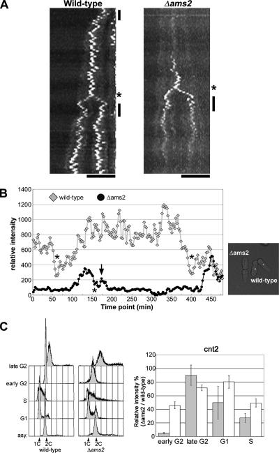
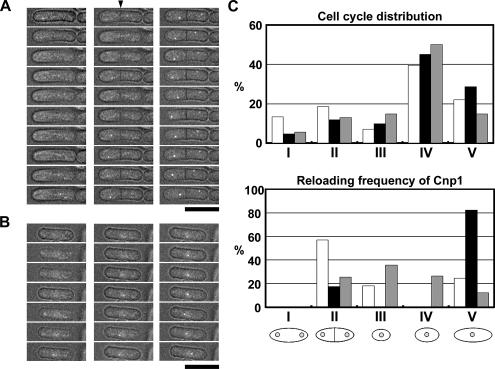
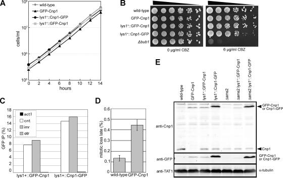
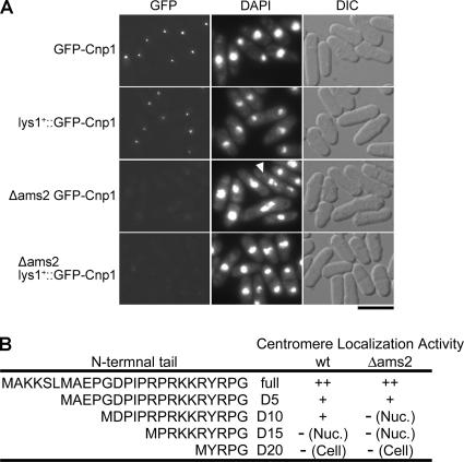
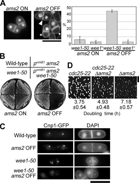
CENP-A is a centromere-specific histone H3 variant that is essential for kinetochore formation. Here, we report that the fission yeast Schizosaccharomyces pombe has at least two distinct CENP-A deposition phases across the cell cycle: S and G2. The S phase deposition requires Ams2 GATA factor, which promotes histone gene activation. In Delta ams2, CENP-A fails to retain during S, but it reaccumulates onto centromeres via the G2 deposition pathway, which is down-regulated by Hip1, a homologue of HIRA histone chaperon. Reducing the length of G2 in Delta ams2 results in failure of CENP-A accumulation, leading to chromosome missegregation. N-terminal green fluorescent protein-tagging reduces the centromeric association of CENP-A, causing cell death in Delta ams2 but not in wild-type cells, suggesting that the N-terminal tail of CENP-A may play a pivotal role in the formation of centromeric nucleosomes at G2. These observations imply that CENP-A is normally localized to centromeres in S phase in an Ams2-dependent manner and that the G2 pathway may salvage CENP-A assembly to promote genome stability. The flexibility of CENP-A incorporation during the cell cycle may account for the plasticity of kinetochore formation when the authentic centromere is damaged.


Figures

Figure 1.
Figure 1.
Cnp1-GFP is accumulated on centromeres during G2, and it is diminished after S in Δams2. (A) Wild-type (SP92) and Δams2 (SP75) cells expressing Cnp1-GFP were cultured in EMM2 at 33°C. Representative time-lapse images of GFP signals were converted to kymographs. Asterisks and vertical lines indicate the positions of mitotic and septated cells, respectively. Bars, 10 μm. (B) Wild-type (SP1055) and Δams2 (SP1698) cells expressing Cnp1-GFP were mixed and cultured in EMM2 at 33°C. Because the wild-type cell carries the Sid4-mRFP gene in the genome, it can be distinguished from the Δams2 cell by detecting monomeric red fluorescent protein signals before starting the live observation. A series of time-lapse images were taken at 2.5-min intervals. The graph shows the corresponding intensity of a centromeric GFP-dot in the nucleus plotted as diamonds for wild-type cell and circles for Δams2 cell. The asterisk indicates the period during which the centromeric GFP signals became weak. This period is presumed to be coincident with phase II (from prometaphase to anaphase A), at which centromere clustering becomes loose. After the M phase, we monitored the centromeric signals in one of two daughter cells, which was correctly on the optical plane. The arrow indicates G1/S phase at which a short uptake of Cnp1 incorporation took place. The corresponding movie is shown as Video 4. (C) ChIP assay to estimate the amount of centromere-bound Cnp1 during the cell cycle. Wild-type and Δams2 cells arrested at G1, S, early G2 or late G2 phase were prepared in the presence (gray bars) or absence (white bars) of the native promoter-driven Cnp1-GFP gene at the lys1 locus (see Materials and Methods). DNA contents of wild-type and Δams2 cells by using ChIP assay were estimated by flow cytometry (left). The positions of 1C and 2C peaks are also indicated. DNAs coprecipitated with Cnp1 by using anti-GFP mAb (gray bars) or anti-Cnp1 polyclonal antibody (white bars) were quantified by real-time PCR by using a cnt2 (central core region of cen2) probe (right). The error bars indicate SD from three independent experiments for early G2 cells or five independent experiments for the others.
Figure 2.
Figure 2.
Ams2 promotes Cnp1 incorporation, whereas Hip1 suppresses the cell cycle-independent loading of Cnp1. (A and B) Wild-type cells containing the Cnp1ts-GFP gene integrated at the lys1 locus (SP1102) were cultured in EMM2 at 36°C, and then they were shifted to 22°C. Representative time-lapse images of GFP signals incorporated into centromeres in S (A) and G2 (B). The arrowhead indicates the position of the septum. A series of time-lapse images were taken at 1.5-min intervals. Bar, 10 μm. (C) Summary of reloading experiments for Cnp1ts-GFP in the wild-type (SP1102), Δams2 (SP1205), or Δhip1 (PHS10) background. Stage I corresponds to M/G1; stage II, G1/S; stage III, S/early G2; stage IV, mid-G2; and stage V, late G2. The cell cycle distribution (top; percentages of cell population) and the reloading frequency of Cnp1 (bottom; percentages of cells in which Cnp1ts-GFP signals occurred at the corresponding stage per total examined cells) are shown. White, black, and gray bars represent data for the wild-type (n = 240), Δams2 (n = 175), and Δhip1 (n = 98) backgrounds, respectively.
Figure 3.
Figure 3.
N-terminal GFP-tagged Cnp1 is functional in normal cell cycle progression in wild-type cells. (A) Increase in number of the cells in culture in YES at 33°C. Cells expressing the authentic Cnp1 gene (wild-type, SP91), the N-terminal tagged GFP-Cnp1 gene (GFP-Cnp1, SP1769), both the authentic Cnp1 gene and the N-terminal tagged GFP-Cnp1 gene additionally integrated at the lys1 locus (lys1+::GFP-Cnp1, SP1468), and both of the authentic Cnp1 gene and the C-terminal tagged Cnp1-GFP gene additionally integrated at the lys1 locus (lys1+::Cnp1-GFP, SP38) were examined. (B) Fivefold serial dilutions of 2 × 10⁴ cells of wild-type (SP91), GFP-Cnp1 (SP1769), lys1+::GFP-Cnp1 (SP1468), lys1+::Cnp1-GFP (SP38), and Δbub1 (SP1339) were spotted on YES plates in the absence or presence of 6 μg/ml CBZ and incubated at 33°C for 3 d. Δbub1 was used as a representative CBZ-sensitive mutant. (C) ChIP assay was performed to confirm that the N-terminal tagged GFP-Cnp1 protein binds correctly to the central core regions of the centromere. Asynchronous cells of GFP-Cnp1 (SP1769) and lys1+::Cnp1-GFP (SP38) strains were prepared by inoculation into YES at 33°C. DNAs coprecipitated with GFP-tagged Cnp1 by using anti-GFP antibody or without antibody (data not shown) from cell extracts were quantified by real-time PCR by using cnt2, imr1, otr (dg), and act1 (background control) probes. (D) The mitotic loss rates of a linear minichromosome, CN2, were examined in wild-type cells (nontagged Cnp1, SP52) and wild-type cells expressing GFP-Cnp1 (GFP-Cnp1, PHS165) cultured in YES (nonselective medium) at 33°C. Error bars indicate SD from six independent experiments. (E) Cell extracts were prepared from wild-type cells expressing authentic Cnp1 (SP91), GFP-Cnp1 (SP1769), lys1+::GFP-Cnp1 (SP1468) and lys1+::Cnp1-GFP (SP92), and Δams2 cells expressing authentic Cnp1 (YTP155), lys1+::GFP-Cnp1 (SP1637) and lys1+::Cnp1-GFP (SP75) cultured in YES at 33°C. Levels of endogenous and exogenous Cnp1 protein expression were examined by immunoblotting by using anti-GFP and anti-Cnp1 antibodies. α-Tubulin detected by TAT1 antibody was used as a loading control.
Figure 4.
Figure 4.
The intact N-terminal tail plays a role in Ams2-dependent retention of Cnp1 at the centromere. (A) N-terminally tagged Cnp1 exerts synthetic lethal effect on Ams2 depletion. Wild-type cells expressing GFP-Cnp1 (SP1769) and lys1+::GFP-Cnp1 (SP1468) and Δams2 cells expressing GFP-Cnp1 (cell samples were prepared from germinated spores) and lys1+::GFP-Cnp1 (SP1637) were cultured in EMM2 at 26°C. Representative images of GFP signals and DAPI staining are shown. Arrowheads indicate the chromosome missegregation observed in Δams2 cells expressing GFP-Cnp1. Bar, 10 μm. (B) The centromeric localization activities of N-terminally deleted derivatives are summarized as indicated by ++ and + for strong and weak centromere-like signals, respectively, and − for noncentromeric, dispersed nuclear (nuc) or cellular (cell) signals.
Figure 5.
Figure 5.
G2 phase acts as the second chance for Cnp1 incorporation. (A) Nuclear morphology (YTP370) stained with DAPI and frequencies of binucleate cells with asymmetric nuclei of pnmt81-ams2 (ams2-shut-off strain, YTP366) or pnmt81-ams2 wee1-50 double (YTP370) mutant cultured in EMM2 in the absence (ams2-ON) or presence (ams2-OFF) of thiamine at 33°C for 4 h. The error bars indicate standard deviations from three independent experiments. Arrowheads indicate cells with large and small daughter nuclei. Septated cells with a single nucleus (asterisks) were counted as cells with asymmetric nuclei. Bar, 10 μm. (B) Synthetic lethality of Ams2-null with wee1-50 mutation. Colony formation of wild-type (SP143), wee1-50 (YTP374), pnmt81-ams2 (YTP366), and pnmt81-ams2 wee1-50 (YTP370) on EMM2 in the absence (ams2-ON) or presence (ams2-OFF) of thiamine at 33°C for 4 d. (C) Cnp1-GFP localization and DAPI staining of representative mitotic wild-type (WT, SP92), pnmt81-ams2 (ams2-OFF, YTP389), wee1-50 (YTP393), and pnmt81-ams2 wee1-50 (ams2-OFF wee1-50, YTP390) cells expressing Cnp1-GFP. Cells were cultured in EMM2 at 33°C for 3 h after addition of thiamine. Bar, 10 μm. (D) Colony formation of cdc25-22 (YTP379), cdc25-22 Δams2 (YTP354), and Δams2 (YTP155) on YES plate at 26°C for 5 d. Bar, 1 cm. The average doubling times of each strain cultured in YES at 26°C are also shown with standard deviations from four independent experiments.

Similar articles

Cited by

References

    1. Ahmad K., Henikoff S. Centromeres are specialized replication domains in heterochromatin. J. Cell Biol. 2001;153:101–110. - PMC - PubMed
    1. Ahmad K., Henikoff S. The histone variant H3.3 marks active chromatin by replication-independent nucleosome assembly. Mol. Cell. 2002;9:1191–1200. - PubMed
    1. Amor D. J., Choo K. H. Neocentromeres: role in human disease, evolution, and centromere study. Am. J. Hum. Genet. 2002;71:695–714. - PMC - PubMed
    1. Black B. E., Foltz D. R., Chakravarthy S., Luger K., Woods V. L., Jr, Cleveland D. W. Structural determinants for generating centromeric chromatin. Nature. 2004;430:578–582. - PubMed
    1. Black B. E., Jansen L. E., Maddox P. S., Foltz D. R., Desai A. B., Shah J. V., Cleveland D. W. Centromere identity maintained by nucleosomes assembled with histone H3 containing the CENP-A targeting domain. Mol. Cell. 2007;25:309–322. - PubMed

Publication types

MeSH terms

Substances

LinkOut - more resources