. 2007 Dec;3(12):e188.
doi: 10.1371/journal.ppat.0030188.

The Legionella pneumophila IcmSW complex interacts with multiple Dot/Icm effectors to facilitate type IV translocation

Affiliations

The Legionella pneumophila IcmSW complex interacts with multiple Dot/Icm effectors to facilitate type IV translocation

Eric D Cambronne et al. PLoS Pathog. 2007 Dec.

Abstract

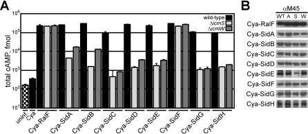
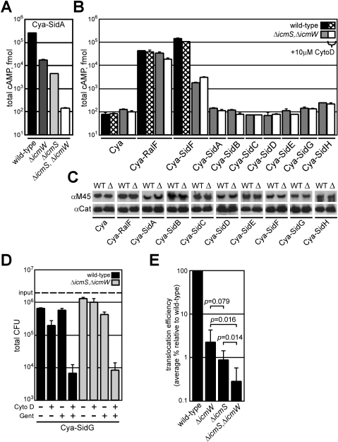
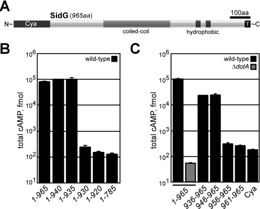
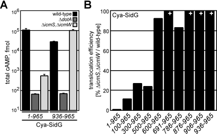
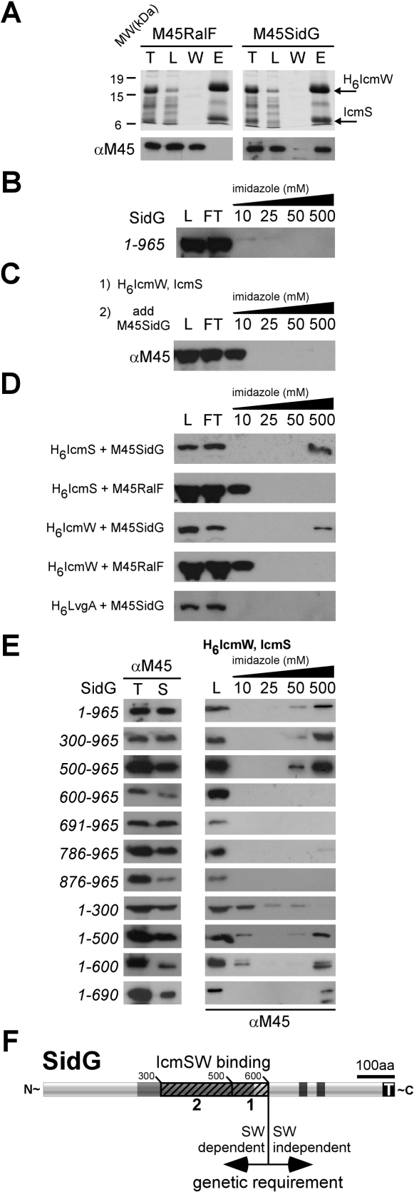
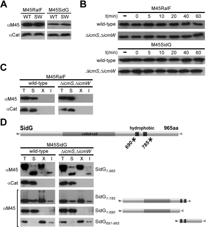
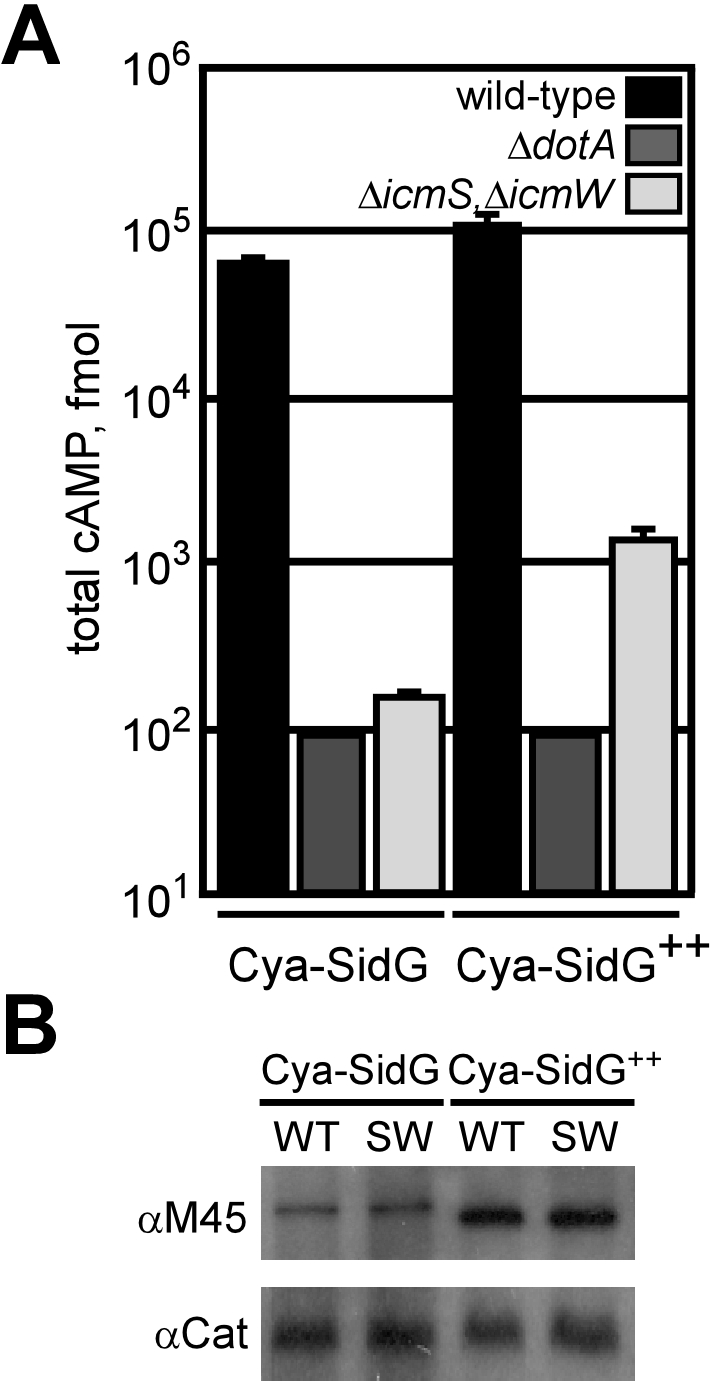
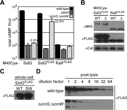
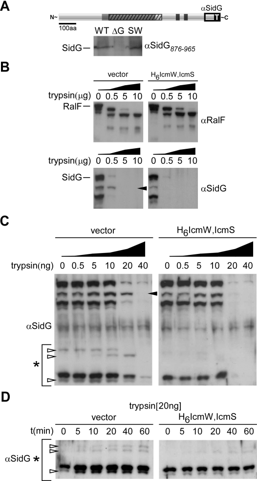
Many gram-negative pathogens use a type IV secretion system (T4SS) to deliver effector proteins into eukaryotic host cells. The fidelity of protein translocation depends on the efficient recognition of effector proteins by the T4SS. Legionella pneumophila delivers a large number of effector proteins into eukaryotic cells using the Dot/Icm T4SS. How the Dot/Icm system is able to recognize and control the delivery of effectors is poorly understood. Recent studies suggest that the IcmS and IcmW proteins interact to form a stable complex that facilitates translocation of effector proteins by the Dot/Icm system by an unknown mechanism. Here we demonstrate that the IcmSW complex is necessary for the productive translocation of multiple Dot/Icm effector proteins. Effector proteins that were able to bind IcmSW in vitro required icmS and icmW for efficient translocation into eukaryotic cells during L. pneumophila infection. We identified regions in the effector protein SidG involved in icmSW-dependent translocation. Although the full-length SidG protein was translocated by an icmSW-dependent mechanism, deletion of amino terminal regions in the SidG protein resulted in icmSW-independent translocation, indicating that the IcmSW complex is not contributing directly to recognition of effector proteins by the Dot/Icm system. Biochemical and genetic studies showed that the IcmSW complex interacts with a central region of the SidG protein. The IcmSW interaction resulted in a conformational change in the SidG protein as determined by differences in protease sensitivity in vitro. These data suggest that IcmSW binding to effectors could enhance effector protein delivery by mediating a conformational change that facilitates T4SS recognition of a translocation domain located in the carboxyl region of the effector protein.


Conflict of interest statement

Competing interests. The authors have declared that no competing interests exist.

Figures

Figure 1
Figure 1. The IcmSW Complex Is Necessary for the Translocation of Multiple Effector Proteins
(A) CHO FcγRII cells were left uninfected (hatch), or infected with wild-type (black), ΔicmS (light grey), or ΔicmW (dark grey) L. pneumophila strains expressing the indicated Cya hybrid proteins. After 1 h of infection, tissue culture cells were lysed and cAMP was extracted from the sample. Total cAMP production induced by translocation of the hybrid was quantified using an enzyme-immunoassay system, indicated as fmol. Results represent average values ± SD of experiments performed in triplicate. (B) Immunoblots of whole-cell bacterial extracts expressing indicated Cya hybrid proteins from wild-type (WT), ΔdotA (A), ΔicmS (S), and ΔicmW (W) probed with monoclonal antibody specific to the M45 epitope.
Figure 2
Figure 2. Translocation Defects Observed for the L. pneumophila ΔicmS, ΔicmW Double Mutant
(A) CHO FcγRII cells were infected with wild-type (black), ΔicmW (dark grey), ΔicmS (light grey), or ΔicmS, ΔicmW (white) strains expressing Cya-SidA. Total cAMP production was determined as in Figure 1. (B) CHO FcγRII cells were infected with wild-type (black) or ΔicmS, ΔicmW (grey) strains expressing the indicated Cya hybrid proteins for 1 h. Wild-type (hatch) or ΔicmS, ΔicmW (white) indicate samples that were conditioned with cytochalasin D prior to infection. Total cAMP production induced by translocation of the hybrid was determined as in (Figure 1). (C) Immunoblots of whole-cell bacterial extracts expressing indicated Cya hybrid proteins from wild-type (WT) or ΔicmS, ΔicmW (Δ) probed with monoclonal antibody specific to the M45 epitope. Immunoblots for chloramphenicol acetyltransferase (Cat) are included as a load control. (D) CHO FcγRII cells were infected with wild-type (black) or ΔicmS, ΔicmW (grey) L. pneumophila strains expressing Cya-SidG. Bacteria (3 × 10⁶; MOI = 30) were added to each well (input line) and subjected to the conditions indicated. +/− Cyto D indicates the addition of cytochalasin D (10 μM) 30 min prior to and during infection. +/− Gent indicates the addition of gentamycin (10 μM) 15 min post-infection. After 1 h of infection, tissue culture cells were washed and osmotically lysed. Lysates were serially diluted, plated, and incubated to determine total colony-forming units (CFU) from each condition. Bars represent average values ± SD of experiments performed in triplicate. (E) Graphical representation of the average translocation efficiency of the icmSW-dependent substrates SidA, SidB, SidC, SidD, SidE, SidG, and SidH. Translocation of each construct in wild-type is considered 100%. The average % defect is plotted for the ΔicmS, ΔicmW, and ΔicmS, ΔicmW strains ± SD of translocation assays performed in triplicate. p-Values from Student's t-test are indicated as comparisons between strains.
Figure 3
Figure 3. SidG Contains a C-Terminal Translocation Signal
(A) Schematic representation of the 965-aa SidG protein, which contains an extensive coiled-coil region (grey) as well as two hydrophobic segments (black). The identified translocation signal is depicted (T). (B and C) CHO FcγRII cells were infected with wild-type (black) or ΔdotA (dark grey) strains expressing the indicated Cya-SidG hybrid proteins for 1 h. Total cAMP production induced by translocation of the hybrid was determined as in Figure 1.
Figure 4
Figure 4. The C-Terminal Translocation Signal of SidG Supports icmSW-Independent Translocation
(A) CHO FcγRII cells were infected with wild-type (black), ΔdotA (dark grey), or ΔicmS, ΔicmW (light grey) strains expressing indicated Cya-SidG hybrid proteins for 1 h. Total cAMP production induced by translocation of the hybrid was determined as in Figure 1. (B) CHO FcγRII cells were infected with wild-type or ΔicmS, ΔicmW strains expressing the indicated Cya-SidG hybrid proteins for 1 h. Total cAMP production induced by translocation of the hybrid was determined for each strain. % values represent translocation efficiency as a measure of (ΔicmS, ΔicmW) / wild-type for the indicated Cya-SidG hybrid proteins. + indicates that cAMP production induced by translocation of the hybrid in ΔicmS, ΔicmW exceeded levels produced in wild-type.
Figure 5
Figure 5. IcmSW Complex Binds to a Region of SidG That Is Distinct from the C-Terminal Translocation Domain
(A) E. coli expressing H6IcmW/IcmS, and either M45-RalF or M45-SidG were lysed (T), clarified, and loaded (L) onto affinity columns, washed (W), and eluted (E). Fractions were resolved on SDS-PAGE, where H6IcmW/IcmS were detected with Coomassie staining, and RalF or SidG was detected by immunoblotting with M45-specific antisera. MW indicators for the gel are shown on the left (kDa). (B) E. coli expressing M45-SidG alone was subjected to affinity purification as in (A). M45-SidG was detected as in (A). (C) Lysate from E. coli expressing H6IcmW/IcmS was pre-charged onto affinity column (1), washed, and subjected to lysate from E. coli expressing M45-SidG (2) (L). Flow-through (FT) was collected and the column was washed and eluted with buffer containing the indicated concentration of imidazole. M45-SidG was detected as in (A). (D) E. coli strains expressing H6IcmS, H6IcmW, or H6LvgA and M45-SidG or M45-RalF were subjected to affinity purification as in (B). M45-SidG or M45-RalF was detected as in (A). (E) E. coli expressing H6IcmW/IcmS, and indicated M45-tagged SidG truncations were lysed (T), clarified (S), and loaded (L) onto affinity columns, and washed and eluted with buffer containing imidazole in the indicated concentrations. Purified M45-SidG fragments were resolved on SDS-PAGE and detected as in (A). (F) Schematic representation of the genetic (arrows) and biochemical (hatch) contribution of IcmSW on SidG translocation. At least one IcmSW binding site resides between residues 500 and 600 of SidG [1], with additional binding sites located between residues 300 and 500 [2]. The IcmSW binding region is distinct from the C-terminal translocation domain (T).
Figure 6
Figure 6. IcmSW Complex Does Not Influence the Stability, Solubility, or Sub-Cellular Localization of SidG
(A) Immunoblots of whole-cell bacterial extracts expressing M45-SidG or M45-RalF proteins from wild-type (WT) or ΔicmS, ΔicmW (SW) probed with monoclonal antibody specific to the M45 epitope. Immunoblots for chloramphenicol acetyltransferase (Cat) are included as a load control. (B) Aliquots containing total protein from stationary phase wild-type or ΔicmS, ΔicmW L. pneumophila strains were precipitated (−). Cultures were harvested, resuspended in media containing kanamycin, and allowed to incubate at 37 °C for times indicated. Aliquots were mixed in sample buffer, resolved on SDS-PAGE, and detected with immunoblotting. (C) Wild-type or ΔicmS, ΔicmW mutant L. pneumophila expressing M45-RalF was cultured to stationary phase, harvested, lysed, and clarified (T). Ultracentrifugation separated a soluble (S) fraction from insoluble material. Inner membrane protein extraction with TritonX-100 (X) and subsequent ultracentrifugation separated this population from insoluble material (I). Protein contained in each fraction was detected with SDS-PAGE and immunoblotting. Immunoblots for (Cat) are included as a soluble protein control. (D) Wild-type or ΔicmS, ΔicmW mutant L. pneumophila expressing M45-SidG or indicated (*) SidG truncation proteins were cultured and subjected to sub-cellular fractionation as in (C). Protein contained in each fraction was detected with SDS-PAGE and immunoblotting. Immunoblots for (Cat) are included as a soluble protein control.
Figure 7
Figure 7. Overproduction of SidG Can Bypass the icmSW Requirement
(A) CHO FcγRII cells were infected with wild-type (black), ΔdotA (dark grey), or ΔicmS, ΔicmW (light grey) L. pneumophila strains expressing the indicated Cya-SidG proteins for 1 h. Cultures that were grown in the presence of 1 mM IPTG prior to and during infection are indicated (Cya-SidG++). Total cAMP production induced by translocation of the hybrid was determined as in Figure 1. (B) Immunoblots of whole-cell bacterial extracts expressing Cya-SidG cultured in the absence or presence of 1 mM IPTG (Cya-SidG++). Proteins from wild-type (WT) or ΔicmS, ΔicmW (SW) were probed with monoclonal antibody specific to the M45 epitope. Immunoblots for chloramphenicol acetyltransferase (Cat) are included as a load control.
Figure 8
Figure 8. A C-Terminal FLAG Epitope Alters the Requirement for icmSW in SidG Translocation
(A) CHO FcγRII cells were infected with wild-type (black), ΔdotA (dark grey), or ΔicmS, ΔicmW (light grey) strains expressing the indicated Cya hybrid proteins. Total cAMP production induced by translocation of the hybrid was determined as in (Figure 1). % values represent translocation efficiency as a measure of (ΔicmS, ΔicmW) / wild-type. (B) Immunoblots of whole-cell bacterial extracts expressing Cya-SidGFLAG or Cya-RalFFLAG proteins from wild-type (WT) or ΔicmS, ΔicmW (Δ) probed with monoclonal antibodies specific to the M45 or FLAG epitopes. Immunoblots for chloramphenicol acetyltransferase (Cat) are included as a load control. (C) Immunoblots of whole-cell bacterial extracts expressing SidGFLAG from wild-type (WT) or ΔicmS, ΔicmW (SW) probed with monoclonal antibody specific to the FLAG epitope. (D) SidGFLAG was expressed in wild-type or ΔicmS, ΔicmW L. pneumophila strains to stationary phase. Cultures were harvested, lysed in the absence of protease inhibitors, and clarified. Lysates were subjected to serial dilutions that were mixed with sample buffer and analyzed by SDS-PAGE and immunoblotting with monoclonal antisera specific for the FLAG epitope.
Figure 9
Figure 9. IcmSW Binding to SidG Promotes Conformational Changes in the Effector Protein
(A) Schematic representation of the SidG polypeptide, with the IcmSW binding domain shown (hatch) as well as the indicated C-terminal domain (876–965) used for generation of polyclonal antisera. Full-length SidG was detected at equivalent levels from wild-type (WT) or ΔicmS, ΔicmW mutant L. pneumophila (SW) and was not detected in the sidG deletion strain (ΔG). (B) Recombinant RalF or SidG were co-expressed in the absence (vector) or presence of the IcmSW complex (H6IcmW,IcmS), and subjected to lysis and clarification. Aliquots of soluble whole-cell lysates were subjected to the indicated concentrations of trypsin for 1 h. Samples were subjected to SDS-PAGE and immunoblotting, where proteolytic fragments of RalF (RalF−) or SidG (SidG−) were detected with specific antisera raised against the indicated polypeptides. The black arrow indicates predominant cleavage product. (C) Lysates containing SidG were processed as in (B), except that trypsin was diluted 1,000-fold. Proteolytic cleavage fragments (white arrows) emerge in the absence of IcmSW in the molecular size range of 50–60 kDa as indicated by the asterisk. (D) Proteolytic time course experiment performed at indicated concentration (20 ng) on SidG-containing lysates. Immunoblots show differential cleavage products (arrows) in the 50–60 kDa molecular size range (*).

Similar articles

Cited by

References

    1. Cascales E, Christie PJ. The versatile bacterial type IV secretion systems. Nat Rev Microbiol. 2003;1:137–149. - PMC - PubMed
    1. Berger KH, Isberg RR. Two distinct defects in intracellular growth complemented by a single genetic locus in Legionella pneumophila . Mol Microbiol. 1993;7:7–19. - PubMed
    1. Marra A, Blander SJ, Horwitz MA, Shuman HA. Identification of a Legionella pneumophila locus required for intracellular multiplication in human macrophages. Proc Natl Acad Sci U S A. 1992;89:9607–9611. - PMC - PubMed
    1. Vogel JP, Andrews HL, Wong SK, Isberg RR. Conjugative transfer by the virulence system of Legionella pneumophila . Science. 1998;279:873–876. - PubMed
    1. Roy CR, Berger KH, Isberg RR. Legionella pneumophila DotA protein is required for early phagosome trafficking decisions that occur within minutes of bacterial uptake. Mol Microbiol. 1998;28:663–674. - PubMed

Publication types