. 2008 Jan;11(1):109-17.
doi: 10.1038/nn2028. Epub 2007 Dec 9.

A versatile prion replication assay in organotypic brain slices

Affiliations

A versatile prion replication assay in organotypic brain slices

Jeppe Falsig et al. Nat Neurosci. 2008 Jan.

Abstract

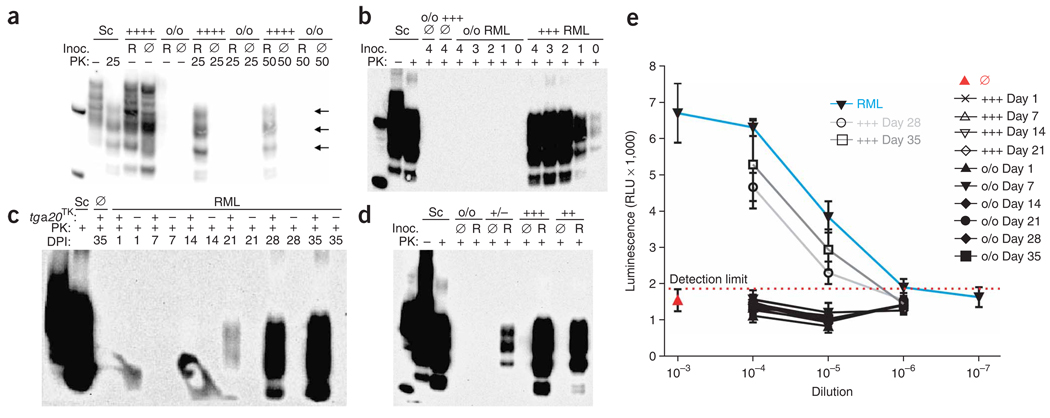
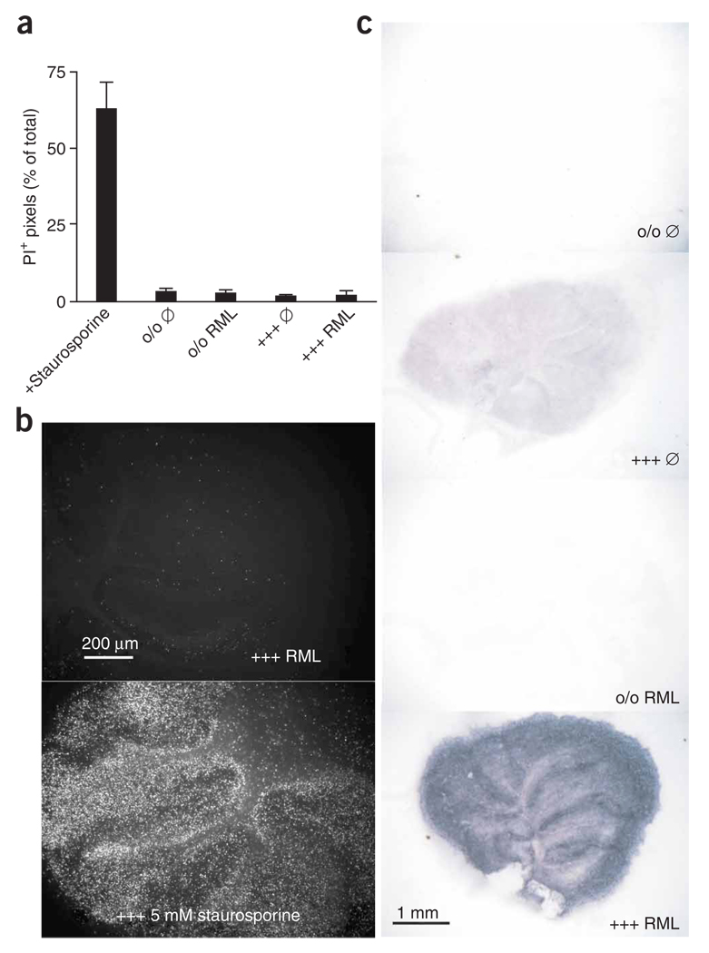
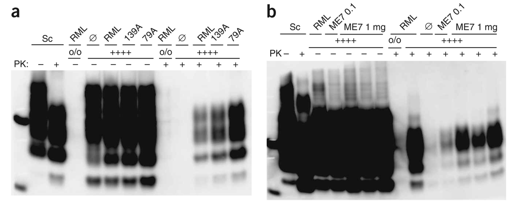
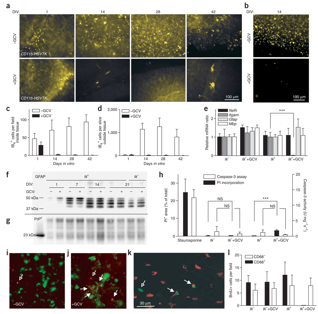
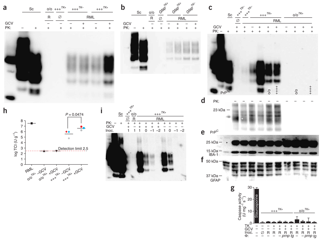
Methods enabling prion replication ex vivo are important for advancing prion studies. However, few such technologies exist, and many prion strains are not amenable to them. Here we describe a prion organotypic slice culture assay (POSCA) that allows prion amplification and titration ex vivo under conditions that closely resemble intracerebral infection. Thirty-five days after contact with prions, mouse cerebellar slices had amplified the abnormal isoform of prion protein, PrP(Sc), >10(5)-fold. This is quantitatively similar to amplification in vivo, but fivefold faster. PrP(Sc) accumulated predominantly in the molecular layer, as in infected mice. The POSCA detected replication of prion strains from disparate sources, including bovines and ovines, with variable detection efficiency. Pharmacogenetic ablation of microglia from POSCA slices led to a 15-fold increase in prion titers and PrP(Sc) concentrations over those in microglia-containing slices, as well as an increase in susceptibility to infection. This suggests that the extensive microglial activation accompanying prion diseases represents an efficacious defensive reaction.


Figures

Figure 1
Figure 1
Prions in slice cultures. (a) Immunoblot from tga20TK and Prnpo/o slices exposed to RML-infected (R) or uninfected (Ø) brain homogenate, cultured for 35 d, optionally digested with PK, and probed with antibody POM1 to PrP. Here and in all following figures, sample labels indicate the PrPC expression dictated by the slice genotype: o/o, Prnpo/o; +/−, hemizygous Prnp+/o; +/+, wild type; ++, hemizygous tga20 and Prnpo/o; +++, hemizygous for the tga20 allele and Prnp+/o; ++++, homozygous tga20 and Prnpo/o; Sc, positive-control homogenate from brain of a mouse with scrapie; inoc., inoculum. Left lane on all blots, molecular weight marker spiked with recombinant PrPC, yielding a PrP signal at 23 kDa with a cleavage product at 15 kDa. Arrows, PrPSc glycoforms. (b) Sensitivity of the POSCA. Immunoblot from tga20TK and Prnpo/o slices inoculated with decadal dilutions (104–100 µg; lane label, log(µg)) of prion-infected (RML) or uninfected (Ø) brain homogenate and cultured for 35 d before harvesting. (c) Time course of PrPSc accumulation in infected slices. Immunoblot from tga20TK (+) and Prnpo/o (−) slices inoculated with 100 µg prion-infected (RML) or uninfected (Ø) brain homogenate. (d) Immunoblot from Tga20TK, tga20+, Prnp+/o or Prnpo/o slices inoculated with 1 mg brain homogenate and harvested 35 dpi. (e) Time-dependent buildup of prion titers in slices. RML-infected or uninfected brain homogenate and slice homogenates from the experiment reported in panel c were serially tenfold diluted in uninfected brain homogenate, and infectivity was determined by transfer to PK1 cells. Means of three independent biological replicas ± s.d. RLU, relative light units.
Figure 2
Figure 2
Localization and impact of prion replication. (a) PI incorporation at 35 dpi in Tga20TK and Prnpo/o cultures inoculated with 100 µg RML or with uninfected homogenate (Ø). Average of 12 slices ± s.d. (b) Representative examples of images used for PI incorporation. Owing to its high cellularity, the granule cell layer of staurosporine-treated slices is prominently stained by PI. (c) Histoblot analysis of PrPSc deposition in POSCA slices. Tga20TK and Prnpo/o cultures were inoculated with 100 µg RML or uninfected homogenate (Ø). PrPSc was detected by immunoblotting with POM1 antibody. PrPSc was found to predominantly accumulate in the molecular layer.
Figure 3
Figure 3
A broad range of strains is detectable by POSCA. (a,b) Immunoblots from cultures from 10-d-old tga20+/+ (++++) and Prnpo/o (o/o) pups, inoculated with various prion strains (100 µg) and harvested after 35 d. Ø, noninfectious brain homogenate. (a) Transmission of mouse-adapted scrapie strains RML, 139A, 79A and Ø. (b) Western blotting performed under less stringent conditions on 30 µg protein digested with 25 µg ml−1 PK (+) or 10 µg undigested protein (−) and detected with POM1. Cultures were from 10-d-old tga20+/+ and Prnpo/o pups, and were inoculated with 0.1 and 1 mg of RML, ME7 or Ø and cultured for 35 d.
Figure 4
Figure 4
Microglial depletion in organotypic slice cultures. (a,b) Slices from 10-d-old CD11b-HSVTK+ mice cultured for 1, 14, 28 or 42 d (DIV) in the optional presence of GCV and stained with IB4 (a) or anti-CD68 (b). (c,d) Quantification of cells resident within (c) or outside (d) of slices. (e) Real-time RT-PCR of CD11b-HSVTK+ (tk+) and CD11b-HSVTK (tk) slices cultured for 14 d in presence or absence of GCV (n = 3). mRNAs encoding neurofilament (Nefh), CD11b (Itgam), GFAP (Gfap) and MBP (Mbp) are shown. (f,g) Western blotting from cultures prepared from 10-d-old tk+ and tk mice cultured with or without GCV, performed on 10 µg protein and detected with mouse IgG1 anti-GFAP (clone GA5) (f) or mouse IgG1 anti-PrPC (POM1) (g). (h) PI incorporation in cultures prepared as in e and treated with GCV for 5 weeks (n = 12). Positive control: tk tissue treated with 5 µM staurosporine for 24 h. Caspase-3 enzymatic activity was measured and normalized to protein contents (n = 3 pools of four slices). (i,j) Tk+ slices untreated (i) or treated (j) with GCV for 10 d and then incubated while still alive with IB4 (green) and PI (red). Open arrows, IB4PI+ cells; closed arrows, IB4+PI+ cells. Scale bar in k applies to i,j as well. (k,l) BrdU incorporation in samples prepared as in e after 13 dpi. (k) Example of stained cells. Green, IgG1 anti-BrdU; red, IgG2a anti-CD68. Closed arrows, CD68+BrdU+ cells; open arrows, BrdU+CD68 cells. (l) Counts of CD68+BrdU+ and CD68BrdU+ cells (n = 6, each the average of five individual ×40 images). Averages of n replicas ± s.d.
Figure 5
Figure 5
Impact of microglial depletion on prion replication. Cultures were prepared from 10-d-old mice either CD11b-HSVTK+ (tga20TK+ or Prnpo/o/TK+) or CD11b-HSVTK (tga20TK−). (a) Western blot from infected tga20TK− and tga20TK+ slices treated with GCV. Samples were PK-digested and PrP detected with POM1. (b) Western blot from cultures from 10-d-old GFAP-HSVTK (GfapTK−), GFAP-HSVTK+ (GfapTK+) or Prnpo/o mice treated as in a. (c–h) Macrophage reconstitution of microglia-depleted tissue. Antigen detected is indicated in lower left corner of immunoblots. (c) Experiment performed as in a, showing marked increase in PrPSc accumulation upon GCV treatment of RML-infected tga20TK+ slices, which was counteracted by Prnpo/o or tga20+/+ macrophages (genotype at bottom of lanes). (d–f) Undigested sample detected with POM1 (d), rabbit anti-Iba1 (e; *unidentified cross-reacting protein) or mouse anti-GFAP (f). (g) Caspase-3 enzymatic activity, normalized to protein content. Averages of four biological replicas ± s.d. (NS, P > 0.05). (h) SCEPA of homogenates of tga20TK+ or Prnpo/o/TK+ slices treated as in a. Three independent biological replicas of tga20TK+ and single replicas of Prnpo/o/TK+ slices or RML were analyzed in tenfold dilution steps using 6–12 replica PK1-containing wells per dilution. Data are indicated as the number of infectious tissue culture units per gram of slice culture protein and are the averages of biological replicas ± s.d. Slices from the same animal (pairs of −GCV and +GCV samples) are represented by the same color. (i) Immunoblot from cultures from 10-d-old tga20TK+ or Prnpo/o/TK+ mice uninfected (Ø) or infected with RML in tenfold dilution steps from 1.7 to −1.3 log SCI50; log(µg) inoculum is indicated above each lane. Each sample was divided into two pools (−GCV and +GCV) and harvested 35 dpi.

Similar articles

Cited by

References

    1. Aguzzi A, Polymenidou M. Mammalian prion biology. One century of evolving concepts. Cell. 2004;116:313–327. - PubMed
    1. Büeler H, et al. Mice devoid of PrP are resistant to scrapie. Cell. 1993;73:1339–1347. - PubMed
    1. Prusiner SB, et al. Measurement of the scrapie agent using an incubation time interval assay. Ann. Neurol. 1982;11:353–358. - PubMed
    1. Klohn PC, Stoltze L, Flechsig E, Enari M, Weissmann C. A quantitative, highly sensitive cell-based infectivity assay for mouse scrapie prions. Proc. Natl. Acad. Sci. USA. 2003;100:11666–11671. - PMC - PubMed
    1. Solassol J, Crozet C, Lehmann S. Prion propagation in cultured cells. Br. Med. Bull. 2003;66:87–97. - PubMed

Publication types

MeSH terms