. 2007 Dec 3;179(5):935-50.
doi: 10.1083/jcb.200706034.

Site-specific ubiquitination exposes a linear motif to promote interferon-alpha receptor endocytosis

Affiliations

Site-specific ubiquitination exposes a linear motif to promote interferon-alpha receptor endocytosis

K G Suresh Kumar et al. J Cell Biol. .

Abstract

Ligand-induced endocytosis and lysosomal degradation of cognate receptors regulate the extent of cell signaling. Along with linear endocytic motifs that recruit the adaptin protein complex 2 (AP2)-clathrin molecules, monoubiquitination of receptors has emerged as a major endocytic signal. By investigating ubiquitin-dependent lysosomal degradation of the interferon (IFN)-alpha/beta receptor 1 (IFNAR1) subunit of the type I IFN receptor, we reveal that IFNAR1 is polyubiquitinated via both Lys48- and Lys63-linked chains. The SCF(betaTrcp) (Skp1-Cullin1-F-box complex) E3 ubiquitin ligase that mediates IFNAR1 ubiquitination and degradation in cells can conjugate both types of chains in vitro. Although either polyubiquitin linkage suffices for postinternalization sorting, both types of chains are necessary but not sufficient for robust IFNAR1 turnover and internalization. These processes also depend on the proximity of ubiquitin-acceptor lysines to a linear endocytic motif and on its integrity. Furthermore, ubiquitination of IFNAR1 promotes its interaction with the AP2 adaptin complex that is required for the robust internalization of IFNAR1, implicating cooperation between site-specific ubiquitination and the linear endocytic motif in regulating this process.


Figures
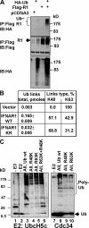
Figure 1.
Figure 1.
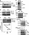
Both K48- and K63-linked polyubiquitin chains are present on IFNAR1 in cells and can be formed by SCFβTrcp in vitro. (A) In vivo ubiquitination of IFNAR1 expressed in 293T cells as indicated, purified via denaturing immunoprecipitation and analyzed by immunoblotting using the indicated antibodies. (B) Amount of IFNAR1-conjugated polyubiquitin chain links (in pmoles) and relative percentage of K48 and K63 linkages found in these preparations were measured by the absolute quantification of ubiquitin adducts technique. (C) In vitro formation of polyubiquitin chains by the SCFβTrcp E3 ligase. Recombinant SCFβTrcp (absent in lane 1) was incubated with E1, E2 (UbcH5c or Cdc34 as indicated), biotinylated ubiquitin (wt, lanes 1–3 and 7–8; R48K, lanes 4, 6, and 9; R63K, lanes 5, 6, and 10), and ATP. The reaction was analyzed by immunoblotting using HRP-conjugated streptavidin.
Figure 2.
Figure 2.
Polyubiquitination is required for IFNAR1 degradation but is dispensable for the phosphorylation of IFNAR1 and its interaction with βTrcp as well as for assembly of the SCFβTrcp E3 ubiquitin ligase. (A) Treatment of cells with IFN stimulates the degradation of endogenous IFNAR1, which was measured as IFNAR1 levels in lysates from cells treated by cycloheximide (a cycloheximide chase) detected by immunoprecipitation/immunoblotting using anti-IFNAR1 antibodies. Control precipitation (NS) was performed using an isotype monoclonal antibody. Positions of fully glycosylated mature IFNAR1 (Ragimbeau et al., 2003) and heavy chain of Igs are indicated by the arrows. (B) Degradation of endogenous IFNAR1 in the presence of IFN-α was analyzed by cycloheximide chase in as in A. (C) Degradation of exogenous IFNAR1 in cells coexpressed with either wild-type ubiquitin or K0 mutant analyzed by cycloheximide chase in the presence of IFN-α using anti-Flag antibody. Results are graphed as a percentage of IFNAR1 remaining at the indicated time points of a chase. (D) Ubiquitination of endogenous IFNAR1 in the IFN-α–treated cells expressing either wild-type or K0 ubiquitin mutant was assessed as in Fig. 1 A. (E) Ser535 phosphorylation of endogenous IFNAR1 in cells expressing either wild-type or K0 ubiquitin mutant and treated or not treated with IFN-α was assessed by immunoprecipitation followed by immunoblotting using the indicated antibodies. (F and G) Effects of K0 expression on the recruitment of myc-tagged βTrcp to either Flag-tagged IFNAR1 (F) or Flag-tagged Cullin1 (G) were analyzed by coimmunoprecipitation followed by immunoblotting using the indicated antibodies. WCE, whole cell extract.
Figure 3.
Figure 3.
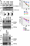
Role of specific polyubiquitin chains in IFNAR1 ubiquitination and degradation. (A, B, and D) Effect of expression of various ubiquitin mutants on the degradation of endogenous (A) and exogenous (B and D) IFNAR1 was assessed via cycloheximide chase in the presence of IFN-α as in Fig. 2 C. Data are represented as the average ± SEM (error bars). (C and E) Effect of these ubiquitin mutants' expression on the extent of IFNAR1 ubiquitination was measured similarly to the experiment shown in Fig. 1 A using antiubiquitin antibody. Ratios between ubiquitin and IFNAR1 signals were calculated as the percentage of such for wild-type ubiquitin (assigned a value of 100%).
Figure 4.
Figure 4.
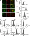
Role of IFNAR1 polyubiquitination in mediating the efficient postinternalization sorting of IFNAR1. (A) Localization of internalized Flag-tagged IFNAR1wt and IFNAR1KR. IFNAR1 proteins were labeled for 1 h in the presence of IFN-α, anti-Flag antibody, and TRITC-conjugated secondary Fab and were washed and chased (allowed to internalize) for an additional 30 min. Lysosomes and recycling endosomes were labeled with FITC-dextran and FITC-transferrin (Tfr), respectively. Dextran completely colocalized with Lamp1 (Fig. S2, available at http://www.jcb.org/cgi/content/full/jcb.200706034/DC1). Single optical sections of representative cells obtained by laser confocal fluorescence microscopy are depicted. A similar localization of IFNAR1KR was also observed when chase time was increased to 90 min. Bars, 10 μm. (B) Vesicular pH (pHv) of Flag-tagged IFNAR1-containing endosomes/lysosomes in transiently transfected HEK293T cells. Endosomal pH was measured by FRIA upon labeling and 30-min chase, and the frequency distribution of the pHv among the indicated number of analyzed vesicles (n) was plotted. After calibration, the ratios are expressed in pH values. A similar distribution for IFNAR1KR was also observed at 90-min chase. The IFNAR1KR-RPT mutant contains an additional ubiquitination repeat module and is ubiquitination competent (see Fig. 6). (C) Postendocytotic sorting of Flag-tagged IFNAR1wt in cells that coexpress ubiquitin constructs (as indicated, upon labeling and chase for 30 min) was monitored by pHv measurements as described in B. Data are expressed as the frequency of pHv and mean ± SEM (n = 3).
Figure 5.
Figure 5.
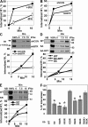
Role of ubiquitination in IFNAR1 internalization. (A) Effect of IFN-α (black squares) on the internalization of endogenous IFNAR1 measured by a fluorescence assay. All other experiments were performed in the presence of IFN-α. (B and C) Effect of βTrcp2 knockdown (shBTR) on IFNAR1 internalization observed by fluorescence assay (B; white squares) or biotinylation assay (C). (C) The internalized, biotinylated IFNAR1 analyzed by immunoblotting using anti-Flag antibody appears at the time points indicated in minutes on the top. 100% indicates the sample that represents the total amount of biotinylated IFNAR1. Quantification of these panels was calculated as (signal at time point X – signal at time point 0)/signal at 100%. (D and E) Comparison of the internalization of wild-type IFNAR1 (black squares) and its ubiquitination-deficient mutants IFNAR1SA and IFNAR1SA-RPT (D; white squares and diamonds, respectively) and IFNAR1KR (E; triangles) that was assessed by biotinylation assays. NB, nonbiotinylated control. (F) Effect of expression of the indicated ubiquitin mutants on the internalization of IFNAR1 assessed by fluorescent assay. Data are depicted as the percentage of IFNAR1 internalization detected in cells expressing wild-type ubiquitin (100%) at 15 min. *, P < 0.05 (t test; differences compared with wild-type ubiquitin). Error bars represent SEM.
Figure 6.
Figure 6.
Site-specific ubiquitination is required for efficient internalization of IFNAR1. (A) Diagram of the intracellular domains of IFNAR1 proteins, including generated IFNAR1 mutants. Positions of S535 and S539 residues within the phosphodegron, of ubiquitin-acceptor cluster K501/525/526, and of Y466 within the linear endocytic motif are shown. (B) Ser535 phosphorylation of Flag-tagged IFNAR1 proteins expressed in IFN-α–treated 293T cells was analyzed by immunoblotting as in Fig. 1 F. Vec, empty vector. (C) Interaction of HA-tagged βTrcp with coexpressed Flag-tagged IFNAR1 proteins in IFN-α–treated 293T cells was analyzed by immunoprecipitation/immunoblotting using the indicated antibodies. (D) Ubiquitination of IFNAR1 proteins in IFN-α–treated 293T cells was analyzed as in Fig. 3 C. Ratios between ubiquitin and IFNAR1 signals were calculated as the percentage of such for wild-type IFNAR1. (E and F) Internalization of Flag-tagged IFNAR1 proteins was measured similar to the experiment shown in Fig. 5 A using anti-Flag antibodies. Error bars represent SEM.
Figure 7.
Figure 7.
Cooperation between IFNAR1 ubiquitination and exposure of the linear Tyr-based endocytic motif. (A) Internalization of IFNAR1wt and IFNAR1Y466F was assessed by biotinylation assay as in Fig. 5 D. (B) Degradation of the indicated IFNAR1 proteins was analyzed by cycloheximide chase as in Fig. 2 C. (C–E) Interaction of endogenous (C) or exogenous (D and E) IFNAR1 proteins with AP50 under the indicated conditions was analyzed by immunoprecipitation/immunoblotting using the indicated antibodies. The fraction of AP50 bound to IFNAR1 (calculated as a thousandth) is shown. NS, immunoprecipitation with irrelevant nonspecific monoclonal antibody. (F) Effect of AP2 knockdown on the internalization of endogenous IFNAR1 was analyzed as in Fig. 5 B. Control siRNA (siGL3) were targeted against luciferase. Error bars represent SEM.

Similar articles

Cited by

References

    1. Barriere, H., C. Nemes, D. Lechardeur, M. Khan-Mohammad, K. Fruh, and G.L. Lukacs. 2006. Molecular basis of oligoubiquitin-dependent internalization of membrane proteins in mammalian cells. Traffic. 7:282–297. - PubMed
    1. Blondel, M.O., J. Morvan, S. Dupre, D. Urban-Grimal, R. Haguenauer-Tsapis, and C. Volland. 2004. Direct sorting of the yeast uracil permease to the endosomal system is controlled by uracil binding and Rsp5p-dependent ubiquitylation. Mol. Biol. Cell. 15:883–895. - PMC - PubMed
    1. Bonifacino, J.S., and L.M. Traub. 2003. Signals for sorting of transmembrane proteins to endosomes and lysosomes. Annu. Rev. Biochem. 72:395–447. - PubMed
    1. Bonifacino, J.S., and A.M. Weissman. 1998. Ubiquitin and the control of protein fate in the secretory and endocytic pathways. Annu. Rev. Cell Dev. Biol. 14:19–57. - PMC - PubMed
    1. Brzovic, P.S., and R.E. Klevit. 2006. Ubiquitin transfer from the E2 perspective: why is UbcH5 so promiscuous? Cell Cycle. 5:2867–2873. - PubMed

Publication types

Substances