. 2007 Nov;3(11):e183.
doi: 10.1371/journal.ppat.0030183.

Secreted NS1 of dengue virus attaches to the surface of cells via interactions with heparan sulfate and chondroitin sulfate E

Affiliations

Secreted NS1 of dengue virus attaches to the surface of cells via interactions with heparan sulfate and chondroitin sulfate E

Panisadee Avirutnan et al. PLoS Pathog. 2007 Nov.

Abstract

Dengue virus (DENV) nonstructural protein-1 (NS1) is a secreted glycoprotein that is absent from viral particles but accumulates in the supernatant and on the plasma membrane of cells during infection. Immune recognition of cell surface NS1 on endothelial cells has been hypothesized as a mechanism for the vascular leakage that occurs during severe DENV infection. However, it has remained unclear how NS1 becomes associated with the plasma membrane, as it contains no membrane-spanning sequence motif. Using flow cytometric and ELISA-based binding assays and mutant cell lines lacking selective glycosaminoglycans, we show that soluble NS1 binds back to the surface of uninfected cells primarily via interactions with heparan sulfate and chondroitin sulfate E. DENV NS1 binds directly to the surface of many types of epithelial and mesenchymal cells yet attaches poorly to most peripheral blood cells. Moreover, DENV NS1 preferentially binds to cultured human microvascular compared to aortic or umbilical cord vein endothelial cells. This binding specificity was confirmed in situ as DENV NS1 bound to lung and liver but not intestine or brain endothelium of mouse tissues. Differential binding of soluble NS1 by tissue endothelium and subsequent recognition by anti-NS1 antibodies could contribute to the selective vascular leakage syndrome that occurs during severe secondary DENV infection.


Conflict of interest statement

Competing interests. The authors have declared that no competing interests exist.

Figures

Figure 1
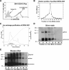
Figure 1. Purification of DENV NS1 from BHK DENV-2 Rep Cells
(A) A flow cytometry histogram of expression of DENV NS1 in BHK DENV-2 Rep cells. Cells were fixed and stained with anti-DENV-2 NS1 mAbs or isotype control Abs. One representative experiment of three is shown. (B) Elution profile of purified NS1 after immunoaffinity chromatography of serum-free supernatants of BHK DENV-2 Rep cells. Peak fractions (19–21) were combined and passed over an ion-exchange column, and purified NS1 was eluted (C) with a linear NaCl gradient (diagonal line). (D) SDS-PAGE of purified soluble DENV NS1 from BHK DENV-2 Rep cells. Purified NS1 was boiled prior to nonreducing 12% SDS-PAGE and silver staining. Molecular weight markers are shown at the left. (E) Immunoreactivity of purified DENV-2 NS1. Western blot analysis of purified DENV-2 NS1 after nonreducing 12% SDS-PAGE was performed using the 2G6 anti-DENV NS1 mAb.
Figure 2
Figure 2. DENV NS1 Binds to Cells of Specific Lineages
Dose-dependent binding of DENV NS1 to cell surfaces. Purified DENV NS1 (5, 10, 20, and 40 μg/ml) was added to different cell types at 4 °C for 1 h. After washing, bound NS1 was detected by anti-NS1 antibody staining and flow cytometry. An example of DENV NS1 binding to CHO-K1 cells using supernatant from BHK-DENV-2 Rep cells is depicted in the inset at the upper right corner of the top left histogram. Examples of histogram profiles of DENV NS1 binding to epithelial, fibroblast, hematopoietic, and endothelial cells are depicted. One representative of three experiments is shown. No specific staining was observed after BSA binding and incubation with an NS1 antibody or NS1 binding and incubation with an isotype-matched control antibody (unpublished data).
Figure 3
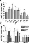
Figure 3. Binding of DENV NS1 to Cell Surface Is GAG-Dependent
(A) DENV NS1 binding to wild type (K1) and seven mutant CHO cell lines that overexpress (K1.5, K1.N7, PPP6, and 3OST5–1) or are defective (M1, H8, and 745) for specific enzymes in GAG synthesis. Binding and staining were performed at 4 °C and analyzed by flow cytometry. Data are expressed as the specific MFI of staining after subtraction of background antibody binding in the absence of NS1. The error bars indicate standard deviation (SD) corresponding to three independent experiments. Asterisks note NS1 binding that is statistically different from the CHO-K1 wild-type cells (* p < 0.05, ** p < 0.005). (B) Cell surface HS and CS were enzymatically removed from BHK or wild-type CHO-K1 cells after treatment with a mixture of heparin lyases I, II, III (H. lyases) or chondroitinase ABC (ABCase) and assayed for binding of DENV NS1. A GAG-negative mutant cell (CHO-745) was used as a negative control. Analysis was performed by flow cytometry. Data are expressed as the specific MFI of staining after subtraction of background antibody binding in the absence of NS1. The error bars indicate SD corresponding to three independent experiments. Asterisks indicate DENV-2 NS1 binding that is statistically different from untreated cells (* p < 0.05, ** p < 0.005).
Figure 4
Figure 4. Soluble HP and CS-E Block DENV NS1 Binding to the Surface of BHK and HMEC
DENV NS1 was incubated with three concentrations of indicated soluble GAG prior to incubation with (A–C) BHK or (D–F) HMEC. DENV NS1 was detected by flow cytometry (as described in Figure 2). Data are expressed as the specific MFI of staining as described in Figure 3. The error bars indicate SD corresponding to three independent experiments. Asterisks denote NS1 binding that is statistically different from untreated cells (* p < 0.05, ** p < 0.005). Examples of histogram profiles in which soluble HP and CS-E at 1 μg/ml concentration block DENV NS1 binding to the surface of (B–C) BHK and (E–F) HMEC are depicted.
Figure 5
Figure 5. Direct Binding of DENV NS1 with HS and CS-E
Microtiter plates were coated with indicated soluble GAG overnight at 4 °C. After incubation with supernatants from BHK-DENV-2-Rep cells or control BHK cells, bound DENV NS1 was detected using DENV NS1–specific mAbs, followed by biotin-conjugated anti-mouse IgG and HRP-conjugated streptavidin. Plates were read at an optical density of 450 nm on a 96-well plate reader. Data are the mean ± SD from four independent experiments. Asterisks denote NS1 binding that is statistically different from binding to background (* p < 0.05, ** p < 0.005). O. D., optical density.
Figure 6
Figure 6. Sulfation Is Required for Soluble DENV NS1 Binding to Cells
(A–B) BHK, wild-type CHO-K1, and GAG-deficient mutant CHO-745 were cultured overnight in sulfate-free medium in the presence of indicated amounts of sodium chlorate. Cells were harvested and tested for NS1 binding by flow cytometry as described in Figure 2. No appreciable cell death was observed by this treatment as judged by exclusion of propidium iodide. Data (A) are expressed as the specific MFI of staining as described in Figure 3. The error bars indicate SD corresponding to three independent experiments. Asterisks denote NS1 binding that is statistically different from untreated cells (** p < 0.005) (C–D) Addition of sodium sulfate (10 mM) restores binding of NS1 to cells. Cells were cultured overnight in sulfate-free medium and 25 mM sodium chlorate with or without 10 mM sodium sulfate (as indicated). Cells were processed for DENV NS1 binding and analyzed by flow cytometry (see Figure 2). Data (C) are expressed as the specific MFI of staining as described in Figure 3. The error bars indicate SD corresponding to three independent experiments. Asterisks denote NS1 binding that is statistically different from untreated cells (** p < 0.005).
Figure 7
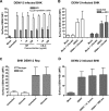
Figure 7. Expression of DENV NS1 on the Surface of Infected Cells Is Not Dependent on GAG
(A) BHK cells were infected with DENV-2 at the MOI of 3 and cultured for 24 h in the presence of indicated concentrations of soluble HP or CS-C. Cells were then harvested for analysis of surface NS1 expression. (B) BHK cells were infected with DENV-2 at the MOI of 3 and cultured for 24 h in the presence of 25 mM sodium chlorate. DENV NS1 expression was determined and analyzed by flow cytometry using anti-DENV NS1 mAb (2G6) or polyclonal anti-DENV NS1. (C) BHK DENV-2 Rep cells were cultured in medium containing 25 mM sodium chlorate for 48 h. Expression of NS1 on the cell surface was determined by using anti-DENV NS1 mAb (2G6) and analyzed by flow cytometry. (D) BHK cells were infected with DENV-2 at the MOI of 3. At 24 h after infection, the cells were treated with a mixture of heparin lyases I, II, III (H. lyases) or chondroitinase ABC (ABCase). NS1 expression was analyzed by flow cytometry using the 2G6 anti-DENV NS1 mAb. Data are the mean ± SD of three independent experiments as described in Figure 6.
Figure 8
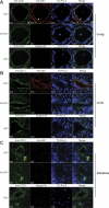
Figure 8. Binding of DENV NS1 to Mouse Tissues
Cryo-sections of mouse (A) lung, (B) liver, and (C) intestine were incubated with serum-free supernatants from BHK DENV-2 Rep or BHK cells for 1 h at room temperature. After extensive washing, bound NS1 was detected by a mixture of NS1 mAbs (1A4, 1F11, 2G6, 1B2) followed by Cy3-conjugated goat anti-mouse IgG. Co-staining with endothelial cell marker was subsequently performed by incubating the sections with rat anti-mouse CD31 (PECAM-1) followed by Alexa Fluor 488-conjugated goat anti-rat IgG. Nuclei were stained with a DNA-specific dye TO-PRO-3. Sections incubated with DENV NS1 followed by an isotype control Ab served as a negative control. Analysis was performed by confocal microscopy. White arrow and yellow arrowhead denote the layer of endothelial cells in the lumen and the outer layer of the adventitia of pulmonary vessel, respectively.
Figure 9
Figure 9. Binding of DENV NS1 to Human Lung
Cryo-sections of human lung tissue were incubated with 20 μg/ml of purified DENV-2 NS1 or BSA for 1 h at room temperature. Bound NS1 was detected by staining with a mixture of DENV NS1 mAbs (1A4, 1F11, 2G6, 1B2) followed by Alexa Fluor 488 conjugated with goat anti-mouse IgG. Subsequently, the sections were co-stained with rabbit anti-human CD31 (PECAM-1) followed by Cy3-conjugated donkey anti-rabbit IgG. Nuclei were stained with a DNA-specific dye (Hoechst) and the sections were analyzed by confocal microscopy. Sections incubated with purified DENV NS1 and stained with isotype mAbs served as a negative control.

Similar articles

Cited by

References

    1. Gubler DJ. Epidemic dengue/dengue hemorrhagic fever as a public health, social, and economic problem in the 21st century. Trends Microbiol. 2002;10:100–103. - PubMed
    1. Sangkawibha N, Rojanasuphot S, Ahandrik S, Viriyapongse S, Jatanasen S, et al. Risk factors in dengue shock syndrome: a prospective epidemiologic study in Rayong, Thailand. I. The 1980 outbreak. Am J Epidemiol. 1984;120:653–669. - PubMed
    1. Guzman MG, Kouri G, Valdes L, Bravo J, Alvarez M, et al. Epidemiologic studies on dengue in Santiago de Cuba, 1997. Am J Epidemiol. 2000;152:793–799. 804. discussion. - PubMed
    1. Nimmannitya S. Clinical spectrum and management of dengue haemorrhagic fever. Southeast Asian J Trop Med Public Health. 1987;18:392–397. - PubMed
    1. Rothman AL, Ennis FA. Immunopathogenesis of dengue hemorrhagic fever. Virology. 1999;257:1–6. - PubMed

Publication types

Substances