

Syndecan-3 is a dendritic cell-specific attachment receptor for HIV-1
- PMID: 18040049
- PMCID: PMC2148312
- DOI: 10.1073/pnas.0703747104
Syndecan-3 is a dendritic cell-specific attachment receptor for HIV-1
Abstract
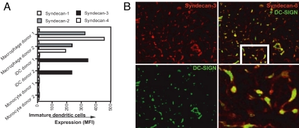
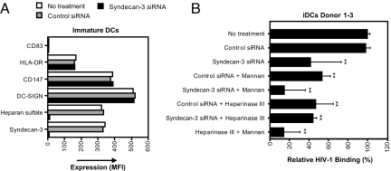
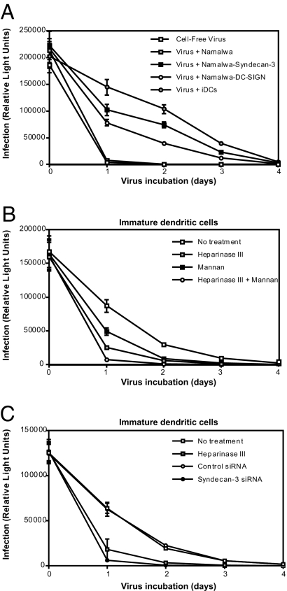
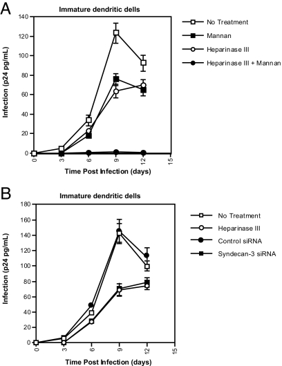
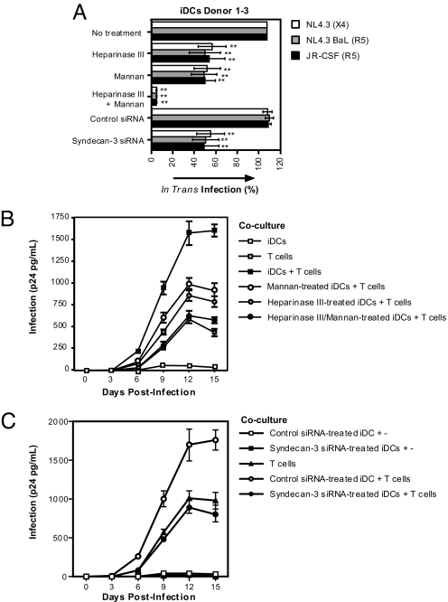
Dendritic cells (DCs) efficiently capture HIV-1 and mediate transmission to T cells, but the underlying molecular mechanism is still being debated. The C-type lectin DC-SIGN is important in HIV-1 transmission by DCs. However, various studies strongly suggest that another HIV-1 receptor on DCs is involved in the capture of HIV-1. Here we have identified syndecan-3 as a major HIV-1 attachment receptor on DCs. Syndecan-3 is a DC-specific heparan sulfate (HS) proteoglycan that captures HIV-1 through interaction with the HIV-1 envelope glycoprotein gp120. Syndecan-3 stabilizes the captured virus, enhances DC infection in cis, and promotes transmission to T cells. Removal of the HSs from the cell surface by heparinase III or by silencing syndecan-3 by siRNA partially inhibited HIV-1 transmission by immature DCs, whereas neutralizing both syndecan-3 and DC-SIGN completely abrogated HIV-1 capture and subsequent transmission. Thus, HIV-1 exploits both syndecan-3 and DC-SIGN to mediate HIV-1 transmission, and an effective microbicide should target both syndecan-3 and DC-SIGN on DCs to prevent transmission.
Conflict of interest statement
The authors declare no conflict of interest.
Figures
Similar articles
-
Lentivirus-mediated RNA interference of DC-SIGN expression inhibits human immunodeficiency virus transmission from dendritic cells to T cells.J Virol. 2004 Oct;78(20):10848-55. doi: 10.1128/JVI.78.20.10848-10855.2004. J Virol. 2004. PMID: 15452205 Free PMC article.
-
DC-SIGN-mediated infectious synapse formation enhances X4 HIV-1 transmission from dendritic cells to T cells.J Exp Med. 2004 Nov 15;200(10):1279-88. doi: 10.1084/jem.20041356. J Exp Med. 2004. PMID: 15545354 Free PMC article.
-
C-type lectin Mermaid inhibits dendritic cell mediated HIV-1 transmission to CD4+ T cells.Virology. 2008 Sep 1;378(2):323-8. doi: 10.1016/j.virol.2008.05.025. Epub 2008 Jul 1. Virology. 2008. PMID: 18597806
-
The interaction of immunodeficiency viruses with dendritic cells.Curr Top Microbiol Immunol. 2003;276:1-30. doi: 10.1007/978-3-662-06508-2_1. Curr Top Microbiol Immunol. 2003. PMID: 12797441 Review.
-
DC-SIGN: a novel HIV receptor on DCs that mediates HIV-1 transmission.Curr Top Microbiol Immunol. 2003;276:31-54. doi: 10.1007/978-3-662-06508-2_2. Curr Top Microbiol Immunol. 2003. PMID: 12797442 Review.
Cited by
-
Syndecans and Pancreatic Ductal Adenocarcinoma.Biomolecules. 2021 Feb 25;11(3):349. doi: 10.3390/biom11030349. Biomolecules. 2021. PMID: 33669066 Free PMC article. Review.
-
Spermatozoa capture HIV-1 through heparan sulfate and efficiently transmit the virus to dendritic cells.J Exp Med. 2009 Nov 23;206(12):2717-33. doi: 10.1084/jem.20091579. Epub 2009 Oct 26. J Exp Med. 2009. PMID: 19858326 Free PMC article.
-
TNF-alpha and TLR agonists increase susceptibility to HIV-1 transmission by human Langerhans cells ex vivo.J Clin Invest. 2008 Oct;118(10):3440-52. doi: 10.1172/JCI34721. J Clin Invest. 2008. PMID: 18776939 Free PMC article.
-
Syndecans in chronic inflammatory and autoimmune diseases: Pathological insights and therapeutic opportunities.J Cell Physiol. 2018 Sep;233(9):6346-6358. doi: 10.1002/jcp.26388. Epub 2018 Mar 25. J Cell Physiol. 2018. PMID: 29226950 Free PMC article. Review.
-
Cataloguing of Potential HIV Susceptibility Factors during the Menstrual Cycle of Pig-Tailed Macaques by Using a Systems Biology Approach.J Virol. 2015 Sep;89(18):9167-77. doi: 10.1128/JVI.00263-15. Epub 2015 Jun 24. J Virol. 2015. PMID: 26109722 Free PMC article.
References
-
- Royce RA, Sena A, Cates W, Jr, Cohen MS. N Engl J Med. 1997;336:1072–1078. - PubMed
-
- Cameron PU, Freudenthal PS, Barker JM, Gezelter S, Inaba K, Steinman RM. Science. 1992;257:383–387. - PubMed
-
- Pope M, Betjes MG, Romani N, Hirmand H, Cameron PU, Hoffman L, Gezelter S, Schuler G, Steinman RM. Cell. 1994;78:389–398. - PubMed
-
- Cameron PU, Lowe MG, Crowe SM, O'Doherty U, Pope M, Gezelter S, Steinman RM. J Leukoc Biol. 1994;56:257–265. - PubMed
Publication types
MeSH terms
Substances
Grants and funding
LinkOut - more resources
Full Text Sources
Other Literature Sources
