. 2007 Dec;9(12):1381-91.
doi: 10.1038/ncb1657. Epub 2007 Nov 18.

Arf6 and microtubules in adhesion-dependent trafficking of lipid rafts

Affiliations

Arf6 and microtubules in adhesion-dependent trafficking of lipid rafts

Nagaraj Balasubramanian et al. Nat Cell Biol. 2007 Dec.

Abstract

Integrin-mediated adhesion regulates membrane binding sites for Rac1 within lipid rafts. Detachment of cells from the substratum triggers the clearance of rafts from the plasma membrane through caveolin-dependent internalization. The small GTPase Arf6 and microtubules also regulate Rac-dependent cell spreading and migration, but the mechanisms are poorly understood. Here we show that endocytosis of rafts after detachment requires F-actin, followed by microtubule-dependent trafficking to recycling endosomes. When cells are replated on fibronectin, rafts exit from recycling endosomes in an Arf6-dependent manner and return to the plasma membrane along microtubules. Both of these steps are required for the plasma membrane targeting of Rac1 and for its activation. These data therefore define a new membrane raft trafficking pathway that is crucial for anchorage-dependent signalling.


Figures
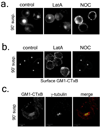
Figure 1
Figure 1. The cytoskeleton in raft endocytosis
(a) Stably adherent MEFs pretreated with 1µM latrunculin A (LatA), 10µM nocodazole (NOC) or DMSO were surface labeled with CTxB-Alexa 488, then detached and held in suspension in the continued presence of drugs for the indicated times. (b) Unlabeled cells suspended for 90 min were surface labeled with CTxB-Alexa 594. Cells with no visible surface GM1 are marked by an asterix. (c) Cells pre-labeled with CTxB-Alexa 488 (as in a) (GM1-CTxB) were suspended for 90 min then fixed and stained for γ-tubulin.
Figure 2
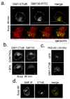
Figure 2. Endocytosed rafts do not localise to the Golgi or smooth ER
(a) Stably adherent MEFS surface labelled with CTxB-Alexa 594 (GM1-CTxB) were suspended for 90min then fixed and stained for the Golgi marker GM130 (GM130-FITC). Lower panel shows an enlarged view of the cell center with the intensity of GM130 (green) increased stepwise while the CTxB-Alexa 594 (red) is held constant. (b) Suspended MEFs with endocytosed CTxB-Alexa 594 (GM1-CTxB) were untreated (CONTROL) or treated with 10µg/ml brefeldin A (+BFA) for 30min. Cells were fixed and stained for GM130. (c) MEFs expressing GFP-tagged PKD-KD mutant (K618N) were surface labeled with CTxB-Alexa 594, detached for 90 min to allow endocytosis, then replated on FN (10µg/ml) for 30min. (d) MEFs surface labelled with CTxB–Alexa 594 (CTxB) and incubated for 90 min in suspension were stained for autocrine motility factor receptor (AMF-R).
Figure 3
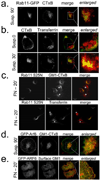
Figure 3. Endocytosed rafts target to recycling endosomes (RE)
(a) MEFs expressing Rab11-GFP were surface labeled with CTxB-Alexa 594 (CTxB) and incubated in suspension for 90 min. The central region is enlarged in the rightmost panel (b) Adherent MEFs labeled with transferrin-Alexa 594 (Transferrin) for 30 min were detached and surface labeled with CTxB-Alexa 488 on ice. Cells were fixed immediately (Susp 0’) or warmed to 37°C for 30min (Susp 30’). The central region is enlarged in the rightmost panel (c) MEFs expressing Rab11 (S25N) -GFP were surface labelled with CTxB-Alexa 594 (GM1-CTxB), held in suspension for 90 min and replated on FN (10µg/ml) for 15min. Re-adherent cells were labeled with transferrin-Alexa 594 (transferrin) for 60min at 37°C (lower panels). Untransfected control cells marked by asterisks. (d) MEFs expressing GFP-WT Arf6 were surface labeled with CTxB-Alexa 594 (GM1-CTxB) and incubated in suspension for 90 min. The central region is enlarged in the rightmost panel (e) Unlabeled GFP-WT Arf6 expressing cells suspended for 90 min were replated on FN (10µg/ml) for 15min and surface GM1 labeled with CTxB-Alexa594. Membrane ruffles are enlarged in the rightmost panel
Figure 4
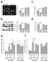
Figure 4. Inhibition of Arf6 function
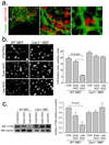
(a) Stably adherent WT and Cav1−/− MEFs expressing HA-tagged WTArf6 (WT) or Arf6T27N (T27N) were suspended for 90min and replated on FN (2µg/ml) for 15min. Cells were then chilled, surface labeled with CTBx-Alexa 594, and images taken. Graph: surface areas were then measured; values are mean ± S.E in arbitary units. (n=25 cells). Figure is representative of three independent experiments. (b) Cells suspended for 90min were replated on FN (2µg/ml) for 15 min. Suspended vs. replated cells were surface labeled with CTxB and bound CTxB detected by Western blotting (CTxB), using tubulin as a loading control (tubulin). Bands were quantified, values (means ± SE, n=3, normalized for tubulin) were calculated relative to WT Arf6-expressing WT MEFs. (c) Cells transfected with a vector coding for both GFP and Arf6 shRNA (KD) or scrambled control RNA (CON) were suspended, replated on FN (2µg/ml) for 15min, and labeled with CTxB-Alexa 594 (as in 4a). Graph: surface areas quantified; values are mean ± S.E in arbitary units. (n=55 cells). Figure is representative of two independent experiments (d) Bound CTxB detected by Western blotting were quantified. Values (means ± SE, n=3, normalized for tubulin) were calculated relative to control WT MEFs. (e) WT and Cav1−/− MEFs expressing HA-tagged WTArf6 (WT) or Arf6T27N (T27N) were suspended for 90min and replated on FN (2µg/ml) for 15 or 45min. Cells were lysed and fractionated as described in Methods. Rac1 in the membrane fraction was determined as described in Methods. Values are means ± SE in arbitary units, n=3. (f) Active Rac1 was measured using the GST-PBD pull down assay and its percentage relative to total Rac1 calculated. Values are means ± SE, n=3.
Figure 5
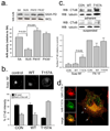
Figure 5. Adhesion dependent activation of ARF6 promotes raft exocytosis
(a) WTMEFs that were stably adherent (SA), suspended for 90min (SUS), or replated on FN (10µg/ml) for 15 or 30min (FN15’, FN30’) were lysed and active ARF6 pulled down on GST-GGA3 beads (GGA-PD). GGA-PD and respective whole cell lysates (WCL) were Western blotted for Arf6 (WB: ARF6). Graph: bands were quantified and normalized intensities calculated relative to SA. Values are means ± SE, n=3. (b) MEFs expressing GFP WTARF6 (WT), HA-T157A Arf6 (T157A) or untransfected (control) were detached, surface labeled with CTxB-Alexa 594 (GM1-CTxB) and held in suspension for 90min. Images of cells with endocytosed GM1 were analyzed to determine the intensity in the central RE pool vs. the rest of the cell. Values are means ± SE, n=15, three independent experiments. (c) Suspended untransfected (CON), WT and T157A Arf6-expressing cells were replated on FN (2µg/ml) for 15min. Cells were surface labeled with CTxB and bound CTxB detected by Western blotting (WB:CTxB). Tubulin served as loading control (WB:tubulin). CTxB band intensity was quantified, normalized to tubulin and calculated relative to adherent control cells. Values are means ± SE, n=3. (d) HA-T157A Arf6-expressing cells with endocytosed GM1-CTxB (as in 5b) were replated on FN (2µg/ml) for 15 min, fixed and stained for HA. Arrows indicate areas of co-labeling suggestive of transport vesicles and ruffling edges.
Figure 6
Figure 6. Microtubules in raft exocytosis
(a) WTMEFs pre-labelled with CTxB-Alexa 594 (GM1-CTxB) were held in suspension for 90 min and replated on FN (2µg/ml) for 15min. Fixed cells were stained for β tubulin. Confocal images from multiple planes are shown as 3D reconstructions. Right panel shows boxed regions of interest (1, 2). (b) WTMEFs and Cav1−/− MEFs were untreated (control), treated with nocodazole (10µm) before detachment (early NOC) or after being suspended for 90min (late NOC). Cells were replated on FN (2µg/ml) for 15 min, fixed and stained for F-actin. Cell surface areas were determined from images. Values are means ± SE in arbitary units; n=25 cells; three independent experiments gave similar results. (c) Re-adherent cells were incubated with CTxB and bound CTxB detected by Western blotting (WB:CTxB). Bands were quantified and normalized for tubulin (WB:tubulin). Values are means ± SE relative to WT MEF control cells, n=3.
Figure 7
Figure 7. Summary
Cell detachment triggers actin-dependent endocytosis of rafts through caveolae. Vesicles are transported along microtubules to recycling endosomes (RE). Replating triggers activation of Arf6, which drives movement of rafts from RE along the microtubules to the cortical region of the cell. Other, unknown adhesion-dependent events complete the return of rafts to the PM.

Similar articles

Cited by

References

    1. Caroni P. New EMBO members' review: actin cytoskeleton regulation through modulation of PI(4,5)P(2) rafts. Embo J. 2001;20:4332–4336. - PMC - PubMed
    1. del Pozo MA, Alderson NB, Kiosses WB, Chiang Hui-Hsien, Anderson RGW, Schwartz MA. Integrins Regulate Rac Targeting by Internalization of Membrane Domains. Science. 2004;303:839–842. - PubMed
    1. del Pozo MA, Balasubramanian N, Alderson NB, Kiosses WB, Grande-Garcia A, Anderson RGW, Schwartz MA. Phospho-caveolin-1 mediates integrin-regulated membrane domain internalization. 2005;7:901–908. - PMC - PubMed
    1. Palazzo AF, Eng CH, Schlaepfer DD, Marcantonio EE, Gundersen GG. Localized stabilization of microtubules by integrin- and FAK-facilitated Rho signaling. Science. 2004;303:836–839. - PubMed
    1. Vasanji A, Ghosh PK, Graham LM, Eppell SJ, Fox PL. Polarization of plasma membrane microviscosity during endothelial cell migration. Dev Cell. 2004;6:29–41. - PubMed

Publication types