

Structure of an archaeal heterotrimeric initiation factor 2 reveals a nucleotide state between the GTP and the GDP states
- PMID: 18000047
- PMCID: PMC2141796
- DOI: 10.1073/pnas.0706784104
Structure of an archaeal heterotrimeric initiation factor 2 reveals a nucleotide state between the GTP and the GDP states
Abstract
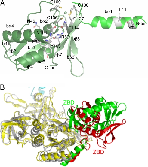
Initiation of translation in eukaryotes and in archaea involves eukaryotic/archaeal initiation factor (e/aIF)1 and the heterotrimeric initiation factor e/aIF2. In its GTP-bound form, e/aIF2 provides the initiation complex with Met-tRNA(i)(Met). After recognition of the start codon by initiator tRNA, e/aIF1 leaves the complex. Finally, e/aIF2, now in a GDP-bound form, loses affinity for Met-tRNA(i)(Met) and dissociates from the ribosome. Here, we report a 3D structure of an aIF2 heterotrimer from the archeon Sulfolobus solfataricus obtained in the presence of GDP. Our report highlights how the two-switch regions involved in formation of the tRNA-binding site on subunit gamma exchange conformational information with alpha and beta. The zinc-binding domain of beta lies close to the guanine nucleotide and directly contacts the switch 1 region. As a result, switch 1 adopts a not yet described conformation. Moreover, unexpectedly for a GDP-bound state, switch 2 has the "ON" conformation. The stability of these conformations is accounted for by a ligand, most probably a phosphate ion, bound near the nucleotide binding site. The structure suggests that this GDP-inorganic phosphate (Pi) bound state of aIF2 may be proficient for tRNA binding. Recently, it has been proposed that dissociation of eIF2 from the initiation complex is closely coupled to that of Pi from eIF2gamma upon start codon recognition. The nucleotide state of aIF2 shown here is indicative of a similar mechanism in archaea. Finally, we consider the possibility that release of Pi takes place after e/aIF2gamma has been informed of e/aIF1 dissociation by e/aIF2beta.
Conflict of interest statement
The authors declare no conflict of interest.
Figures


Similar articles
-
Start Codon Recognition in Eukaryotic and Archaeal Translation Initiation: A Common Structural Core.Int J Mol Sci. 2019 Feb 21;20(4):939. doi: 10.3390/ijms20040939. Int J Mol Sci. 2019. PMID: 30795538 Free PMC article. Review.
-
Conformational transitions in the γ subunit of the archaeal translation initiation factor 2.Acta Crystallogr D Biol Crystallogr. 2014 Mar;70(Pt 3):658-67. doi: 10.1107/S1399004713032240. Epub 2014 Feb 15. Acta Crystallogr D Biol Crystallogr. 2014. PMID: 24598735
-
Free energy simulations of a GTPase: GTP and GDP binding to archaeal initiation factor 2.J Phys Chem B. 2011 May 26;115(20):6749-63. doi: 10.1021/jp201934p. Epub 2011 May 2. J Phys Chem B. 2011. PMID: 21534562 Free PMC article.
-
Structure of the ternary initiation complex aIF2-GDPNP-methionylated initiator tRNA.Nat Struct Mol Biol. 2012 Mar 25;19(4):450-4. doi: 10.1038/nsmb.2259. Nat Struct Mol Biol. 2012. PMID: 22447243
-
Electrostatic free energies in translational GTPases: Classic allostery and the rest.Biochim Biophys Acta. 2015 May;1850(5):1006-1016. doi: 10.1016/j.bbagen.2014.07.006. Epub 2014 Jul 15. Biochim Biophys Acta. 2015. PMID: 25047891 Review.
Cited by
-
The mechanism of eukaryotic translation initiation: new insights and challenges.Cold Spring Harb Perspect Biol. 2012 Oct 1;4(10):a011544. doi: 10.1101/cshperspect.a011544. Cold Spring Harb Perspect Biol. 2012. PMID: 22815232 Free PMC article. Review.
-
Translation initiation factor 2gamma mutant alters start codon selection independent of Met-tRNA binding.Mol Cell Biol. 2008 Nov;28(22):6877-88. doi: 10.1128/MCB.01147-08. Epub 2008 Sep 15. Mol Cell Biol. 2008. PMID: 18794367 Free PMC article.
-
Roles of yeast eIF2α and eIF2β subunits in the binding of the initiator methionyl-tRNA.Nucleic Acids Res. 2013 Jan;41(2):1047-57. doi: 10.1093/nar/gks1180. Epub 2012 Nov 27. Nucleic Acids Res. 2013. PMID: 23193270 Free PMC article.
-
Start Codon Recognition in Eukaryotic and Archaeal Translation Initiation: A Common Structural Core.Int J Mol Sci. 2019 Feb 21;20(4):939. doi: 10.3390/ijms20040939. Int J Mol Sci. 2019. PMID: 30795538 Free PMC article. Review.
-
The Jigsaw Puzzle of mRNA Translation Initiation in Eukaryotes: A Decade of Structures Unraveling the Mechanics of the Process.Annu Rev Biophys. 2018 May 20;47:125-151. doi: 10.1146/annurev-biophys-070816-034034. Epub 2018 Mar 1. Annu Rev Biophys. 2018. PMID: 29494255 Free PMC article.
References
-
- Maag D, Fekete CA, Gryczynski Z, Lorsch JR. Mol Cell. 2005;17:265–275. - PubMed
Publication types
MeSH terms
Substances
Associated data
- Actions
- Actions
LinkOut - more resources
Full Text Sources
Molecular Biology Databases
Research Materials
Miscellaneous
