. 2007 Nov;12(5):432-44.
doi: 10.1016/j.ccr.2007.10.014.

Role of nucleosomal occupancy in the epigenetic silencing of the MLH1 CpG island

Affiliations

Role of nucleosomal occupancy in the epigenetic silencing of the MLH1 CpG island

Joy C Lin et al. Cancer Cell. 2007 Nov.

Abstract

Epigenetic silencing of tumor suppressor genes is generally thought to involve DNA cytosine methylation, covalent modifications of histones, and chromatin compaction. Here, we show that silencing of the three transcription start sites in the bidirectional MLH1 promoter CpG island in cancer cells involves distinct changes in nucleosomal occupancy. Three nucleosomes, almost completely absent from the start sites in normal cells, are present on the methylated and silenced promoter, suggesting that epigenetic silencing may be accomplished by the stable placement of nucleosomes into previously vacant positions. Activation of the promoter by demethylation with 5-aza-2'-deoxycytidine involves nucleosome eviction. Epigenetic silencing of tumor suppressor genes may involve heritable changes in nucleosome occupancy enabled by cytosine methylation.


Figures
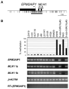
Figure 1
Figure 1. Correlation of the Methylation Status of the Bidirectional Promoter and the Expression Patterns of EPM2AIP1 and MLH1
(A) The bidirectional promoter and the CpG island. Horizontal arrows show the transcription start sites as established by 5′-RACE analysis, and black boxes show the respective first exons. Black tick marks indicate CpG dinucleotides. The horizontal bar underneath the tick marks represents the CpG island. (B) The average methylation levels of three CpGs in the EPM2AIP1/MLH1 promoter region were analyzed by Ms-SNuPE, and the expression of both genes was determined by RT-PCR in a normal LD419 human bladder fibroblast cell line and various human cancer cell lines. β-ACTIN expression served as a control for the input amount of cDNA. RT served as a negative control for the intronless gene, EPM2AIP1.
Figure 2
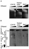
Figure 2. Detection of Hypersensitive Sites by DNaseI Digestion
(A) Genomic naked DNA and nuclei from LD419 and RKO cells were treated with increasing concentrations of DNaseI, DNA from each sample was then purified and treated with DraI, and the digestion products were analyzed by Southern blot. Naked DNA, as a control, was used to confirm the lack of sequence specificity of the enzyme. Digested samples prior to DraI treatment were resolved by gel electrophoresis as shown. (B) Southern blot analysis revealed DNaseI hypersensitivity in the EPM2AIP1/MLH1 promoter. On the left, drawn to scale, the 1399 bp DNA fragment generated by DraI digestion, transcription start sites (arrows), and the probe fragment (black box) are indicated. Numbers show the distance in bp.
Figure 3
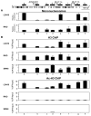
Figure 3. Nucleosomal Depletion by Mononucleosomal DNA and ChIP Analyses
(A) Mononucleosomal DNA analysis. Nuclei from LD419 and RKO were digested partially with MNase, and the reaction mixture was run on a sucrose gradient to isolate mononucleosomal DNA. Enrichment of mononucleosomal DNA was analyzed by real-time PCR by using primers specific for eight regions (a–h), shown as black rectangles on the top of the figure. (B) Distinct chromatin structures at the EPM2AIP1/MLH1 promoter in expressing LD419 and nonexpressing RKO and SW48 cells. ChIP analysis performed with antibodies against histone H3 and acetylated histone H3. Immunoprecipitated DNA was analyzed by real-time PCR as described in (A). The fraction of immunoprecipitated DNA was calculated as a percentage of input DNA. Results are shown as the mean (bar) ± SD of two or three experiments from two independent chromatin preparations.
Figure 4
Figure 4. Accessibility of Native Chromatin to M.SssI at the EPM2AIP1/MLH1 Promoter Region in Expressing LD419 Cells
(A–C) Nuclei were extracted from expressing unmethylated LD419 cells and were then treated with M.SssI for 15 min, followed by bisulfite genomic sequencing. Two independent bisulfite-sequencing reactions were done to avoid introducing a bias in the analyses. Four PCR products of different sizes, indicated by the blue, dotted lines, were included in the analysis. (A) Untreated nuclei. (B) Naked DNA treated with M.SssI. (C) Nuclei treated with M.SssI. Horizontal lines with circles indicate individual molecules that were sequenced after PCR amplification and cloning of bisulfite-treated DNA. Solid circles, methylated CpG dinucleotides; open circles, unmethylated CpG dinucleotides. Pink bars indicate areas or patches that are inaccessible to M.SssI, suggesting the presence of nucleosomes. Patches are defined as at least two consecutively unmethylated sites flanked on each side by at least two consecutively methylated CpG sites (Fatemi et al., 2005). Blue bars show the putative protein-binding regions.
Figure 5
Figure 5. Chromatin Structural Changes upon Gene Reactivation by 5-Aza-CdR
RKO cells were treated with 5-aza-CdR and then harvested 72 hr and 44 days after drug addition for RT-PCR and ChIP analyses. (A) Quantitative RT-PCR. Expression levels were normalized with β-ACTIN, which served as a control for the input cDNA. A minus-RT control served as a negative control for the intronless gene, EPM2AIP1 (data not shown). Results are shown as the mean (bar) ± SD of two PCRs from two independent cDNA preparations. (B) Histone H3 acetylation patterns by ChIP. Immunoprecipitated DNA was analyzed by real-time PCR as described (Figure 3B). The fraction of immunoprecipitated DNA was calculated as a percentage of input DNA. Results are shown as the mean (bar) ± SD of two or three experiments from two independent chromatin preparations.
Figure 6
Figure 6. Eviction of Nucleosomes by 5-Aza-CdR Treatment
(A–E) RKO cells were treated with 5-aza-CdR for 24 hr and were harvested 72 hr and 44 days after drug treatment started. Both DNA and nuclei from drug-treated RKO cells were extracted and then subjected to M.SssI treatment, followed by bisulfite genomic sequencing. (A and D) Demethylation of the promoter in RKO cells (A) 72 hr and (D) 44 days after addition of 5-aza-CdR. (B) Accessibility of native chromatin to M.SssI at the EPM2AIP1/MLH1 1a promoter region in RKO cells 72 hr after drug addition. To filter out extensively methylated molecules that were not suitable for M.SssI treatment, PCR analyses were done with selective primers that only anneal to unmethylated molecules. See the main text for rationale. (C and E) M.SssI treatments on nuclei from drug-treated RKO cells (C) 72 hr and (E) 44 days after drug addition, followed by bisulfite genomic sequencing with selective primers. Please refer to Figure 4 for descriptions of molecules and patches. The DNA molecules with nucleosome depletion in the promoter region are boxed in blue.
Figure 7
Figure 7. Correlation between the Expression Level and the Percentage of Nucleosome-Depleted Molecules
Data were graphed based on the results from Figures 5, 6, and 7. A decrease in expression is associated with a reduced nucleosome-depleted population.
Figure 8
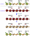
Figure 8. A Simplified Model for the Epigenetic Silencing of the MLH1 Gene
The active promoters of 1a and 1b are depleted of at least one nucleosome just upstream of each of the transcription start sites. In the silenced state, the inactive promoters of 1a and 1b are occupied by nucleosomes. Treatment with 5-aza-CdR causes substantial DNA demethylation of the promoter region, although some molecules remain methylated. While some hemimethylated promoter molecules may still be occupied by nucleosomes, the nucleo-some-free zone is established in some of the promoter molecules. The activation of genes is probably derived from these molecules with a nucleosome-free zone in the promoter, although we could not determine this definitively. Green represents nucleosomes bearing active marks. Red represents nucleosomes bearing repressive marks. Gray represents nucleosomes occupying the region of hemimethylation. Open circles indicate unmethylated CpG dinucleotides, and filled circles represent methylated CpG dinucleotides. Hemimethylated DNA is shown as half-filled circles. Ac refers to acetylated histone tails.

Comment in

Similar articles

Cited by

References

    1. Adachi N, Lieber MR. Bidirectional gene organization: a common architectural feature of the human genome. Cell. 2002;109:807–809. - PubMed
    1. Bachman KE, Park BH, Rhee I, Rajagopalan H, Herman JG, Baylin SB, Kinzler KW, Vogelstein B. Histone modifications and silencing prior to DNA methylation of a tumor suppressor gene. Cancer Cell. 2003;3:89–95. - PubMed
    1. Bender CM, Gonzalgo ML, Gonzales FA, Nguyen CT, Robertson KD, Jones PA. Roles of cell division and gene transcription in the methylation of CpG islands. Mol. Cell. Biol. 1999;19:6690–6698. - PMC - PubMed
    1. Bernstein BE, Liu CL, Humphrey EL, Perlstein EO, Schreiber SL. Global nucleosome occupancy in yeast. Genome Biol. 2004;5:R62. - PMC - PubMed
    1. Bernstein E, Allis CD. RNA meets chromatin. Genes Dev. 2005;19:1635–1655. - PubMed

Publication types

LinkOut - more resources