. 2007 Dec;117(12):3786-99.
doi: 10.1172/JCI32285.

IL-1R1/MyD88 signaling and the inflammasome are essential in pulmonary inflammation and fibrosis in mice

Affiliations

IL-1R1/MyD88 signaling and the inflammasome are essential in pulmonary inflammation and fibrosis in mice

Pamela Gasse et al. J Clin Invest. 2007 Dec.

Abstract

The molecular mechanisms of acute lung injury resulting in inflammation and fibrosis are not well established. Here we investigate the roles of the IL-1 receptor 1 (IL-1R1) and the common adaptor for Toll/IL-1R signal transduction, MyD88, in this process using a murine model of acute pulmonary injury. Bleomycin insult results in expression of neutrophil and lymphocyte chemotactic factors, chronic inflammation, remodeling, and fibrosis. We demonstrate that these end points were attenuated in the lungs of IL-1R1- and MyD88-deficient mice. Further, in bone marrow chimera experiments, bleomycin-induced inflammation required primarily MyD88 signaling from radioresistant resident cells. Exogenous rIL-1beta recapitulated a high degree of bleomycin-induced lung pathology, and specific blockade of IL-1R1 by IL-1 receptor antagonist dramatically reduced bleomycin-induced inflammation. Finally, we found that lung IL-1beta production and inflammation in response to bleomycin required ASC, an inflammasome adaptor molecule. In conclusion, bleomycin-induced lung pathology required the inflammasome and IL-1R1/MyD88 signaling, and IL-1 represented a critical effector of pathology and therapeutic target of chronic lung inflammation and fibrosis.


Figures

Figure 1
Figure 1. Reduced neutrophil and lymphocyte recruitment in the bronchoalveolar space of BLM-challenged MyD88–/– and IL-1R1–/– mice.
(A) Total cell counts were augmented at day 1 in BLM-treated WT (B6) mice (15 mg/kg i.n.) and further increased at days 7 and 11 after BLM administration in WT mice, but less so in MyD88–/– and IL-1R1–/– mice. (B) Neutrophils were recruited into BALF in WT mice within 24 hours, persisted over 7 days, and normalized on day 11, while only few neutrophils were detected in the absence of MyD88 or IL-1R1. (C) Lymphocytes were found in BALF at day 7 and persisted until day 11 in WT mice and were less prominent in MyD88–/– and IL-1R1–/– mice. (D) Alveolar macrophages were augmented at day 11 in WT but not MyD88–/– or IL-1R1–/– mice. Data are from 1 experiment representative of 3 independent experiments (n = 4 mice per group; *P < 0.05; **P < 0.01).
Figure 2
Figure 2. Reduced BLM-induced acute lung inflammation in IL-1R1– and MyD88-deficient mice.
IL-1R1– and MyD88-deficient mice showed reduced neutrophil recruitment in BALF (A) and lung tissue (B), whereas IL-18R–deficient mice had neutrophil counts comparable to WT mice (C). BALs were performed 24 hours after BLM instillation (15 mg/kg), and MPO activity was analyzed at day 7. IL-1β (D), KC (E), and IL-6 (F) in the lung at 24 hours were reduced in IL-1R1– and MyD88-deficient mice as compared with WT mice. Cytokine and chemokine quantification in lung homogenates were performed by multiplex ELISA cytokine arrays (detection limit at 1 pg/ml). Data represent mean values ± SD from 2 independent experiments (n = 4 mice per group; *P < 0.05; **P < 0.01).
Figure 3
Figure 3. Resident radioresistant cells mediate BLM-induced acute inflammation into lung and bronchoalveolar space.
Bone marrow chimeras were prepared by lethal irradiation of either WT or MyD88–/– mice, followed by bone marrow cell reconstitution. WT bone marrow reconstitution of irradiated MyD88–/– mice (WT→KO) did not restore neutrophil recruitment to the BAL (A) or to the lung parenchyma (B) after BLM administration (15 mg/kg i.n.). Irradiated WT mice reconstituted with MyD88–/– bone marrow (KO→WT) displayed reduced neutrophil influx in BAL (A) and lung parenchyma (B) compared with WT controls (WT→WT). WT bone marrow reconstitution of irradiated MyD88–/– mice (WT→KO) did not restore IL-1β (C), KC (D), or IL-6 (E) productions in lung parenchyma. Neutrophil recruitment into the BAL, MPO activity (measured as optical density [OD]), and cytokine and chemokine lung levels were evaluated 24 hours after BLM administration, as described above. Data represent mean values ± SD from 2 independent experiments (n = 4 mice per group; *P < 0.05; **P < 0.01; ns, not significant).
Figure 4
Figure 4. BLM-induced lung tissue remodeling is IL-1R1 and MyD88 dependent.
pro-MMP-9 (99 kDa) and MMP-2 (71 kDa) activities in BALF were analyzed by zymography 1 and 11 days after administration of BLM (15 mg/kg i.n.). Pro-MMP-9 was upregulated in the BALF of WT mice 24 hours after BLM administration and returned to normal levels at day 11, whereas MMP-2 was upregulated only at day 11 after BLM administration (data not shown). (A) Pro-MMP-9 activity was not upregulated in the BALF of MyD88–/– mice and only partially in IL-1R1–/– mice 24 hours after BLM administration. (B) MMP-2 was upregulated by BLM in the BALF of WT mice, but less so in MyD88–/– and IL-1R1–/– mice on day 11. (C) TIMP-1 as an indicator of a fibrotic process was upregulated in the lungs of WT but not MyD88–/– or IL-1R1–/– mice at 24 hours after BLM administration (15 mg/kg). (D) TIMP-1 concentrations were also increased in TLR2 and TLR4 double-deficient mice. TIMP-1 levels were assessed by ELISA, and data represent mean values ± SD from 2 independent experiments (n = 4 mice per group; *P < 0.05; **P < 0.01).
Figure 5
Figure 5. Reduced pulmonary fibrosis in IL-1R1– and MyD88-deficient mice.
Lung microscopic sections showed extensive fibrotic areas at day 11 with collagen deposition in WT mice (B) and TLR2 and TLR4 double-deficient mice (C) treated with BLM (15 mg/kg i.n.) in comparison with saline control mice (A). Fibrosis was significantly reduced in IL-1R1–/– mice (D) and in MyD88–/– mice (E) treated with BLM. Chromotrope Aniline Blue staining; magnification, ×200; n = 8 mice). Pulmonary collagen content was increased in WT mice, but not in IL1-R1–/– mice (F), as determined by Sircol assay. The latent form of TGF-β1 was present in BALF from WT mice 7 days after BLM but was undetectable in BALF from MyD88- and IL-1R1–deficient mice as determined by ELISA assay (G). Data represent mean values ± SD from 3 independent experiments (n = 4 mice per group; *P < 0.05; **P < 0.01).
Figure 6
Figure 6. Exogenous IL-1β induces acute lung inflammation and lung tissue remodeling comparable to BLM.
One i.n. rmIL-1β administration (1 μg per mouse, i.e., 0.05 mg/kg) induced acute neutrophil recruitment into BALF (A) and lung (B) in WT, TLR2/4–/–, and TNF-α–/– mice within 24 hours but was absent in IL-1R1–/– and MyD88–/– mice. rmIL-1β induced lung KC (C), IL-6 (D), and TARC (E) in WT, TLR2/4–/–, and TNF-α–/– mice but not in IL-1R1–/– and MyD88–/– mice. Pro-MMP-9 (103 kDa) (F) and TIMP-1 (G) activities were upregulated 24 hours after IL-1β instillation in the BALF of WT, TLR2/4–/–, and TNF-α–/– mice but not of IL-1R1–/– and MyD88–/– mice. Data represent mean values ± SD from 2 independent experiments (n = 4 mice per group; *P < 0.05; **P < 0.01).
Figure 7
Figure 7. IL-1β induced lymphocyte inflammation, collagen deposition, and fibrotic disease.
(A) Lymphocyte recruitment into BALF was induced at day 7 in WT mice after by 1 i.n. rmIL-1β administration (1 μg per mouse, i.e., 0.05 mg/kg), as for BLM (15 mg/kg i.n.). (B) Pulmonary collagen content (Sircol assay) was increased 7 days after rmIL-1β, similar to results from BLM administration. Microscopic analysis revealed tissue injury with disruption of alveolar architecture and repair in lung 7 days after IL-1β administration (D), similar to that induced by BLM (15 mg/kg) (E), which was absent in control WT mice (C). H&E staining (magnification, ×200). Tissue and subpleural accumulation of collagen was observed 14 days after IL-1β (G) or BLM (H) administration, in comparison with saline-treated control mice (F). Chromotrope Aniline Blue staining (magnification, ×400). The biochemical results represent mean values ± SD from 2 independent experiments (n = 4 mice per group; *P < 0.05; **P < 0.01).
Figure 8
Figure 8. Blockade of IL-1R1 reduces acute lung inflammation.
One administration of IL-1Ra (10 mg/kg i.p.) at the time of BLM challenge (15 mg/kg i.n., day 0), dramatically prevented neutrophil recruitment in BALF (A) as well as IL-1β (C), KC (D), and IL-6 (E) production and TIMP-1 expression (F) in the lung measured at day 1. Several administrations of IL-1Ra (1 mg/kg i.p.) on days 0, 2, 4, 7, 9 and 11 after BLM challenge (15 mg/kg i.n., day 0) induced a prolonged reduction of neutrophil recruitment in BALF, documented on day 14 (B). Data represent mean values ± SD from 2 independent experiments (n = 4 mice per group; *P < 0.05; **P < 0.01).
Figure 9
Figure 9. Reduced inflammation upon BLM administration in ASC-deficient mice.
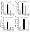
ASC-deficient mice showed reduced total cell (A) and neutrophil (B) recruitment in BALF in comparison with littermate control mice after BLM administration (15 mg/kg i.n.). (C) ASC-deficient mice also showed reduced neutrophil influx into the lung tissue in comparison with littermate controls. BALs and MPO activity were analyzed 24 hours after BLM instillation. IL-1β (D) and IL-6 (E) levels in the lung were reduced at 24 hours in ASC-deficient mice as compared with littermate controls. Cytokine and chemokine quantification in lung homogenates were performed by multiplex ELISA cytokine arrays (detection limit at 1 pg/ml). Data represent mean values ± SD from 2 independent experiments (n = 4 mice per group; *P < 0.05; **P < 0.01).
Figure 10
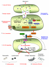
Figure 10. Schematic diagram illustrating the signaling pathways, cellular sources and the specific cascade after BLM-induced lung injury.
(i) BLM induces pulmonary cell injury. (ii) Danger/stress signals generated by dying cells are sensed by the inflammasome rather than TLRs in antigen-presenting cells (alveolar macrophages, dendritic cells, and/or epithelial cells). (iii) Danger signals induce membrane signals, leading probably to the activation of a NALP protein and to the recruitment and activation of the adaptor ASC and caspase-1, known to interact via PYR-PYR and CARD-CARD homotypic interactions. (iv) Inflammasome activation results in the processing and maturation of pro-IL-1β into its biologically active form IL-1, and to IL-1β secretion. The production of pro-IL-1β seems independent of TLRs and could be attributed to sensing by other NACHT-LRRs. (v) IL-1β then activates the IL-1R1 complex in tissue-resident cells (probably pulmonary epithelial cells), leading to the recruitment of MyD88 via TIR-TIR homotypic interactions. Triggering of the IL-1R1/MyD88 pathway results in the activation of transcription factors such as NF-κB, which will turn on the transcription of neutrophil-attracting chemokines such as KC, lymphocyte-attracting chemokines such as TARC, and the transcription of inflammatory cytokines such as IL-6, resulting in lung inflammation (vi). IL-1R1/MyD88-dependent IL-1β production by tissue-resident cells may contribute to an autocrine/paracrine amplification loop. IL-1R1 blockade with IL-1Ra (anakinra) attenuates lung inflammation induced by BLM. IL-1β will also activate the IL-1R1/MyD88 pathway in fibroblasts, leading to metalloproteinase/TIMP-1 imbalance in favor of TIMP-1 and to fibroblast proliferation, resulting in collagen deposition and pulmonary fibrosis (vii).

Similar articles

Cited by

References

    1. Gross T.J., Hunninghake G.W. Idiopathic pulmonary fibrosis. N. Engl. J. Med. 2001;345:517–525. - PubMed
    1. Piguet P.F. Cytokines involved in pulmonary fibrosis. Int. Rev. Exp. Pathol. 1993;34:173–181. - PubMed
    1. Keane M.P., Strieter R.M., Belperio J.A. Mechanisms and mediators of pulmonary fibrosis. Crit. Rev. Immunol. 2005;25:429–463. - PubMed
    1. Beeh K.M., Beier J., Kornmann O., Buhl R. Sputum matrix metalloproteinase-9, tissue inhibitor of metalloprotinease-1, and their molar ratio in patients with chronic obstructive pulmonary disease, idiopathic pulmonary fibrosis and healthy subjects. Respir. Med. 2003;97:634–639. - PubMed
    1. Manoury B., et al. The absence of reactive oxygen species production protects mice against bleomycin-induced pulmonary fibrosis. Respir. Res. 2005;6:11. - PMC - PubMed

Publication types

MeSH terms