. 2007 Dec 15;405(1-2):65-78.
doi: 10.1016/j.gene.2007.09.010. Epub 2007 Sep 19.

Direct inhibition of CDK9 blocks HIV-1 replication without preventing T-cell activation in primary human peripheral blood lymphocytes

Affiliations

Direct inhibition of CDK9 blocks HIV-1 replication without preventing T-cell activation in primary human peripheral blood lymphocytes

Dominic Salerno et al. Gene. .

Abstract

HIV-1 transcription is essential for the virus replication cycle. HIV-1 Tat is a viral transactivator that strongly stimulates the processivity of RNA polymerase II (RNAPII) via recruitment of the cyclin T1/CDK9 positive transcription elongation factor, which phosphorylates the C-terminal domain (CTD) of RNAPII. Consistently, HIV-1 replication in transformed cells is very sensitive to direct CDK9 inhibition. Thus, CDK9 could be a potential target for anti-HIV-1 therapy. A clearer understanding of the requirements for CDK9 activity in primary human T cells is needed to assess whether the CDK9-dependent step in HIV-1 transcription can be targeted clinically. We have investigated the effects of limiting CDK9 activity with recombinant lentiviruses expressing a dominant-negative form of CDK9 (HA-dnCDK9) in peripheral blood lymphocytes (PBLs) and other cells. Our results show that direct inhibition of CDK9 potently inhibits HIV-1 replication in single-round infection assays with little to undetectable effects on RNAPII transcription, RNA synthesis, proliferation and viability. In PBLs purified from multiple donors, direct inhibition of CDK9 activity blocks HIV-1 replication/transcription but does not prevent T-cell activation, as determined via measurement of cell surface and cell cycle entry and progression markers, and DNA synthesis. We have also compared the effects of HA-dnCDK9 to flavopiridol (FVP), a general CDK inhibitor that potently inhibits CDK9. In contrast to HA-dnCDK9, FVP interferes with key cellular processes at concentrations that inhibit HIV-1 replication with potency similar to HA-dnCDK9. In particular, FVP inhibits several T-cell activation markers and DNA synthesis in primary PBLs at the minimal concentrations required to inhibit HIV-1 replication. Our results imply that small pharmacological compounds targeting CDK9 with enhanced selectivity could be developed into effective anti-HIV-1 therapeutic drugs.


Figures

Fig. 1
Fig. 1. Generation of recombinant lentiviruses and HIV-1 reporter viruses
(A) For lentiviral production, 293T cells are transfected via the calcium phosphate method, with a packaging construct that directs the expression of viral proteins minus the Env and Vpu, a plasmid encoding the VSV-G envelope and a transfer plasmid that contains the genes of interest under the control of CMV promoters (expression cassettes) similarly as described previously (Naldini et al., 1996; Hasham and Tsygankov, 2004). In the transfer constructs (pCPP or pCEIII), the indicated expression cassettes are flanked by HIV-1 LTRs and contain the packaging signal, as well as other indicated elements required for efficient lentiviral packaging. Supernatants are collected 48 and 60 hours later and concentrated by ultracentrifugation. cDNAs encoding HA-dnCDK9, HA-CDK9, and HA-cyclin T1 were subcloned in pCPP. Additionally, HA-dnCDK9 and Flag-Hexim1 were subcloned in pCEIII transfer vectors as described in the Methods section. Gene X represents HA-dnCDK9, HA-CDK9, HA-cyclin T1 or Flag-Hexim1. (B) HIV-1-luc viruses pseudotyped with either HXB2 or VSV-G envelopes are generated by cotransfection of the HIV-1 molecular clone mutant NL4-3.Luc.R-E- and the indicated envelope plasmids in 293T cells. These viruses are fully infectious and are replication defective as they exhibit a mutation in the Env and Vpr genes. Luciferase expression is directly proportional to transcription of full-length HIV-1 genome and, thus, reflects replication (Connor et al., 1995; He et al., 1995).
Fig 2
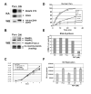
Fig 2. Direct inhibition of CDK9 activity with HA-dnCDK9 in 293T cells inhibits RNAPII phosphorylation on CTD Ser-2 and HIV-1 transcription, but fails to affect the overall rates of cellular transcription and cell proliferation and viability
293 cells stably expressing HA-dnCDK9, HA-CDK9 or HA-cyclin T1 and control puromycin resistant (Puro) cells were generated by lentiviral infection followed by selection with puromycin. Lentiviruses were generated as described in Fig 1A and the Methods section using the corresponding pCPP transfer construct for each transgene. Clones expressing the highest transgene levels were selected for further experiments. (A) Ectopic expression of dnCDK9 inhibits CDK9-associated kinase activity. HA-dnCDK9 and endogenous CDK9 expression was determined by Western blot analysis of whole cell lysates of control (Puro) and HA-dnCDK9 (DN) expressing cells (WB). Cellular CDK9 was immunoprecipitated with anti-CDK9 antibodies. Kinase reactions were performed by incubating the immunoprecipitates with GST-RNAPII-CTD (exogenous substrate) as described in the Materials and Methods section. RNAPII-CTD phosphorylation and CDK9 autophosphorylation is shown (KA). (B) Ectopic expression of HA-dnCDK9 inhibits phosphorylation of RNAPII on Ser-2 in vivo. The effects of HA-dnCDK9 expression on RNAPII Ser-2 phosphorylation were determined by Western blot analysis. The expression of total RNAPII is also shown. Cross-reacting bands detected with the anti-Ser-2 antibody are shown as loading control. (C) Cell proliferation and viability of exponentially growing cells was measured by trypan blue staining and cell counting. No significant increase in cell death was detected in cells expressing HA-dnCDK9, HA-CDK9 or HA-cyclin T1. (D) The rates of cellular transcription were determined via run-on assays in 293T and HA-dnCDK9 (DN) cells as described in the Methods section. The effects of FVP (300 nM) and/or α-amanitin (Amt) were also determined where indicated. (E) RNA synthesis in HA-dnCDK9 (DN), HA-CDK9, Puro cells and Puro cells treated with the indicated concentrations of FVP was determined as by measuring [3H]-uridine incorporation into cellular RNA as described in the Methods section. (F) Single-round HIV-1 transcription assays were performed by infecting HA-dnCDK9 (DN), Puro and FVP treated Puro cells with HIV-1-luc viruses pseudotyped with a VSV-G envelope. HIV-1 transcription was measured by determining luciferase activity 48 h post infection in duplicate samples (see Methods section).
Fig. 3
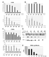
Fig. 3. Direct inhibition of CDK9 with dnCDK9 or Hexim1 is as efficient as FVP in inhibiting HIV-1 replication in single-round infection assays, but dnCDK9 is not a potent inducer of apoptosis and does not inhibit cellular RNA synthesis
(A and B) Effects of inhibition of CDK9 activity via transduction with HA-dnCDK9 (DN), Flag1-Hexim1 (HEX) or control (EGFP) lentiviruses on HIV-1 replication by single-round HIV-1 infection assays in MAGI (A) and Jurkat (B) cells. C MAGI cells transduced with the indicated lentiviruses were infected with HIV-1-luc viruses pseudotyped with a HXB2 envelope. All cells were harvested 48 hours post-infection. HIV-1 replication was determined by measuring luciferase activity in duplicate samples. (C and D) Comparison of dose-dependent effects of FVP and increased transduction with HA-dnCDK9 (DN) or control (EGFP) lentiviruses (increased MOIs) on (C) HIV-1 replication (as in A) and (D) cellular markers of transcription and apoptosis in MAGI cells via Western blot analysis. (E) Effects of transduction with HA-dnCDK9 lentiviruses or FVP in RNA synthesis in duplicate samples. RNA synthesis was determined as described in Fig. 2E.
Fig. 4
Fig. 4. Treatment of PBLs with FVP inhibits the expression of markers of T cell activation, cell cycle entry and transcription as well as DNA synthesis
FVP inhibits the expression of the T cell activation markers CD25, CD62L and CD69 (A-C). T cell surface markers were measured 24 hours following mitogenic stimulation by flow cytometric analysis. PBLs were treated as described in the Methods section. All treatments were done in triplicate. Bars on upper panels represent relative numbers of positive cells, whereas lower panels represent the mean fluorescent intensity corresponding to each marker. Experimental variability is represented by error bars. Similar results were obtained using PBLs obtained from other donors. (D) The expression of cell cycle entry (p107), transcription (Mcl1) and apoptosis (PARP cleavage) markers was determined by Western blot analysis of whole cell lysates of PBLs collected 48 h following mitogenic stimulation. (E) FVP inhibits DNA synthesis. DNA synthesis was determined by measuring incorporation of [3H]-thymidine into DNA for 24 hrs. [3H]-thymidine was added 24 hrs post mitogenic stimulation. All treatments were done in duplicate.
Fig. 5
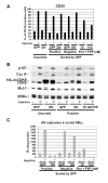
Fig. 5. Expression of dnCDK9 in PBLs inhibits HIV-1 replication in single round assays, but does not affect T cell activation as determined by the expression of T cell activation and cell cycle markers
PBLs were transduced with HA-dnCDK9/EGFP (DN) or EGFP (GFP) lentiviruses and sorted 48 hrs later to separate a population of cells enriched with EGFP expressing cells (Positive) from a population with no detectable EGFP expression (Negative). Sorted EGFP-transduced control PBLs were also treated with FVP as indicated (Pos + FVP). Sorted and unsorted PBLs were then stimulated with PMA/PHA and processed for analysis of CD25 surface marker expression (A) or WB (B) analysis as in Fig. 4. In contrast to FVP, HA-dnCDK9 does not affect CD25 expression at the T cell surface (A) or cell cycle markers (B). (C) The cellular fractions described above were infected with HXB2 pseudotyped HIV-luc viruses and HIV-1 transcription/replication was determined at 48 hrs post-stimulation by measuring luciferase activity. HA-dnCDK9 inhibits HIV-1 replication more potently that 30 nM FVP without the cellular effects associated with FVP.
Fig. 6
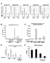
Fig. 6. Direct CDK9 activity downregulation inhibits HIV-1 replication in multiple donors, over extended time periods and independently of the T cell activation stage
(A) dnCDK9 inhibits HIV-1 replication in PBLs from multiple donors (duplicate samples). (B) comparison of the effects of FVP and inhibition of CDK9 activity via transduction with HA-dnCDK9 lentiviruses on HIV-1 replication by single-round HIV-1 infection assays in PBLs up to 5 days post-mitogenic stimulation. Cells were transduced as described above. PBLs were stimulated with irradiated PBMCs, IL-2 (20 U/ml) and PHA (1 μg/ml) and collected at 2 and 5 days and HIV-1 replication was determined via luciferase assays in duplicate samples. (C) HA-dnCDK9 inhibits HIV-1 replication when expressed in post-stimulated PBLs (D) HA-dnCDK9 expression inhibits HIV-1 replication in PBLs previously infected with HIV-1-luc viruses. Cells were first infected with HIV-1-luc overnight, and subsequently infected with EGFP or HA-dnCDK9 lentiviruses. Sixteen h later, PBLs were activated with PMA/PHA. PBLs were collected and prepared for luciferase assays 48 h following stimulation (duplicate samples). (E) HA-dnCDK9 expression does not inhibit DNA synthesis in PBLs. DNA synthesis was determined by measuring incorporation of [3H]-thymidine into DNA for 24 hrs. [3H]-thymidine was added 24 hrs post activation in duplicate samples.

Similar articles

Cited by

References

    1. Blagosklonny MV. Flavopiridol, an inhibitor of transcription: implications, problems and solutions. Cell Cycle. 2004;3:1537–42. - PubMed
    1. Chackerian B, Long EM, Luciw PA, Overbaugh J. Human immunodeficiency virus type 1 coreceptors participate in postentry stages in the virus replication cycle and function in simian immunodeficiency virus infection. J Virol. 1997;71:3932–9. - PMC - PubMed
    1. Chao SH, Fujinaga K, Marion JE, Taube R, Sausville EA, Senderowicz AM, Peterlin BM, Price DH. Flavopiridol inhibits P-TEFb and blocks HIV-1 replication. J Biol Chem. 2000;275:28345–8. - PubMed
    1. Chao SH, Price DH. Flavopiridol inactivates P-TEFb and blocks most RNA polymerase II transcription in vivo. J Biol Chem. 2001;276:31793–9. - PubMed
    1. Chiu YL, Cao H, Jacque JM, Stevenson M, Rana TM. Inhibition of human immunodeficiency virus type 1 replication by RNA interference directed against human transcription elongation factor P-TEFb (CDK9/CyclinT1) J Virol. 2004;78:2517–29. - PMC - PubMed

Publication types

Substances