. 2007 Nov 14;26(22):4709-19.
doi: 10.1038/sj.emboj.7601893. Epub 2007 Oct 18.

Dysfunctional telomeres activate an ATM-ATR-dependent DNA damage response to suppress tumorigenesis

Affiliations

Dysfunctional telomeres activate an ATM-ATR-dependent DNA damage response to suppress tumorigenesis

Xiaolan Guo et al. EMBO J. .

Abstract

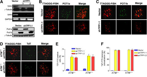
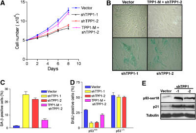
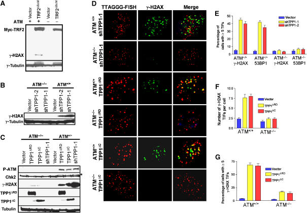
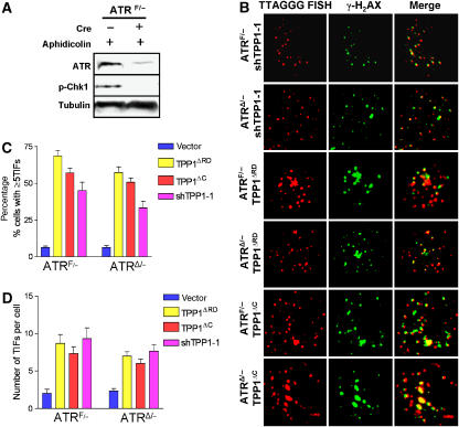
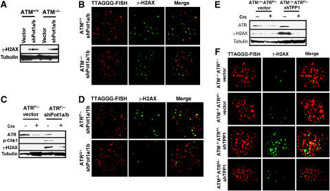
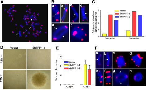
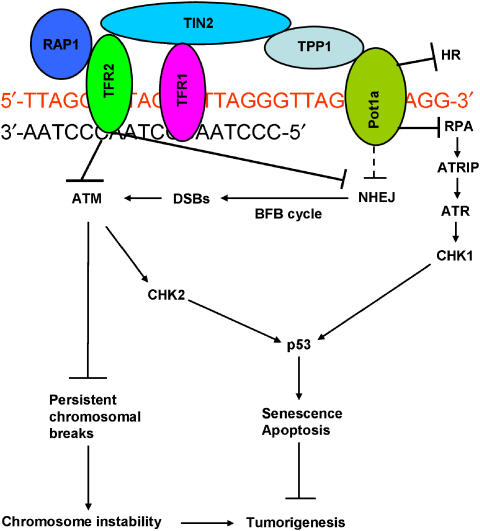
The POT1 (protection of telomeres) protein binds the single-stranded G-rich overhang and is essential for both telomere end protection and telomere length regulation. Telomeric binding of POT1 is enhanced by its interaction with TPP1. In this study, we demonstrate that mouse Tpp1 confers telomere end protection by recruiting Pot1a and Pot1b to telomeres. Knockdown of Tpp1 elicits a p53-dependent growth arrest and an ATM-dependent DNA damage response at telomeres. In contrast to depletion of Trf2, which activates ATM, removal of Pot1a and Pot1b from telomeres initiates an ATR-dependent DNA damage response (DDR). Finally, we show that telomere dysfunction as a result of Tpp1 depletion promotes chromosomal instability and tumorigenesis in the absence of an ATM-dependent DDR. Our results uncover a novel ATR-dependent DDR at telomeres that is normally shielded by POT1 binding to the single-stranded G-overhang. In addition, our results suggest that loss of ATM can cooperate with dysfunctional telomeres to promote cellular transformation and tumor formation in vivo.


Figures

Figure 1
Figure 1
Tpp1 mediates Pot1a and Pot1b telomeric localization and protection. (A) Top panel: immunoblot showing reduced expression of exogenous HA-tagged Tpp1 upon introduction of Tpp1 shRNAs-1 and -2. Middle panel: RT–PCR of MEFs infected with the control vector or Tpp1 shRNAs-encoding retroviruses was processed to detect Tpp1 and GAPDH mRNAs. Bottom panel: western blot of control vector or Tpp1-knockdown p53−/−MEFs expressing Myc-tagged Pot1a or Flag-tagged Pot1b. Tubulin serves as loading control. (B) Tpp1 knockdown reduced telomeric localization of Pot1a. p53−/− MEFs infected with control vector or shTpp1 were costained with PNA-TTAGGG to detect telomeres (red), and with anti-Myc antibody (green) to detect Myc-tagged Pot1a. Merged image is shown on right. (C) Tpp1 knockdown reduced Pot1b telomeric localization. MEFs were treated as in panel B, with PNA-TTAGGG to detect telomeres (red) and anti-Flag antibody (green) to detect Flag-tagged Pot1b. (D) Knockdown of Tpp1 induces accessible telomere ends. PNA-TTAGGG repeats (red) were used to detect telomeres, while TdT-FITC (green) was used as a marker of uncapped telomeres in ATM+/+ and ATM−/− Tpp1-knockdown MEFs. Merged signals are on the right. (E) Quantification of the percentage of TdT–FITC-positive cells in ATM+/+ and ATM−/− Tpp1-knockdown cells. (F) Quantification of the percentage of colocalization of telomeric signals with TdT–FITC signals in ATM+/+ and ATM−/− Tpp1-knockdown cells. In panels E and F, a minimum of 100 nuclei was scored, and error bars represent standard error of the mean (s.e.m).
Figure 2
Figure 2
Tpp1 knockdown initiates premature cellular senescence in primary mouse fibroblasts. (A) Diminished cell proliferation upon inhibition of Tpp1. Early-passage (P2) primary MEFs were infected with control vectors, shRNA-1, -2 or RNAi-resistant mutant retroviruses, and subjected to puromycin selection. Subsequently, cells numbers were measured at the indicated time points, with day 0 representing the first day after puromycin selection. Error bars represent s.e.m. (B) MEFs with reduced Tpp1 expression display a senescence phenotype and stain for SA-β-galactosidase. (C) Quantification of SA-β-gal-positive cells. Error bars represent s.e.m. (D) Quantification of BrdU incorporation in p53+/+ or p53−/− primary MEFs expressing vector, shTpp1-1, -2 or shRNA-resistant Tpp1 mutant. Error bars represent s.e.m. (E) Induction of p53-ser18 phosphorylation and p21 upon Tpp1 inhibition. Immunoblot of extracts from cells expressing vector or shTpp1-1 and -2.
Figure 3
Figure 3
Stable knockdown of Tpp1 or overexpression of Tpp1 mutants initiates an ATM-dependent DDR at telomeres. (A) Western blot showing overexpression of myc-TRF2ΔBΔM-induced γ-H2AX phosphorylation in ATM+/+ but not in ATM−/− MEFs. Trf2ΔBΔM was detected by anti-myc antibody and tubulin serves as a loading control. (B) Western analysis revealing that knockdown of Tpp1 with shTpp1-1 and -2 induced γ-H2AX phosphorylation in ATM+/+ but not in ATM−/− MEFs. Tubulin serves as loading control. (C) Knockdown of Tpp1 or overexpression of HA-Tpp1ΔRD or HA-Tpp1ΔC mutants induced phosphorylation of ATM, γ-H2AX and Chk 2 in ATM+/+ but not in ATM−/− MEFs. Tpp1ΔRD and Tpp1ΔC were determined by anti-HA antibody and tubulin serves as loading control. *, phospho-Chk2. (D) Expression of shTpp1-1 or Tpp1 mutants induced ATM-dependent phosphorylation of γ-H2AX at telomeres. ATM+/+ or ATM−/−MEFs infected by the indicated retroviral constructs were analyzed by telomere PNA-FISH (red) and antibody to γ-H2AX (green). Representative images are shown. (E) Percentage of cells containing five or more γ-H2AX or 53BP1 TIFs in ATM+/+ or ATM−/−, Tpp1-depleted MEFs. Error bars represent s.e.m. (F) Quantitation of the number of γ-H2AX positive TIFs per cell expressing Tpp1ΔRD or Tpp1ΔC mutants. Error bars represent s.e.m. (G) Percentage of cells containing five or more γ-H2AX-positive TIFs in ATM+/+ or ATM−/−MEFs stably expressing Tpp1ΔRD or TPP1ΔC mutants. Error bars represent s.e.m.
Figure 4
Figure 4
Expression of Tpp1 mutants does not engage an ATR-dependent DDR at telomeres. (A) ATR is required for Chk1 phosphorylation. ATRF/− MEFs were infected with the AdE empty vector (−) or AdCre (+) at an MOI of 200, and treated with aphidicolin to induce Chk1 phosphorylation at Serine 345. Tubulin served as loading control. (B) Stable shRNA-mediated depletion of Tpp1 or expression of Tpp1 mutants induced ATR-independent phosphorylation of γ-H2AX at telomeres. ATRF/− MEFs were treated with either AdE, or with AdCre at an MOI of 200 to generate ATRΔ/− MEFs. Cells were infected with the indicated retroviral constructs and analyzed by telomere PNA-FISH (red) and with antibody to γ-H2AX (green) to detect TIF formation. The images were merged to evaluate colocalization. Representative images are shown. (C) Quantitation of percentage of cells with ⩾5 γ-H2AX-positive TIFs in ATRF/− or ATRΔ/−MEFs expressing shTpp1, Tpp1ΔRD or Tpp1ΔC mutants. Error bars represent s.e.m. (D) Quantitation of the number of γ-H2AX-positive TIFs per cell expressing shTPP1, TPP1ΔRD or TPP1ΔC mutants. Error bars represent s.e.m.
Figure 5
Figure 5
Removal of Pot1a and Pot1b from telomeres initiates an ATR-dependent, ATM-independent DDR. (A) ATM+/+ and ATM−/− MEFs were infected with control vector or shRNAs targeting both Pot1a and Pot1b. Pot1a and Pot1b knockdown induced γ-H2AX phosphorylation. Tubulin served as loading control. (B) Depletion of Pot1a and Pot1b induced the formation of γ-H2AX-positive TIFs in the absence of ATM. TIF formation in ATM+/+ or ATM−/−MEFs were analyzed following shPot1a and shPot1b retroviral infection with telomere PNA-FISH (red) and an antibody to γ-H2AX (green). (C) Lysates from ATRF/− or ATRΔ/−MEFs treated with vector or shPot1a and shPot1b were probed for ATR, Chk1 and γ-H2AX. Tubulin served as a loading control. (D) ATR is required for TIF formation in Pot1a- and Pot1b-knockdown MEFs. ATRF/− or ATRΔ/− MEFs were infected with shPot1a and shPot1b and analyzed by telomere PNA-FISH (red) and with an antibody to γ-H2AX (green). (E) Transient knockdown of Tpp1 elicits an ATR-dependent DDR at telomeres. ATR and γ-H2AX phosphorylation levels were monitored by western blotting in ATM−/−ATRF/− and ATM−/−ATRΔ/− MEFs transiently treated with vector or shTPP1. Tubulin served as a loading control. (F) ATR is required for TIF formation in Tpp1-depleted MEFs. ATM−/−ATRF/− or ATM−/−ATRΔ/− MEFs was transiently infected with vector or shTpp1-1 and analyzed by telomere PNA-FISH (red) and with an antibody to γ-H2AX (green) for the presence of TIFs (merge).
Figure 6
Figure 6
Knockdown of Tpp1 in ATM−/− cells induces chromosome instability, cellular transformation and tumorigenesis in vivo. (A) Telomeric FISH on metaphases derived from ATM−/− cells expressing shTpp1-1. Telomeric hybridization signal is shown in red and DAPI-counterstained chromosomes in blue. Arrows indicate chromosomal aberrations. (B) Knockdown of Tpp1 induced multiple chromosomal aberrations, including p–p arm fusions with TTAGGG repeats at the fusion sites (a, b); p–p arm fusions without TTAGGG repeats at the fusion sites (c, d); q–q arm chromosomal fusions without TTAGGG repeats at the fusion sites (e) and diplochromosomes (f). In all panels, arrowheads point to the site of fusion. (C) The frequency of cytogenetic aberrations is quantitated; fusions+telo, fusions with telomere signal at the site of fusion; fusions–telo, fusions without telomere signals at the site of fusion. (D) ATM−/−, p53−/− MEFs stably expressing shTpp1-1 readily formed colonies in anchorage-independent assay. Representative images are shown. (E) Quantitation of the number of colonies formed when ATM−/−, p53−/− MEFs were infected with vector and shTpp1-1 and -2 siRNAs. Error bars represents s.e.m. (F) Tumor cells from Tpp1-knockdown ATM−/− MEFs possess multiple chromosomal aberrations, including p–p arm fusions without TTAGGG repeats at the fusion sites (a, b); p–p arm fusions with TTAGGG repeats at fusion sites (e, f); diplochromosomes (c, d); q–q arm fusion without TTAGGG repeats at the sites of fusion (g) and isochromatid rings without TTAGGG repeats at the sites of fusion (h).
Figure 7
Figure 7
Speculative model of how the shelterin complex represses ATM/ATR-dependent DDR at telomeres. We envision Pot1a as the main repressor of ATR, while ATM is mainly repressed by Trf2. Repression of ATR prevents the activation of Chk1 and p53, while ATM is required to prevent the accumulation of chromosomal breaks and subsequent genomic instability. NHEJ at telomeres is repressed mainly by Trf2, while aberrant telomere HR is repressed mainly by Pot1a/Pot1b.

Similar articles

Cited by

References

    1. Bartek J, Lukas J (2003) Chk1 and Chk2 kinases in checkpoint control and cancer. Cancer Cell 3: 421–429 - PubMed
    1. Bartkova J, Horejsi Z, Koed K, Kramer A, Tort F, Zieger K, Guldberg P, Sehested M, Nesland JM, Lukas C, Orntoft T, Lukas J, Bartek J (2005) DNA damage response as a candidate anticancer barrier in early human tumorigenesis. Nature 434: 864–870 - PubMed
    1. Bassing CH, Suh H, Ferguson DO, Chua KF, Manis J, Eckersdorff M, Gleason M, Bronson R, Lee C, Alt FW (2003) Histone H2AX: a dosage-dependent suppressor of oncogenic translocations and tumors. Cell 114: 359–370 - PubMed
    1. Brown EJ, Baltimore D (2003) Essential and dispensable roles of ATR in cell cycle arrest and genome maintenance. Genes Dev 17: 615–628 - PMC - PubMed
    1. Brummelkamp TR, Bernards R, Agami R (2002) A system for stable expression of short interfering RNAs in mammalian cells. Science 296: 550–553 - PubMed

Publication types

MeSH terms