. 2007 Oct 12;3(10):1459-69.
doi: 10.1371/journal.ppat.0030146.

HMBA releases P-TEFb from HEXIM1 and 7SK snRNA via PI3K/Akt and activates HIV transcription

Affiliations

HMBA releases P-TEFb from HEXIM1 and 7SK snRNA via PI3K/Akt and activates HIV transcription

Xavier Contreras et al. PLoS Pathog. .

Abstract

Hexamethylene bisacetamide (HMBA) is a potent inducer of cell differentiation and HIV production in chronically infected cells. However, its mechanism of action remains poorly defined. In this study, we demonstrate that HMBA activates transiently the PI3K/Akt pathway, which leads to the phosphorylation of HEXIM1 and the subsequent release of active positive transcription elongation factor b (P-TEFb) from its transcriptionally inactive complex with HEXIM1 and 7SK small nuclear RNA (snRNA). As a result, P-TEFb is recruited to the HIV promoter to stimulate transcription elongation and viral production. Despite the continuous presence of HMBA, the released P-TEFb reassembles rapidly with 7SK snRNA and HEXIM1. In contrast, a mutant HEXIM1 protein that cannot be phosphorylated and released from P-TEFb and 7SK snRNA via the PI3K/Akt pathway antagonizes this HMBA-mediated induction of viral production. Thus, our studies reveal how HIV transcription is induced by HMBA and suggest how modifications in the equilibrium between active and inactive P-TEFb could contribute to cell differentiation.


Conflict of interest statement

Competing interests. The authors have declared that no competing interests exist.

Figures

Figure 1
Figure 1. HMBA Transiently Disrupts the LC
(A) U1, Ach-2, and JΔK cells were stimulated with increasing concentrations of HMBA (1, 5, 10, and 20 mM) and viral production was assessed at 48 h in the supernatant using p24 ELISA. (B) Resting CD4+ T cells, which were isolated from PBMCs that were infected with HIV-1LAI and rested for 11 d were treated or not with HMBA (1 mM) for 3 d. Viral production was assessed using p24 ELISA. (C) Total cell lysates of Jurkat cells stimulated or not with HMBA (5 mM) for 30 min, 1 h, 2 h, 6 h, and 24 h, were subjected to glycerol gradient sedimentation analyses (10%–30%), and the fractions were analyzed by western blotting using HEXIM1, HEXIM2, and Cdk9 antibodies. Numbers below the western blots correspond to fractions from glycerol gradient analyses. (D) LC/SC represents the ratio of cumulated intensities of fractions 5, 6, and 7 (LC) over intensities of fractions 1, 2, and 3 (SC). Values were normalized to ratios obtained in unstimulated cells.
Figure 2
Figure 2. HMBA Induces HIV Production from Latently Infected Cell Lines via the PI3K/Akt Pathway
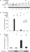
(A) Total cell lysates of Jurkat cells preincubated or not with inhibitors of Akt or PI3K (AI8 or LY294002) and stimulated or not by HMBA (5 mM) for 30 min, 1 h, 6 h, and 24 h were analyzed by western blotting using anti-phospho-Akt antibodies, or Anti-Akt antibodies for loading controls. (B) U1 cells were cultured in the presence or absence of LY294002 (10 μM) or AI8 (1 μM) prior to stimulation with HMBA (5 mM). Concentrations of p24 antigens were then measured from supernatants at 24 and 48 h after stimulation to assess viral production. (C) U1 cells were transfected with a plasmid expressing a dominant negative mutant Akt protein (DN-Akt) or with an empty plasmid vector. After 24 h, cells were stimulated or not with HMBA (5 mM) and viruses in the supernatant were quantified 24 h later using p24 ELISA. Lower panels represent the levels of DN-Akt expressed in cells as assessed using glu-glu tag antibodies.
Figure 3
Figure 3. HMBA Disrupts the LC via the PI3K/Akt Pathway
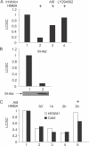
Jurkat cells were stimulated or not with HMBA (5 mM) for 1 h in the presence or absence of LY294002 (10 μM) or AI8 (1 μM) in (A) or were electroporated with the empty plasmid vector or a vector expressing M-Akt for 24 h in (B). Total cell lysates were then subjected to glycerol gradient sedimentation and fractions were analyzed as in Figure 1. Western blotting was performed using anti-HEXIM1 antibodies. Results present the ratio LC/SC as in Figure 1. In (C), a similar analysis was performed using total cell lysates of non-adherent PBMCs pre-treated or not with AI8 and stimulated or not with HMBA (5 mM) for 30 min, 1 h, and 2 h.
Figure 4
Figure 4. HMBA Induces the Recruitment of P-TEFb to the HIV Promoter
(A) Schematic representation of the HIV promoter (LTR) and coding region (Gag). Lack of NF-κB sites is denoted by ΔκB. LTR: long terminal repeat; Sp1: 3 Sp1 binding sites; T: TATA box; I: initiator; TAR, transactivation response element; Gag: group specific antigen. (B) JΔK cells, chronically infected with HIV-1 lacking NF-κB binding sites, were pre-treated or not with AI8 prior to the stimulation with HMBA (5 mM, 1 h) and proteins were fixed onto DNA with formaldehyde. Following sonication, the anti-CycT1 or anti-RNAPII antibodies were added to the chromatin solution for immunoprecipitation of DNA–protein complexes. PCR was performed with the indicated primers (A) to analyze amounts of DNA that were associated with CycT1 or RNAPII at the promoter (primer 1) or 1,000 nt downstream in the Gag gene (primer 2). Quantitative PCRs were performed with anti-RNAPII or anti-CycT1-immunoprecipitated samples, as well as with input DNA before immunoprecipitation, which served as controls for the amplification efficiency of individual sets of PCR primers. Immunoprecipitates obtained without using any antibody were used as negative controls.
Figure 5
Figure 5. Mutant f:Hex1(4D) Protein Is Functionally Inactive
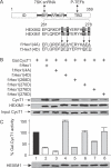
(A) Schematic representation of HEXIM1. Sequences are representative of mutant f:Hex1(4A) and f:Hex1(4D) proteins. Mutations are underlined. ID: inhibitory domain; BR: basic region; AR: acidic region; TBD, CycT-binding domain. (B) Extracts prepared from Jurkat cells transfected with an empty plasmid vector or those expressing f:Hex1, mutant f:Hex1(4A), or f:Hex1(4D) proteins, or point mutant f:Hex1(268D), f:Hex1(270D), f:Hex1(276D), or f:Hex1(278D) proteins, were subjected to anti-Flag immunoprecipitation. Levels of endogenous CycT1 protein bound to Flag epitope-tagged mutant HEXIM1 proteins are indicated in the upper panel. The middle panel represents levels of Flag epitope-tagged mutant HEXIM1 proteins expressed in Jurkat cells and the lower panel shows the input of CycT1. (C) Jurkat cells were co-transfected with the reporter plasmid pG6TAR (0.4 μg) and, where indicated, with the plasmids expressing Gal4.CycT1 (0.6 μg), f:Hex1 (0.8 μg), f:Hex1(4A) (0.8 μg), f:Hex1(4D) (0.8 μg), f:Hex1(268D) (0.8 μg), f:Hex1(270D) (0.8 μg), f:Hex1(276D) (0.8 μg), or f:Hex1(278D) (0.8 μg). Error bars represent the mean +/− SD. Lower panel presents the levels of Flag epitope-tagged HEXIM1 proteins expressed in Jurkat cells.
Figure 6
Figure 6. T270 and S278 in the TBD of HEXIM1 Are Phosphorylated following the Activation of Akt In Vivo
f:Hex1 protein and mutant f:Hex1(4A) protein in (A), and mutant f:Hex1(268A), f:Hex1(270A), f:Hex1(276A), or f:Hex1(278A) proteins in (B) were expressed in Jurkat cells transfected with an empty plasmid vector or a vector expressing M-Akt. After 24 h, protein extracts were subjected to immunoprecipitation using anti-Flag antibodies, and levels of phospho-Akt substrates (pAktSub) and Flag-tagged expressed proteins were measured by western blotting.
Figure 7
Figure 7. Mutation of the Conserved Threonines and Serines Renders HEXIM1 Insensitive to HMBA Treatment and Antagonizes the Induction of HIV Production by HMBA
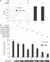
(A) Jurkat cells were transfected with plasmids expressing wild-type f:Hex1 or mutant f:Hex1(4A) proteins and then stimulated or not with HMBA (5 mM) for 1 h in the presence or absence of AI8 (1 μM). Total lysates were then subjected to glycerol gradient sedimentation and fractions were analyzed as in Figure 2. Western blot was performed using anti-Flag antibodies and the ratio LC/SC measured as in Figure 1. (B) U1 cells were transfected with an empty plasmid vector or those expressing mutant f:Hex1, f:Hex1(2A), f:Hex1(4A), f:Hex1(268A), f:Hex1(270A), f:Hex1(276A), or f:Hex1(278A) proteins. After 24 h, cells were stimulated or not with HMBA (5 mM), supernatant collected after another 24 h, and virus quantified using p24 ELISA.

Similar articles

Cited by

References

    1. Contreras X, Lenasi T, Peterlin BM. HIV latency: present knowledge and future directions. Future Virology. 2006;1:733–745. - PMC - PubMed
    1. Lassen K, Han Y, Zhou Y, Siliciano J, Siliciano RF. The multifactorial nature of HIV-1 latency. Trends Mol Med. 2004;10:525–531. - PubMed
    1. Chun TW, Engel D, Mizell SB, Hallahan CW, Fischette M, et al. Effect of interleukin-2 on the pool of latently infected, resting CD4+ T cells in HIV-1-infected patients receiving highly active anti-retroviral therapy. Nat Med. 1999;5:651–655. - PubMed
    1. Stellbrink HJ, van Lunzen J, Westby M, O'Sullivan E, Schneider C, et al. Effects of interleukin-2 plus highly active antiretroviral therapy on HIV-1 replication and proviral DNA (COSMIC trial) AIDS. 2002;16:1479–1487. - PubMed
    1. Biancotto A, Grivel JC, Gondois-Rey F, Bettendroffer L, Vigne R, et al. Dual role of prostratin in inhibition of infection and reactivation of human immunodeficiency virus from latency in primary blood lymphocytes and lymphoid tissue. J Virol. 2004;78:10507–10515. - PMC - PubMed

Publication types

MeSH terms