

Glutamate receptor expression in multiple sclerosis lesions
- PMID: 17924980
- PMCID: PMC8095601
- DOI: 10.1111/j.1750-3639.2007.00101.x
Glutamate receptor expression in multiple sclerosis lesions
Abstract
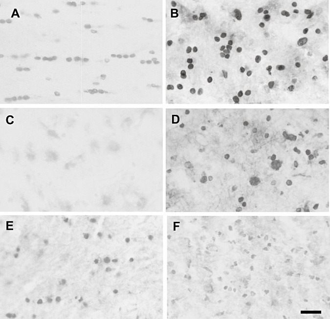
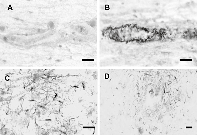
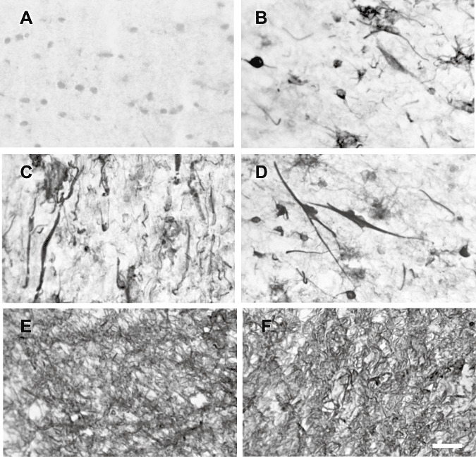
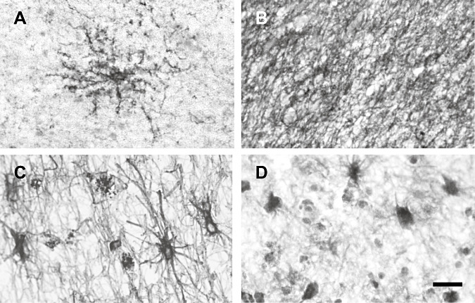
Blockade of receptors for the excitatory neurotransmitter glutamate ameliorates neurological clinical signs in models of the CNS inflammatory demyelinating disease multiple sclerosis (MS). To investigate whether glutamate excitoxicity may play a role in MS pathogenesis, the cellular localization of glutamate and its receptors, transporters and enzymes was examined. Expression of glutamate receptor (GluR) 1, a Ca(++)-permeable ionotropic AMPA receptor subunit, was up-regulated on oligodendrocytes in active MS lesion borders, but Ca(++)-impermeable AMPA GluR2 subunit levels were not increased. Reactive astrocytes in active plaques expressed AMPA GluR3 and metabotropic mGluR1, 2/3 and 5 receptors and the GLT-1 transporter, and a subpopulation was immunostained with glutamate antibodies. Activated microglia and macrophages were immunopositive for GluR2, GluR4 and NMDA receptor subunit 1. Kainate receptor GluR5-7 immunostaining showed endothelial cells and dystrophic axons. Astrocyte and macrophage populations expressed glutamate metabolizing enzymes and unexpectedly the EAAC1 transporter, which may play a role in glutamate uptake in lesions. Thus, reactive astrocytes in MS white matter lesions are equipped for a protective role in sequestering and metabolizing extracellular glutamate. However, they may be unable to maintain glutamate at levels low enough to protect oligodendrocytes rendered vulnerable to excitotoxic damage because of GluR1 up-regulation.
Figures
Similar articles
-
The link between excitotoxic oligodendroglial death and demyelinating diseases.Trends Neurosci. 2001 Apr;24(4):224-30. doi: 10.1016/s0166-2236(00)01746-x. Trends Neurosci. 2001. PMID: 11250007 Review.
-
Glutamate uptake by oligodendrocytes: Implications for excitotoxicity in multiple sclerosis.Neurology. 2003 Oct 28;61(8):1113-20. doi: 10.1212/01.wnl.0000090564.88719.37. Neurology. 2003. PMID: 14581674
-
Glutamate receptors on myelinated spinal cord axons: II. AMPA and GluR5 receptors.Ann Neurol. 2009 Feb;65(2):160-6. doi: 10.1002/ana.21539. Ann Neurol. 2009. PMID: 19224531 Free PMC article.
-
AMPA/kainate receptors in mouse spinal cord cell-specific display of receptor subunits by oligodendrocytes and astrocytes and at the nodes of Ranvier.Glia. 2003 Apr 1;42(1):12-24. doi: 10.1002/glia.10136. Glia. 2003. PMID: 12594733
-
Glutamate, T cells and multiple sclerosis.J Neural Transm (Vienna). 2017 Jul;124(7):775-798. doi: 10.1007/s00702-016-1661-z. Epub 2017 Feb 24. J Neural Transm (Vienna). 2017. PMID: 28236206 Review.
Cited by
-
Glial Cell AMPA Receptors in Nervous System Health, Injury and Disease.Int J Mol Sci. 2019 May 17;20(10):2450. doi: 10.3390/ijms20102450. Int J Mol Sci. 2019. PMID: 31108947 Free PMC article. Review.
-
Mechanisms and pharmacology of neuropathic pain in multiple sclerosis.Curr Top Behav Neurosci. 2014;20:75-97. doi: 10.1007/7854_2014_288. Curr Top Behav Neurosci. 2014. PMID: 24590824 Free PMC article.
-
Psychostimulant abuse and neuroinflammation: emerging evidence of their interconnection.Neurotox Res. 2013 Feb;23(2):174-88. doi: 10.1007/s12640-012-9334-7. Epub 2012 Jun 20. Neurotox Res. 2013. PMID: 22714667 Review.
-
Outer Retinal Dysfunction on Multifocal Electroretinography May Help Differentiating Multiple Sclerosis From Neuromyelitis Optica Spectrum Disorder.Front Neurol. 2019 Aug 27;10:928. doi: 10.3389/fneur.2019.00928. eCollection 2019. Front Neurol. 2019. PMID: 31507527 Free PMC article.
-
In vivo imaging reveals rapid astrocyte depletion and axon damage in a model of neuromyelitis optica-related pathology.Ann Neurol. 2016 May;79(5):794-805. doi: 10.1002/ana.24630. Epub 2016 Apr 18. Ann Neurol. 2016. PMID: 26946517 Free PMC article.
References
-
- Aronica E, Van Vliet EA, MayboroDa OA, Troost D, Da Silva FHL, Gorter JA (2000) Upregulation of metabotropic glutamate receptor subtype mGluR3 and mGluR5 in reactive astrocytes in a rat model of mesial temporal lobe epilepsy. Eur J Neurosci 12:2333–2344. - PubMed
-
- Aronica E, Catania MV, Geurts J, Yankaya B, Troost D (2001) Immunohistochemical localization of group I and II metabotropic glutamate receptors in control and amyotrophic lateral sclerosis human spinal cord: upregulation in reactive astrocytes. Neuroscience 105:509–520. - PubMed
-
- Barbour B, Brew H, Attwell D (1988) Electrogenic glutamate uptake in glial‐cells is activated by intracellular potassium. Nature 335:433–435. - PubMed
-
- Behrens PF, Langemann H, Strohschein R, Draeger J, Henning J (2000) Extracellular glutamate and other metabolites in and around RG2 rat glioma: an intracerebral microdialysis study. J Neurooncol 47:11–22. - PubMed
MeSH terms
Substances
LinkOut - more resources
Full Text Sources
Medical
