. 2007 Oct 4;56(1):109-23.
doi: 10.1016/j.neuron.2007.08.006.

The BMP ligand Gbb gates the expression of synaptic homeostasis independent of synaptic growth control

Affiliations

The BMP ligand Gbb gates the expression of synaptic homeostasis independent of synaptic growth control

Carleton P Goold et al. Neuron. .

Abstract

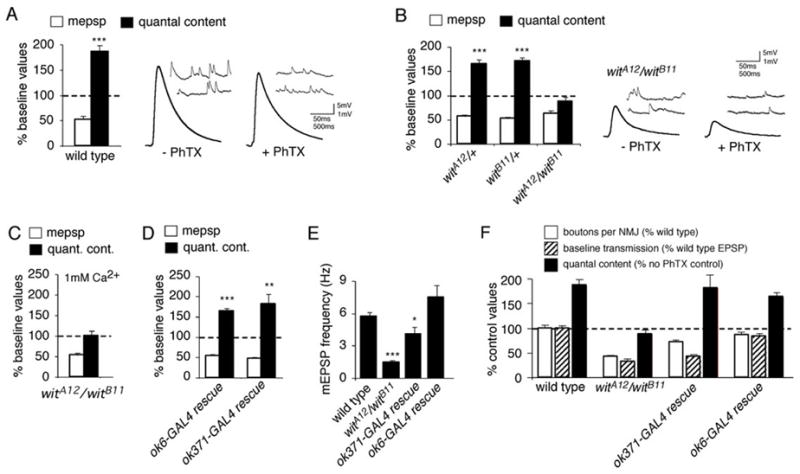
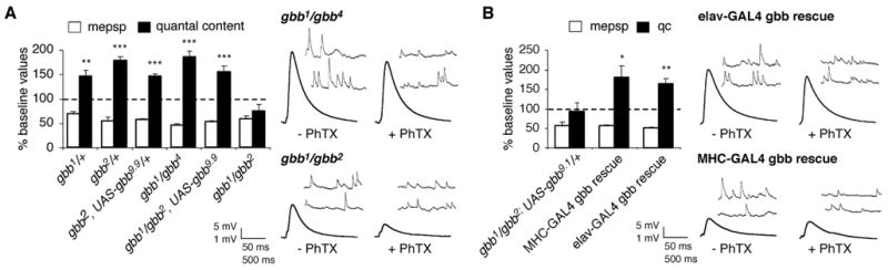
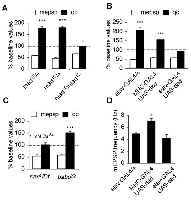
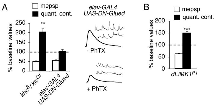
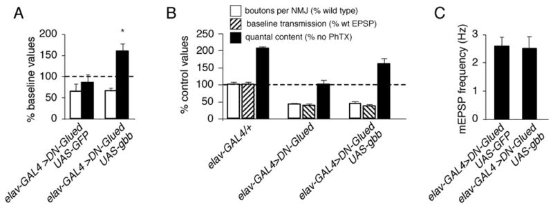
Inhibition of postsynaptic glutamate receptors at the Drosophila NMJ initiates a compensatory increase in presynaptic release termed synaptic homeostasis. BMP signaling is necessary for normal synaptic growth and stability. It remains unknown whether BMPs have a specific role during synaptic homeostasis and, if so, whether BMP signaling functions as an instructive retrograde signal that directly modulates presynaptic transmitter release. Here, we demonstrate that the BMP receptor (Wit) and ligand (Gbb) are necessary for the rapid induction of synaptic homeostasis. We also provide evidence that both Wit and Gbb have functions during synaptic homeostasis that are separable from NMJ growth. However, further genetic experiments demonstrate that Gbb does not function as an instructive retrograde signal during synaptic homeostasis. Rather, our data indicate that Wit and Gbb function via the downstream transcription factor Mad and that Mad-mediated signaling is continuously required during development to confer competence of motoneurons to express synaptic homeostasis.


Figures

Figure 1
Figure 1. The type-II BMP receptor wishful thinking is required presynaptically for the rapid induction of synaptic homeostasis
(A) Quantal content (filled bar) and mEPSP amplitude (open bar) are quantified. The dashed line represents normalized wild type baseline values recorded in the absence of PhTx. Bars represent values recorded after 10 minute PhTx application, normalized to wild type in the absence of PhTx. There is a significant decrease in mEPSP amplitude and a significant, compensatory increase in quantal content. Right, representative traces showing mEPSPs (inset) and EPSPs for control and PhTx-treated wild type animals. (B) Data are presented as in (A). Application of PhTx to heterozygous controls (witA12/+ and witB11/+) induces a decrease in mEPSP amplitude and a compensatory increase in quantal content compared to heterozygous controls in the absence of PhTx (p<0.001). No increase in quantal content is observed in the null mutant (witA12/witB11) animals compared to witA12/witB11 animals in the absence of PhTx (p>0.5). Sample traces are shown for the null witA12/witB11 animals with and without PhTx application for 10 minutes. (C) Data are presented as in (A). Synaptic homeostasis remains blocked in witA12/witB11 animals when recordings are conducted in saline containing 1 mM Ca2+ and 10 mM Mg2+. (D) Data are presented as in (A). Expressing UAS-wit using either of the presynaptic GAL4 drivers OK6-GAL4 or OK371-GAL4 in the wit mutant background (witA12/witB11) restores synaptic homeostasis, as demonstrated by a significant increase in quantal content after PhTx challenge (p < 0.001 and p<0.01 respectively). (E) mEPSP frequency in wild type, wit mutant animals, and wit animals in which UAS-wit is expressed presynaptically using OK6-GAL4 or OK371-GAL4. (F) Quantification of data for bouton number (open; percent wild type bouton number), baseline transmission (hatched; percent wild type EPSP amplitude) and quantal content (filled). Values for quantal content are normalized to recordings in the absence of PhTx for a given genotype as in (A). Wit mutant animals (witA12/witB11) have decreased bouton number, decreased EPSP amplitude and no homeostatic increase in quantal content (as shown in B). Presynaptic expression of UAS-wit in the wit mutant using OK371-GAL4 partially restores bouton number (numbers are significantly less than wild type, p<0.01), does not rescue EPSP amplitude, and completely rescues a homeostatic increase in release (p<0.001). Presynaptic expression of UAS-wit in the wit mutant using OK6-GAL4 restores all aspects of synaptic growth and function. Significance is indicated as follows for this figure and all subsequent figures: *p<0.05, **p < 0.01, ***p < 0.001 Student’s t-test.
Figure 2
Figure 2. The BMP ligand Gbb is required for synaptic homeostasis
(A) Quantal content (filled bar) and mEPSP amplitude (open bar) are quantified and normalized to amplitudes recorded for each genotype in the absence of PhTx (as in figure 1A). A homeostatic increase in quantal content offsets a significant decrease in mEPSP amplitude in all mutant combinations examined except the null gbb combination gbb1/gbb2 which does not show a homeostatic increase in quantal content in response to PhTx treatment (p > 0.2). Representative traces are shown for indicated genotypes at right. (B) Data are quantified as in (A). Either neuronal-specific (elavC155-GAL4) or muscle-specific (MHC-GAL4) expression of UAS-gbb9.1 in the gbb null mutant background restores a homeostatic increase in quantal content. Representative traces are shown at right.
Figure 3
Figure 3. Impaired synaptic growth in gbb mutants does not correlate with the expression of synaptic homeostasis
(A–C) Composite images of anti-Synapsin staining at gbb1/gbb2 and gbb1/gbb2, UAS-gbb9.9 as well as wild type synapses. Images represent the NMJ at muscle 6/7. (D) Quantification of bouton number (open; percent wild type bouton number), baseline transmission (hatched; percent wild type EPSP amplitude), and quantal content (filled). Values for quantal content are normalized to control values recorded for each genotype in the absence of PhTx. Bouton number and baseline transmission are significantly impaired in gbb1/gbb2 (p<0.01) and there is no significant homeostatic increase in quantal content (p>0.2). Bouton numbers are significantly decreased in gbb1/gbb4 (p<0.01). Baseline transmission and bouton number are significantly impaired in gbb1/gbb2, UAS-gbb9.9 (p<0.001). Neuronal-specific rescue of gbb (elav-GAL4 gbb rescue) restores synaptic homeostasis and significantly rescues both NMJ growth and baseline neurotransmission (p<0.001). Muscle-specific rescue of gbb (MHC-GAL4 gbb rescue) restores synaptic homeostasis and significantly rescues NMJ growth (p < 0.001) but does not significantly rescue baseline neurotransmission.
Figure 4
Figure 4. Mad-mediated signaling is required in motoneurons for the expression of synaptic homeostasis
(A) Quantal content (filled bar) and mEPSP amplitude (open bar) are quantified and normalized to amplitudes recorded for each genotype in the absence of PhTx (as in figure 1A). The mad heterozygous animals show a significant decrease in mEPSP amplitude and a significant homeostatic increase in quantal content following PhTx application. The mad null mutant (mad10/mad12) fails to show a homeostatic increase in quantal content in response to decreased mEPSP amplitude. (B) There is no significant increase in quantal content in response to PhTx application in animals that express UAS-dad in neurons using elavC155-GAL4 (p>0.3). A significant, homeostatic increase in quantal content is observed following muscle expression of UAS-dad using MHC-GAL4 (p<0.01). (C) Quantification as in (A) for sax4/Df and babo32/babo32 mutations. Synaptic homeostasis is blocked in the sax4/Df mutants (recorded at elevated calcium as indicated). (D) Quantification of mEPSP frequency.
Figure 5
Figure 5. Impaired retrograde axonal transport blocks the rapid induction of synaptic homeostasis
(A) Quantal content (filled bar) and mEPSP amplitude (open bar) are quantified and normalized to amplitudes recorded for each genotype in the absence of PhTx. Neuronal expression of UAS-DN-Glued (elavC155-GAL4/+; UAS-GluedDNΔ84/+) prevents an increase in quantal content in response to PhTx-challenge (p> 0.9). Animals with a double heterozygous combination of mutations in kinesin heavy chain and kinesin light chain (khc8/+; klcDf/+) show a robust homeostatic increase in presynaptic release following PhTx application. (B) Data are quantified as in (A). A robust homeostatic increase in quantal content is observed in a LIM Kinase mutant (DLIMKP1).
Figure 6
Figure 6. Neuronal expression of Gbb in a background of impaired retrograde transport restores synaptic homeostasis but not growth or synaptic efficacy
(A). Quantal content (filled bar) and mEPSP amplitude (open bar) are quantified and normalized to amplitudes recorded for each genotype in the absence of PhTx. Animals simultaneously expressing UAS-DN-Glued and UAS-GFP in neurons (elavC155-GAL4/+; UAS-GluedDNΔ84/UAS-CD8-GFP) do not show a homeostatic increase in quantal content compared to controls. However, synaptic homeostasis is restored when UAS-gbb is simultaneously overexpressed with UAS-DN-Glued (elavC155-GAL4/+; UAS-GluedDNΔ84/UAS-gbb9.1). (B) Quantification of bouton number (open; percent wild type bouton number), baseline transmission (hatched; % wild type EPSP amplitude), and quantal content (filled). Values for quantal content are normalized to control values recorded for each genotype in the absence of PhTx. UAS-gbb expression in neurons restores a homeostatic increase in quantal content but does not restore synaptic growth or baseline EPSP amplitudes compared to controls. (C) Quantification of mEPSP frequency.
Figure 7
Figure 7. Normal synaptic homeostasis in mutations that disrupt synaptic vesicle release
A) Baseline EPSP amplitude is significantly impaired in both cspU1 and syx/+ mutants (wild type amplitudes are repeated from figure 1). Representative EPSP traces are shown at right. B) The cspU1 mutants show normal homeostatic compensation in response to PhTx application, recorded in 1 mM extracellular calcium to increase absolute EPSP amplitude. Representative traces are shown at right. C) The syx/+ mutants show normal synaptic homeostasis in response to PhTx application (normal saline). Representative traces shown at right.
Figure 8
Figure 8. Continuous BMP signaling is required to sustain the ability of motoneurons to express homeostatic plasticity
(A) Quantification of bouton number at muscles 6/7 in elavGS-UAS-dad animals (elavGS-GAL4/+; UAS-Dad/+) receiving RU486 administration for different durations of time as indicated. Each data point represents bouton numbers normalized to wild type animals that received identical RU486 administration. Time in RU486 refers to the duration of RU486 exposure prior to the end of larval development. (B) Representative traces and average EPSP amplitudes (numbers above traces) for elavGS-UAS-dad animals raised on RU486 for the indicated durations. RU486 feeding does not have a significant effect on baseline EPSP amplitudes in wild type. (C) Quantification of mEPSP frequency for animals in A–C. (D) Quantal content (filled bar) and mEPSP amplitudes (open bar) are quantified for wild type animals, raised on RU486 for indicated times. Data are normalized to amplitudes recorded for wild type in the absence of PhTx. (E) Data are quantified and presented as in (D) for elavGS-UAS-dad animals raised on RU486 for the indicated durations of time prior to dissection at the end of larval development.

Similar articles

Cited by

References

    1. Aberle H, Haghighi AP, Fetter RD, McCabe BD, Magalhaes TR, Goodman CS. wishful thinking encodes a BMP type II receptor that regulates synaptic growth in Drosophila. Neuron. 2002;33:545–558. - PubMed
    1. Allen MJ, Shan X, Caruccio P, Froggett SJ, Moffat KG, Murphey RK. Targeted expression of truncated glued disrupts giant fiber synapse formation in Drosophila. J Neurosci. 1999;19:9374–9384. - PMC - PubMed
    1. Allan DW, St Pierre SE, Miguel-Aliaga I, Thor S. Specification of neuropeptide cell identity by the integration of retrograde BMP signaling and a combinatorial transcription factor code. Cell. 2003;113:73–86. - PubMed
    1. Baines RA. Synaptic strengthening mediated by bone morphogenetic protein-dependent retrograde signaling in the Drosophila CNS. J Neurosci. 2004;24:6904–6911. - PMC - PubMed
    1. Brummel T, Abdollah S, Haerry TE, Shimell MJ, Merriam J, Rafter L, Wrana JL, O’Connor MB. The Drosophila activin receptor baboon signals through dSmad2 and controls cell proliferation but not patterning during larval development. Genes Dev. 1999;13:98–111. - PMC - PubMed

Publication types

MeSH terms

LinkOut - more resources