Altered phosphorylated signal transducer and activator of transcription profile of CD4+CD161+ T cells in asthma: modulation by allergic status and oral corticosteroids
- PMID: 17919711
- PMCID: PMC2679255
- DOI: 10.1016/j.jaci.2007.08.012
Altered phosphorylated signal transducer and activator of transcription profile of CD4+CD161+ T cells in asthma: modulation by allergic status and oral corticosteroids
Abstract
Background: Asthma is a complex immunologic disorder linked to altered cytokine signaling.
Objective: We tested whether asthmatic patients showed any change in cytokine-dependent signal transducer and activator of transcription (STAT) levels, focusing on the central/effector-memory CD4(+)CD161(+) subset, which represents 15% to 25% of circulating T cells.
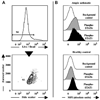
Methods: We quantified intracellular levels of active phosphorylated STAT (phospho-STAT) 1, 3, 5, and 6 by means of flow cytometry, without any activation or expansion.
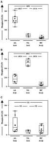
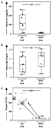
Results: Baseline phospho-STAT1 and phospho-STAT6 levels were increased in CD4(+)CD161(+) T cells from asthmatic patients compared with those from healthy control subjects (by 10- and 8-fold, respectively). This asthma-associated alteration was both subset specific because no change was seen in CD4(+)CD161(-)CD25(+) (regulatory T cells) and CD4(+)CD161(-)CD25(-) subsets and isoform specific because phospho-STAT5 and phospho-STAT3 levels were unchanged. Among asthmatic patients, phospho-STAT1 and phospho-STAT6 levels correlated negatively with each other, suggesting antagonistic regulation. Oral corticosteroid (OCS) treatment significantly decreased phospho-STAT6 and IL-4 levels but not phospho-STAT1 levels. Disease parameters showing significant correlations with phospho-STAT1, phospho-STAT6, or both included age at onset, plasma IgE levels, and levels of the T(H)2 cytokines IL-4 and IL-10 and the T(H)1 cytokine IL-2. Overall, combined phospho-STAT1 and phospho-STAT6 measurements showed excellent predictive value for identifying (1) asthmatic patients versus healthy control subjects, (2) allergic versus nonallergic asthmatic patients, and (3) asthmatic patients taking versus those not taking OCSs.
Conclusion: Baseline changes in phospho-STAT1 and phospho-STAT6 levels in blood CD4(+)CD161(+) T cells identify asthmatic patients and mirror their allergic status and response to OCSs.
Clinical implications: These results confirm the pathologic importance of activated STAT1 and STAT6 in asthma and suggest their potential use as clinical biomarkers.
Conflict of interest statement
Disclosure of potential conflict of interest: The authors have declared that they have no conflict of interest.
Figures


Similar articles
-
Effect of memory CD4+ T cells' signal transducer and activator of transcription (STATs) functional shift on cytokine-releasing properties in asthma.Cell Biol Toxicol. 2017 Feb;33(1):27-39. doi: 10.1007/s10565-016-9357-6. Epub 2016 Aug 31. Cell Biol Toxicol. 2017. PMID: 27581546
-
IL-4 confers resistance to IL-27-mediated suppression on CD4+ T cells by impairing signal transducer and activator of transcription 1 signaling.J Allergy Clin Immunol. 2013 Oct;132(4):912-21.e1-5. doi: 10.1016/j.jaci.2013.06.035. Epub 2013 Aug 16. J Allergy Clin Immunol. 2013. PMID: 23958647 Free PMC article.
-
Identification of a novel subset of human circulating memory CD4(+) T cells that produce both IL-17A and IL-4.J Allergy Clin Immunol. 2010 Jan;125(1):222-30.e1-4. doi: 10.1016/j.jaci.2009.10.012. J Allergy Clin Immunol. 2010. PMID: 20109749
-
IL-5 and IL-5 receptor in asthma.Mem Inst Oswaldo Cruz. 1997;92 Suppl 2:75-91. doi: 10.1590/s0074-02761997000800012. Mem Inst Oswaldo Cruz. 1997. PMID: 9698919 Review.
-
Th9 and other IL-9-producing cells in allergic asthma.Semin Immunopathol. 2017 Jan;39(1):55-68. doi: 10.1007/s00281-016-0601-1. Epub 2016 Nov 17. Semin Immunopathol. 2017. PMID: 27858144 Review.
Cited by
-
Activated Phosphorylated STAT1 Levels as a Biologically Relevant Immune Signal in Schizophrenia.Neuroimmunomodulation. 2016;23(4):224-229. doi: 10.1159/000450581. Epub 2016 Nov 8. Neuroimmunomodulation. 2016. PMID: 27820940 Free PMC article.
-
Janus kinase-3 dependent inflammatory responses in allergic asthma.Int Immunopharmacol. 2010 Aug;10(8):829-36. doi: 10.1016/j.intimp.2010.04.014. Epub 2010 Apr 27. Int Immunopharmacol. 2010. PMID: 20430118 Free PMC article. Review.
-
IL-2 and IL-4 stimulate MEK1 expression and contribute to T cell resistance against suppression by TGF-beta and IL-10 in asthma.J Immunol. 2010 Nov 15;185(10):5704-13. doi: 10.4049/jimmunol.1000690. Epub 2010 Oct 6. J Immunol. 2010. PMID: 20926789 Free PMC article.
-
Increased cytotoxicity of CD4+ invariant NKT cells against CD4+CD25hiCD127lo/- regulatory T cells in allergic asthma.Eur J Immunol. 2008 Jul;38(7):2034-45. doi: 10.1002/eji.200738082. Eur J Immunol. 2008. PMID: 18581330 Free PMC article.
-
A phenotypically and functionally distinct human TH2 cell subpopulation is associated with allergic disorders.Sci Transl Med. 2017 Aug 2;9(401):eaam9171. doi: 10.1126/scitranslmed.aam9171. Sci Transl Med. 2017. PMID: 28768806 Free PMC article. Clinical Trial.
References
-
- Cohn L, Elias JA, Chupp GL. Asthma: mechanisms of disease persistence and progression. Annu Rev Immunol. 2004;22:789–815. - PubMed
-
- Umetsu DT, Dekruyff RH. The regulation of allergy and asthma. Immunol Rev. 2006;212:238–255. - PubMed
-
- O’Byrne PM. Cytokines or their antagonists for the treatment of asthma. Chest. 2006;130:244–250. - PubMed
-
- Bel EH. Clinical phenotypes of asthma. Curr Opin Pulm Med. 2004;10:44–50. - PubMed
-
- Buhl R. Anti-IgE antibodies for the treatment of asthma. Curr Opin Pulm Med. 2005;11:27–34. - PubMed
Publication types
MeSH terms
Substances
Grants and funding
LinkOut - more resources
Full Text Sources
Other Literature Sources
Medical
Research Materials
Miscellaneous

