HSF2 binds to the Hsp90, Hsp27, and c-Fos promoters constitutively and modulates their expression
- PMID: 17915561
- PMCID: PMC1971238
- DOI: 10.1379/csc-250.1
HSF2 binds to the Hsp90, Hsp27, and c-Fos promoters constitutively and modulates their expression
Abstract
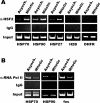
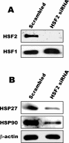
Although the vast majority of genomic DNA is tightly compacted during mitosis, the promoter regions of a number of genes remain in a less compacted state throughout this stage of the cell cycle. The decreased compaction of these promoter regions, which is referred to as gene bookmarking, is thought to be important for the ability of cells to express these genes during the following interphase. Previously, we reported a role for the DNA-binding protein heat shock factor (HSF2) in bookmarking the stress-inducible 70,000-Da heat shock protein (hsp70) gene. In this report, we have extended those studies and found that during mitosis, HSF2 is bound to the HSE promoter elements of other heat shock genes, including hsp90 and hsp27, as well as the proto-oncogene c-fos. The presence of HSF2 is important for expression of these genes because blocking HSF2 levels by RNA interference techniques leads to decreased levels of these proteins. These results suggest that HSF2 is important for constitutive as well as stress-inducible expression of HSE-containing genes.
Figures


Similar articles
-
Mechanism of hsp70i gene bookmarking.Science. 2005 Jan 21;307(5708):421-3. doi: 10.1126/science.1106478. Science. 2005. PMID: 15662014
-
Transcriptional regulation and binding of heat shock factor 1 and heat shock factor 2 to 32 human heat shock genes during thermal stress and differentiation.Cell Stress Chaperones. 2004 Mar;9(1):21-8. doi: 10.1379/481.1. Cell Stress Chaperones. 2004. PMID: 15270074 Free PMC article.
-
Characterization of constitutive HSF2 DNA-binding activity in mouse embryonal carcinoma cells.Mol Cell Biol. 1994 Aug;14(8):5309-17. doi: 10.1128/mcb.14.8.5309-5317.1994. Mol Cell Biol. 1994. PMID: 8035809 Free PMC article.
-
Heat Shock Transcription Factor 2 Is Significantly Involved in Neurodegenerative Diseases, Inflammatory Bowel Disease, Cancer, Male Infertility, and Fetal Alcohol Spectrum Disorder: The Novel Mechanisms of Several Severe Diseases.Int J Mol Sci. 2022 Nov 9;23(22):13763. doi: 10.3390/ijms232213763. Int J Mol Sci. 2022. PMID: 36430241 Free PMC article. Review.
-
Modulatory effects of curcumin on heat shock proteins in cancer: A promising therapeutic approach.Biofactors. 2019 Sep;45(5):631-640. doi: 10.1002/biof.1522. Epub 2019 May 28. Biofactors. 2019. PMID: 31136038 Review.
Cited by
-
Advances of Heat Shock Family in Ulcerative Colitis.Front Pharmacol. 2022 May 12;13:869930. doi: 10.3389/fphar.2022.869930. eCollection 2022. Front Pharmacol. 2022. PMID: 35645809 Free PMC article. Review.
-
HSP90 inhibitor 17-AAG selectively eradicates lymphoma stem cells.Cancer Res. 2012 Sep 1;72(17):4551-61. doi: 10.1158/0008-5472.CAN-11-3600. Epub 2012 Jun 29. Cancer Res. 2012. PMID: 22751135 Free PMC article.
-
Transcriptional response to stress in the dynamic chromatin environment of cycling and mitotic cells.Proc Natl Acad Sci U S A. 2013 Sep 3;110(36):E3388-97. doi: 10.1073/pnas.1305275110. Epub 2013 Aug 19. Proc Natl Acad Sci U S A. 2013. PMID: 23959860 Free PMC article.
-
Heat shock factor 1 confers resistance to Hsp90 inhibitors through p62/SQSTM1 expression and promotion of autophagic flux.Biochem Pharmacol. 2014 Feb 1;87(3):445-55. doi: 10.1016/j.bcp.2013.11.014. Epub 2013 Nov 28. Biochem Pharmacol. 2014. PMID: 24291777 Free PMC article.
-
A dominant-negative mutation of HSF2 associated with idiopathic azoospermia.Hum Genet. 2013 Feb;132(2):159-65. doi: 10.1007/s00439-012-1234-7. Epub 2012 Oct 14. Hum Genet. 2013. PMID: 23064888
References
Publication types
MeSH terms
Substances
Grants and funding
LinkOut - more resources
Full Text Sources
Other Literature Sources
Research Materials
Miscellaneous
