HIV-1 induced activation of CD4+ T cells creates new targets for HIV-1 infection in human lymphoid tissue ex vivo
- PMID: 17909079
- PMCID: PMC2200839
- DOI: 10.1182/blood-2007-05-088435
HIV-1 induced activation of CD4+ T cells creates new targets for HIV-1 infection in human lymphoid tissue ex vivo
Abstract
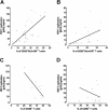
We demonstrate mechanisms by which HIV-1 appears to facilitate its own infection in ex vivo-infected human lymphoid tissue. In this system, HIV-1 readily infects various CD4+ T cells, but productive viral infection was supported predominantly by activated T cells expressing either CD25 or HLA-DR or both (CD25/HLA-DR) but not other activation markers: There was a strong positive correlation (r=0.64, P=.001) between virus production and the number of CD25+/HLA-DR+ T cells. HIV-1 infection of lymphoid tissue was associated with activation of both HIV-1-infected and uninfected (bystanders) T cells. In these tissues, apoptosis was selectively increased in T cells expressing CD25/HLA-DR and p24gag but not in cells expressing either of these markers alone. In the course of HIV-1 infection, there was a significant increase in the number of activated (CD25+/HLA-DR+) T cells both infected and uninfected (bystander). By inducing T cells to express particular markers of activation that create new targets for infection, HIV-1 generates in ex vivo lymphoid tissues a vicious destructive circle of activation and infection. In vivo, such self-perpetuating cycle could contribute to HIV-1 disease.
Figures


Similar articles
-
Bystander CD4+ T lymphocytes survive in HIV-infected human lymphoid tissue.AIDS Res Hum Retroviruses. 2003 Mar;19(3):211-6. doi: 10.1089/088922203763315713. AIDS Res Hum Retroviruses. 2003. PMID: 12689413
-
HLA-DR expression on regulatory T cells is closely associated with the global immune activation in HIV-1 infected subjects naïve to antiretroviral therapy.Chin Med J (Engl). 2011 Aug;124(15):2340-6. Chin Med J (Engl). 2011. PMID: 21933566
-
Interactions between human immunodeficiency virus type 1 and vaccinia virus in human lymphoid tissue ex vivo.J Virol. 2007 Nov;81(22):12458-64. doi: 10.1128/JVI.00326-07. Epub 2007 Sep 5. J Virol. 2007. PMID: 17804502 Free PMC article.
-
Comprehensive Mass Cytometry Analysis of Cell Cycle, Activation, and Coinhibitory Receptors Expression in CD4 T Cells from Healthy and HIV-Infected Individuals.Cytometry B Clin Cytom. 2017 Jan;92(1):21-32. doi: 10.1002/cyto.b.21502. Cytometry B Clin Cytom. 2017. PMID: 27997758 Review.
-
Population biology of HIV-1 infection: viral and CD4+ T cell demographics and dynamics in lymphatic tissues.Annu Rev Immunol. 1999;17:625-56. doi: 10.1146/annurev.immunol.17.1.625. Annu Rev Immunol. 1999. PMID: 10358770 Review.
Cited by
-
Impact of actin polymerization and filopodia formation on herpes simplex virus entry in epithelial, neuronal, and T lymphocyte cells.Front Cell Infect Microbiol. 2023 Nov 24;13:1301859. doi: 10.3389/fcimb.2023.1301859. eCollection 2023. Front Cell Infect Microbiol. 2023. PMID: 38076455 Free PMC article. Review.
-
Resident memory T cells are a cellular reservoir for HIV in the cervical mucosa.Nat Commun. 2019 Oct 18;10(1):4739. doi: 10.1038/s41467-019-12732-2. Nat Commun. 2019. PMID: 31628331 Free PMC article.
-
Wuchereria bancrofti infection is linked to systemic activation of CD4 and CD8 T cells.PLoS Negl Trop Dis. 2019 Aug 19;13(8):e0007623. doi: 10.1371/journal.pntd.0007623. eCollection 2019 Aug. PLoS Negl Trop Dis. 2019. PMID: 31425508 Free PMC article.
-
Preferential Infection of α4β7+ Memory CD4+ T Cells During Early Acute Human Immunodeficiency Virus Type 1 Infection.Clin Infect Dis. 2020 Dec 31;71(11):e735-e743. doi: 10.1093/cid/ciaa497. Clin Infect Dis. 2020. PMID: 32348459 Free PMC article.
-
Targeting strategies for delivery of anti-HIV drugs.J Control Release. 2014 Oct 28;192:271-83. doi: 10.1016/j.jconrel.2014.08.003. Epub 2014 Aug 10. J Control Release. 2014. PMID: 25119469 Free PMC article. Review.
References
-
- Schacker T, Little S, Connick E, et al. Productive infection of T cells in lymphoid tissues during primary and early human immunodeficiency virus infection. J Infect Dis. 2001;183:555–562. - PubMed
-
- Fauci AS. The human immunodeficiency virus: infectivity and mechanisms of pathogenesis. Science. 1988;239:617–622. - PubMed
-
- Fauci AS. Multifactorial nature of human immunodeficiency virus disease: implications for therapy. Science. 1993;262:1011–1018. - PubMed
-
- Pantaleo G, Graziosi C, Fauci AS. Virologic and immunologic events in primary HIV infection. Springer Semin Immunopathol. 1997;18:257–266. - PubMed
-
- Dutton RW, Mishell RI. Lymphocytic proliferation in response to homologous tissue antigens. Fed Proc. 1966;25:1723–1726. - PubMed
Publication types
MeSH terms
Substances
Grants and funding
LinkOut - more resources
Full Text Sources
Other Literature Sources
Medical
Research Materials

