. 2007 Dec;27(23):8296-305.
doi: 10.1128/MCB.00598-07. Epub 2007 Oct 1.

RACK1 targets the extracellular signal-regulated kinase/mitogen-activated protein kinase pathway to link integrin engagement with focal adhesion disassembly and cell motility

Affiliations

RACK1 targets the extracellular signal-regulated kinase/mitogen-activated protein kinase pathway to link integrin engagement with focal adhesion disassembly and cell motility

Tomas Vomastek et al. Mol Cell Biol. 2007 Dec.

Abstract

The extracellular signal-regulated kinase (ERK) cascade is activated in response to a multitude of extracellular signals and converts these signals into a variety of specific biological responses, including cell differentiation, cell movement, cell division, and apoptosis. The specificity of the biological response is likely to be controlled in large measure by the localization of signaling, thus enabling ERK activity to be directed towards specific targets. Here we show that the RACK1 scaffold protein functions specifically in integrin-mediated activation of the mitogen-activated protein kinase/ERK cascade and targets active ERK to focal adhesions. We found that RACK1 associated with the core kinases of the ERK pathway, Raf, MEK, and ERK, and that attenuation of RACK1 expression resulted in a decrease in ERK activity in response to adhesion but not in response to growth factors. RACK1 silencing also caused a reduction of active ERK in focal adhesions, an increase in focal adhesion length, a decreased rate of focal adhesion disassembly, and decreased motility. Our data further suggest that focal adhesion kinase is an upstream activator of the RACK1/ERK pathway. We suggest that RACK1 tethers the ERK pathway core kinases and channels signals from upstream activation by integrins to downstream targets at focal adhesions.


Figures
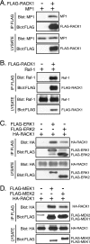
FIG. 1.
FIG. 1.
RACK1 associates with components of the ERK pathway. (A) Coimmunoprecipitation of MP1 with RACK1 from CCL39 cells transiently transfected with either control vector or FLAG-RACK1 and MP1 constructs. RACK1 was precipitated and detected with FLAG M2 antibody. Coprecipitated MP1 was detected with MP1 antibody, and cell lysates were probed with the FLAG and MP1 antibodies to verify comparable expression levels of RACK1 and MP1 protein. (B) Coimmunoprecipitation of Raf-1 with RACK1. CCL39 cells were transfected with either control vector or FLAG-RACK1 and untagged Raf-1. RACK1 precipitation and immunodetection were performed as described for panel A, with the exception that Raf-1 antibody was used to detect Raf-1. (C) Coimmunoprecipitation of RACK1 with ERK1 and ERK2. CCL39 cells were transfected with control vector or FLAG-ERK1 or FLAG-ERK2 constructs and HA-RACK1. ERK precipitation and immunodetection were performed as described for panel A, and RACK1 was detected with HA antibody. (D) Coimmunoprecipitation of RACK1 with MEK1 and MEK2. CCL39 cells were transfected with control vector or FLAG-MEK1 or FLAG-MEK2 constructs and HA-RACK1. MEK precipitation and immunodetection were performed as described for panel C. The upper band in the MEK blot represents the FLAG antibody heavy chain. IP, immunoprecipitate.
FIG. 2.
FIG. 2.
Suppression of RACK1 interferes with ERK, MEK, and Raf-1 activation in response to adhesion. (A) Downregulation of endogenous RACK1 protein inhibits ERK activation in response to adhesion. REF52 cells were transfected with siRNA for 48 h, suspended in serum-free medium for 90 min, and replated on fibronectin-coated dishes for 15 min. Cell lysates were probed with antibody recognizing the doubly phosphorylated active form of ERK and with antibody directed against ERK2 and RACK1 to confirm equal loading of proteins and knockdown efficiency, respectively. Depletion of RACK1 protein by siRNA targeting different regions of RACK1 mRNA also inhibited ERK activation in response to adhesion to fibronectin, confirming the siRNA specificity (data not shown). (B) Downregulation of RACK1 protein inhibits ERK phosphorylation in response to adhesion but not in response to EGF. REF52 cells were treated as described for panel A, with the exception that cells were stimulated in side-by-side comparisons by replating of cells on fibronectin (FN) for 15 min or by stimulation of adherent cells with EGF (2 ng/ml) for 15 min. Cell lysates were probed as described for panel A. (C) Downregulation of RACK1 protein inhibits MEK phosphorylation on serines 218/222. REF52 cells were treated as described for panel A, and cell lysates were probed with antibody recognizing MEK phosphorylated on serines 218/222 (p218/222) required for the MEK activation. (D) Downregulation of RACK1 protein does not inhibit MEK phosphorylation on serine 298. REF52 cells were treated as described for panel A, and cell lysates were probed with antibody recognizing MEK phosphorylated on serine 298 (p298). (E) Cell adhesion to fibronectin induces Raf-1 phosphorylation on serine 338. REF52 cells were treated as described for panel A, and Raf-1 was precipitated as described in Materials and Methods. Immune complexes were probed with antibody recognizing Raf-1 phosphorylated on S338 and with antibody directed against Raf-1 to confirm equal amounts of proteins. (F) Downregulation of RACK1 protein inhibits Raf-1 phosphorylation on serine 338. REF52 cells were treated as described for panel A, and cell lysates were probed with antibody recognizing Raf-1 phosphorylated on S338.
FIG. 3.
FIG. 3.
RACK1 regulates the localization of activated ERK to focal adhesions. (A) RACK1 silencing inhibits active ERK localization to focal adhesions in response to adhesion. REF52 cells were transfected with siRNA for 48 h, suspended, and replated on fibronectin for 15 min. Cells were costained with antibodies recognizing the active form of ERK (red) and paxillin (green). The inset shows a higher magnification of the boxed area. (B) RACK1 silencing inhibits active ERK localization to focal adhesions in adherent cells. REF52 cells were transfected with siRNA for 48 h in the presence of 10% serum. Adherent cells were costained with antibodies recognizing the active form of ERK (red) and paxillin (green). Cell lysates were probed with antibody directed against RACK1 and active ERK2 to confirm knockdown efficiency and equal loading of proteins, respectively (right).
FIG. 4.
FIG. 4.
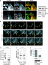
RACK1 regulates focal adhesion disassembly and cell motility. (A) RACK1 regulates the localization of active ERK to focal adhesions in wounded cell monolayers. NIH 3T3 cells were transfected with siRNA, and 48 h after the transfection a wound was made in a confluent monolayer of cells in the presence of 10% serum. Five hours after the wound, cells were costained with antibodies recognizing the active form of ERK (red) and paxillin (green). The inset shows a higher magnification of the boxed area. (B) RACK1 regulates focal adhesion turnover. Series of images from time-lapse TIRF microscopy recording the dynamics of GFP-paxillin adhesions in control and RACK1 knockdown cells. Arrows indicate examples of dynamic focal adhesion at a protrusive region of the cell where both assembly and disassembly were seen. A longer time frame of GFP-paxillin dynamic is shown in RACK1-silenced cells to demonstrate the slower rate of focal adhesion disassembly. NIH 3T3 cells stably expressing GFP-paxillin were transfected and wounded as described for panel A. GFP-paxillin-containing focal adhesions were imaged 5 h after wounding by time-lapse TIRF microscopy by collecting images every 3 min. (C) Rates of focal adhesion disassembly. Rate constants of focal adhesion disassembly were determined from values obtained from 37 focal adhesions in four cells (control cells) or 30 focal adhesions in five cells (RACK1 siRNA-treated cells). Each point represents one focal adhesion analyzed. Only focal adhesions that displayed an initial increase in fluorescence intensity (i.e., focal adhesion assembly) were included in the analysis. (D) Rates of focal adhesion assembly. Rate constants of focal adhesion assembly were determined from values obtained from 16 focal adhesions in four cells (control cells) or 15 focal adhesions in five cells (RACK1 siRNA-treated cells). (E) RACK1 depletion inhibits cell migration toward fibronectin. REF52 cells were transfected with siRNA for 48 h and suspended, and cell migration toward the fibronectin was measured in triplicate in a Boyden chamber assay. The data represent averages ± standard deviations. Cell lysates were probed with antibodies directed against RACK1 and ERK2 to confirm knockdown efficiency and equal loading of proteins, respectively (bottom). Depletion of RACK1 protein by siRNA targeting different regions of RACK1 mRNA also inhibited cell migration toward fibronectin (data not shown).
FIG. 5.
FIG. 5.
RACK1 and ERK regulate focal adhesion length. (A) RACK1 silencing increases focal adhesion length. RAT2 cells were transfected with siRNA, and 48 h after the transfection cells were plated on fibronectin (1 μg/ml)-coated coverslips in the presence of 10% serum. Four hours after the plating, cells were stained with antibody recognizing paxillin. The right panel shows the quantification of focal adhesion length in control and RACK1 siRNA-transfected cells (determined from values obtained from ∼200 focal adhesions in 10 cells). Only peripheral focal adhesions were included in the analysis. Depletion of RACK1 protein by siRNA targeting different regions of RACK1 mRNA also increased the length of the focal adhesions (data not shown). (B) MEK inhibition induces an increase in focal adhesion length. RAT2 cells were plated on fibronectin (1 μg/ml)-coated coverslips and treated with MEK inhibitor UO126 (10 μM) for 48 h. Cell staining and quantification of focal adhesion length were done as described for panel A. (C) RACK1 silencing does not inhibit paxillin Y118 phosphorylation. RAT2 cells were treated as described for panel A, and cell lysates were probed with antibodies directed against paxillin and paxillin pY118. DMSO, dimethyl sulfoxide; Ctrl, control.
FIG. 6.
FIG. 6.
FAK regulates active ERK localization to focal adhesion. (A) Efficiency of FAK knockdown. RAT2 cells were transfected with FAK siRNA for 48 h, suspended in medium with 10% serum, and replated on fibronectin-coated dishes for 4 h. Cell lysates were probed with antibodies recognizing FAK, active FAK [FAK (pY397)], ERK, and active ERK. (B) FAK silencing inhibits active ERK localization to focal adhesions. RAT2 cells were transfected with FAK siRNA for 48 h, suspended in medium with 10% serum, and replated on fibronectin-coated coverslips. Four hours after the plating, cells were costained with antibodies recognizing the active form of ERK (red) and paxillin (green). Ctrl, control.

Similar articles

Cited by

References

    1. Carragher, N. O., M. A. Westhoff, V. J. Fincham, M. D. Schaller, and M. C. Frame. 2003. A novel role for FAK as a protease-targeting adaptor protein: regulation by p42 ERK and Src. Curr. Biol. 13:1442-1450. - PubMed
    1. Chang, B. Y., K. B. Conroy, E. M. Machleder, and C. A. Cartwright. 1998. RACK1, a receptor for activated C kinase and a homolog of the beta subunit of G proteins, inhibits activity of Src tyrosine kinases and growth of NIH 3T3 cells. Mol. Cell. Biol. 18:3245-3256. - PMC - PubMed
    1. Chang, L., and M. Karin. 2001. Mammalian MAP kinase signalling cascades. Nature 410:37-40. - PubMed
    1. Chaudhary, A., W. G. King, M. D. Mattaliano, J. A. Frost, B. Diaz, D. K. Morrison, M. H. Cobb, M. S. Marshall, and J. S. Brugge. 2000. Phosphatidylinositol 3-kinase regulates Raf1 through Pak phosphorylation of serine 338. Curr. Biol. 10:551-554. - PubMed
    1. Chen, Q., M. S. Kinch, T. H. Lin, K. Burridge, and R. L. Juliano. 1994. Integrin-mediated cell adhesion activates mitogen-activated protein kinases. J. Biol. Chem. 269:26602-26605. - PubMed

Publication types

LinkOut - more resources