

Pancreas volumes in humans from birth to age one hundred taking into account sex, obesity, and presence of type-2 diabetes
- PMID: 17879305
- PMCID: PMC2680737
- DOI: 10.1002/ca.20543
Pancreas volumes in humans from birth to age one hundred taking into account sex, obesity, and presence of type-2 diabetes
Abstract
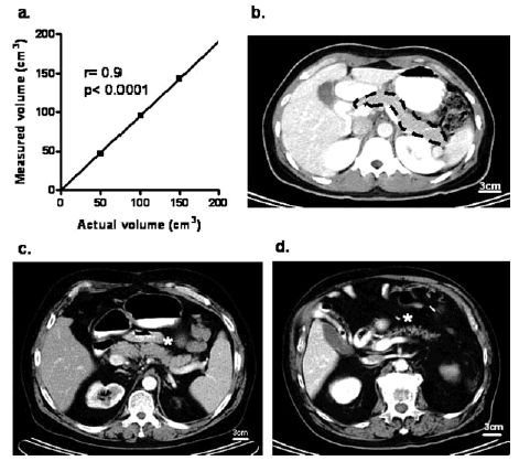
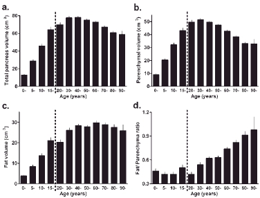
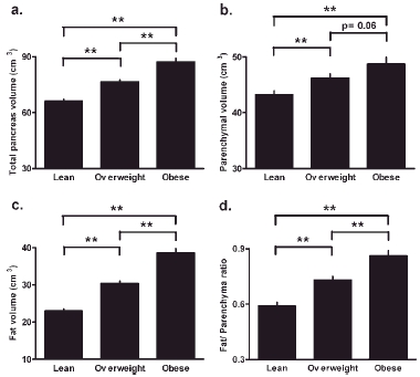
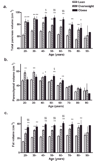
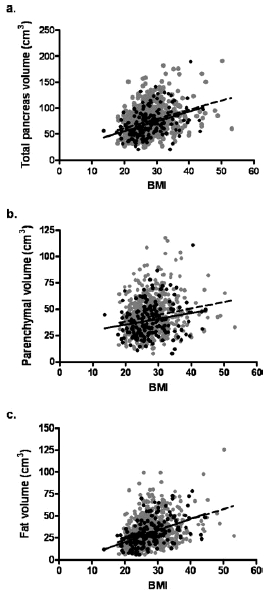
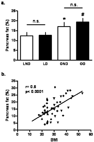
Our aims were (1) by computed tomography (CT) to establish a population database for pancreas volume (parenchyma and fat) from birth to age 100 years, (2) in adults, to establish the impact of gender, obesity, and the presence or absence of type-2 diabetes on pancreatic volume (parenchyma and fat), and (3) to confirm the latter histologically from pancreatic tissue obtained at autopsy with a particular emphasis on whether pancreatic fat is increased in type-2 diabetes. We measured pancreas volume in 135 children and 1,886 adults (1,721 nondiabetic and 165 with type-2 diabetes) with no history of pancreas disease who had undergone abdominal CT scan between 2003 and 2006. Pancreas volume was computed from the contour of the pancreas on each CT image. In addition to total pancreas volume, parenchymal volume, fat volume, and fat/parenchyma ratio (F/P ratio) were determined by CT density. We also quantified pancreatic fat in autopsy tissue of 47 adults (24 nondiabetic and 23 with type-2 diabetes). During childhood and adolescence, the volumes of total pancreas, pancreatic parenchyma, and fat increase linearly with age. From age 20-60 years, pancreas volume reaches a plateau (72.4 +/- 25.8 cm(3) total; 44.5 +/- 16.5 cm(3) parenchyma) and then declines thereafter. In adults, total ( approximately 32%), parenchymal ( approximately 13%), and fat ( approximately 68%) volumes increase with obesity. Pancreatic fat content also increases with aging but is not further increased in type-2 diabetes. We provide lifelong population data for total pancreatic, parenchymal, and fat volumes in humans. Although pancreatic fat increases with aging and obesity, it is not increased in type-2 diabetes.
2007 Wiley-Liss, Inc
Figures
Similar articles
-
Trajectories of body mass index amongst children who develop type 2 diabetes as adults.J Intern Med. 2015 Aug;278(2):219-26. doi: 10.1111/joim.12354. Epub 2015 Mar 16. J Intern Med. 2015. PMID: 25683182
-
Differences in pancreatic volume, fat content, and fat density measured by multidetector-row computed tomography according to the duration of diabetes.Acta Diabetol. 2014 Oct;51(5):739-48. doi: 10.1007/s00592-014-0581-3. Epub 2014 Mar 27. Acta Diabetol. 2014. PMID: 24671510
-
Body Fat Patterning, Hepatic Fat and Pancreatic Volume of Non-Obese Asian Indians with Type 2 Diabetes in North India: A Case-Control Study.PLoS One. 2015 Oct 16;10(10):e0140447. doi: 10.1371/journal.pone.0140447. eCollection 2015. PLoS One. 2015. PMID: 26474415 Free PMC article.
-
Pancreas volume in health and disease: a systematic review and meta-analysis.Expert Rev Gastroenterol Hepatol. 2018 Aug;12(8):757-766. doi: 10.1080/17474124.2018.1496015. Epub 2018 Jul 16. Expert Rev Gastroenterol Hepatol. 2018. PMID: 29972077 Review.
-
Pancreatic size and fat content in diabetes: A systematic review and meta-analysis of imaging studies.PLoS One. 2017 Jul 24;12(7):e0180911. doi: 10.1371/journal.pone.0180911. eCollection 2017. PLoS One. 2017. PMID: 28742102 Free PMC article. Review.
Cited by
-
Fatty Pancreas and Cardiometabolic Risk: Response of Ectopic Fat to Lifestyle and Surgical Interventions.Nutrients. 2022 Nov 17;14(22):4873. doi: 10.3390/nu14224873. Nutrients. 2022. PMID: 36432559 Free PMC article. Review.
-
Current State of Imaging of Pediatric Pancreatitis: AJR Expert Panel Narrative Review.AJR Am J Roentgenol. 2021 Aug;217(2):265-277. doi: 10.2214/AJR.21.25508. Epub 2021 Mar 17. AJR Am J Roentgenol. 2021. PMID: 33728974 Free PMC article. Review.
-
Morphology of the pancreas in type 2 diabetes: effect of weight loss with or without normalisation of insulin secretory capacity.Diabetologia. 2016 Aug;59(8):1753-9. doi: 10.1007/s00125-016-3984-6. Epub 2016 May 14. Diabetologia. 2016. PMID: 27179658 Free PMC article.
-
A non-invasive method of quantifying pancreatic volume in mice using micro-MRI.PLoS One. 2014 Mar 18;9(3):e92263. doi: 10.1371/journal.pone.0092263. eCollection 2014. PLoS One. 2014. PMID: 24642611 Free PMC article.
-
The Pdx1-Bound Swi/Snf Chromatin Remodeling Complex Regulates Pancreatic Progenitor Cell Proliferation and Mature Islet β-Cell Function.Diabetes. 2019 Sep;68(9):1806-1818. doi: 10.2337/db19-0349. Epub 2019 Jun 14. Diabetes. 2019. PMID: 31201281 Free PMC article.
References
-
- Altman PL, Dittmer DS. Growth-including reproduction and morphological development. Washington, DC: Federation of American Societies for Experimental Biology; 1962.
-
- Altobelli E, Blasetti A, Verrotti A, Di Giandomenico V, Bonomo L, Chiarelli F. Size of pancreas in children and adolescents with type 1 (insulin-dependent) diabetes. J Clin Ultrasound. 1998;26:391–395. - PubMed
-
- Alzaid A, Aideyan O, Nawaz S. The size of the pancreas in diabetes mellitus. Diabet Med. 1993;10:759–763. - PubMed
-
- Assimacopoulos-Jeannet F. Fat storage in pancreas and in insulin-sensitive tissues in pathogenesis of type 2 diabetes. Int J Obes Relat Metab Disord. 2004;28(Suppl 4):S53–S57. - PubMed
-
- Butler AE, Janson J, Bonner-Weir S, Ritzel R, Rizza RA, Butler PC. β-Cell deficit and increased β-cell apoptosis in humans with type 2 diabetes. Diabetes. 2003;52:102–110. - PubMed
Publication types
MeSH terms
Grants and funding
LinkOut - more resources
Full Text Sources
Other Literature Sources
Medical
