. 2007 Sep;5(9):e238.
doi: 10.1371/journal.pbio.0050238.

Down-regulation of NF-kappaB target genes by the AP-1 and STAT complex during the innate immune response in Drosophila

Affiliations

Down-regulation of NF-kappaB target genes by the AP-1 and STAT complex during the innate immune response in Drosophila

Lark Kyun Kim et al. PLoS Biol. 2007 Sep.

Abstract

The activation of several transcription factors is required for the elimination of infectious pathogens via the innate immune response. The transcription factors NF-kappaB, AP-1, and STAT play major roles in the synthesis of immune effector molecules during innate immune responses. However, the fact that these immune responses can have cytotoxic effects requires their tight regulation to achieve restricted and transient activation, and mis-regulation of the damping process has pathological consequences. Here we show that AP-1 and STAT are themselves the major inhibitors responsible for damping NF-kappaB-mediated transcriptional activation during the innate immune response in Drosophila. As the levels of dAP-1 and Stat92E increase due to continuous immune signaling, they play a repressive role by forming a repressosome complex with the Drosophila HMG protein, Dsp1. The dAP-1-, Stat92E-, and Dsp1-containing complexes replace Relish at the promoters of diverse immune effector genes by binding to evolutionarily conserved cis-elements, and they recruit histone deacetylase to inhibit transcription. Reduction by mutation of dAP-1, Stat92E, or Dsp1 results in hyperactivation of Relish target genes and reduces the viability of bacterially infected flies despite more efficient pathogen clearance. These defects are rescued by reducing the Relish copy number, thus confirming that mis-regulation of Relish, not inadequate activation of dAP-1, Stat92E, or Dsp1 target genes, is responsible for the reduced survival of the mutants. We conclude that an inhibitory effect of AP-1 and STAT on NF-kappaB is required for properly balanced immune responses and appears to be evolutionarily conserved.


Conflict of interest statement

Competing interests. The authors have declared that no competing interests exist.

Figures

Figure 1
Figure 1. Down-Regulation of Relish Signaling by Stat92E as well as Jra in Response to LPS/PGN
Real-time PCR analysis showing LPS/PGN-induced transcriptional activation in various mutant backgrounds. SL2 cells were incubated with dsRNA, as indicated in the top box, for three days. The levels of the transcripts before (-) and after (+) LPS/PGN treatment (10 μg/ml; 1hr) were measured by real time PCR. The degree of depletion of the corresponding transcript by RNAi is shown in the right panel.
Figure 2
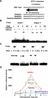
Figure 2. A Stat92E Binding Site on the Attacin-A Promoter Plays a Crucial Role in Down-Regulating Attacin-A
(A) Region Y contains an NF-κB binding site and a STAT binding site. The NF-κB consensus and STAT consensus binding sequences are shown along with the wild-type sequence of region Y. The mutant forms of the NF-κB and/or the STAT binding sites of region Y are designated Relish2m, Stat92Em, and Relish2m-Stat92Em, respectively. The mutated sequences are shown in lower case and underlined. (B) Nuclear extracts of SL2 cells pre-incubated with dsRNA for Luciferase (L), Relish (R), Stat92E (S), or both Relish and Stat92E (RS) and treated with 10 μg/ml of LPS/PGN were assayed by EMSAs with 32P-labeled double-stranded oligonucleotide probes containing Relish1 or region Y. (C) EMSAs with probes containing the wild-type region Y (double arrow), a mutation in the Relish binding site (double arrow with X on the left), a mutation in the Stat92E binding site (double arrow with X on the right) or mutations in both binding sites (double arrow with double X). Black and white arrows indicate the Relish 2 and Stat92E binding sites, respectively, and the mutations are indicated by Xs. Nuclear extracts were as in (B). (D) The dAP-1 and Stat92E promoter elements are required for down-regulation of Attacin-A. SL2 cells transfected with each reporter under the control of a mutant version of the Attacin-A promoter as indicated were treated with 10 μg/ml LPS/PGN for the time indicated on the abscissa. The mean levels of the normalized luciferase activities are shown with standard deviations. These experiments were repeated at least three times independently.
Figure 3
Figure 3. Synergistic Binding of Jra and Stat92E to the Attacin-A Promoter
ChIP assays of the transcription factors indicated below using various mutants. SL2 cells were depleted of the transcripts by dsRNA treatment, as indicated in the top box, for 3 d. Then chromatin extracts were prepared before (−) or after 30 min (+) of LPS/PGN treatment. The amounts of Attacin-A promoter fragments co-precipitated with antibodies against the transcription factors indicated below the data were measured by real-time PCR. The levels were normalized by the input used in each ChIP assay and are shown with standard deviations. These experiments were repeated independently at least three times.
Figure 4
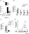
Figure 4. Dsp1 Plays a Crucial Role in the Interactions between Jra and Stat92E
(A) Down-regulation of Attacin-A transcripts by HMG protein. Real-time PCR analysis showing Attacin-A transcript levels after 1 h of LPS/PGN treatment of SL2 cells depleted by RNAi of the transcription factors and HMG proteins indicated below the histograms. The levels were normalized with RpL32 transcripts. The extents of depletion of the corresponding transcripts by RNAi are shown in the top panel. (B) Dsp1 is required for binding of Jra, Stat92E, and dHDAC1 to the Attacin-A promoter. SL2 cells were incubated with Luciferase or Dsp1 dsRNA for three days, then used in ChIP assays with (+) and without (−) LPS/PGN treatment (10 μg/ml for 1 h). The amounts of Attacin-A promoter fragments co-precipitated with the antibodies were normalized for the input used in each assay and are shown with standard deviations. These experiments were repeated at least three times independently. (C) Requirement for Jra and Stat92E for recruitment of Dsp1 to the Attacin-A promoter. SL2 cells were depleted of the protein indicated on the right by RNAi, then used in ChIP assays before (−) and after (+) LPS/PGN treatment. The amounts of Attacin-A promoter co-precipitated with anti-Dsp1 antibody in the ChIP assays are shown with standard deviations. These experiments were repeated at least three times independently. (D) The amounts of chromatin fragments co-precipitated with anti-Jun (solid squares), anti-Dsp1 (open triangles) or anti-Relish (solid circles) antibodies in the indicated regions of the Attacin-A promoter were measured by real-time PCR in a Roche Lightcycler, and the averages and standard deviations of three independent experiments are plotted.
Figure 5
Figure 5. Dsp1 Interacts with Jra and Stat92E to Form a Repressosome Complex
(A) Co-immunoprecipitation of Relish, Jra, Stat92E, and Dsp1. Nuclear extracts prepared from SL2 cells with (right panel) or without (left panel) LPS/PGN (10 μg/ml for 45 min) treatment were immunoprecipitated with the antibodies indicated at the top of the figure, and the amounts of the proteins in the pellets were measured by immunoblot analysis with the antibodies indicated on the left. For co-immunoprecipitation of Relish, Jra, and Stat92E, 10-μg aliquots of nuclear extracts were used, whereas for Dsp1, 3-μg aliquots were used. Five percent of the amount of nuclear extract used in each immunoprecipitation assay is shown as Input. (B) Regulation of Attacin-A transcription by ectopic expression of transcription factors. The N-terminal half of Relish that is competent as a transcriptional activator (Rel-ΔC), epitope-tagged Jra (S-Jra), and Stat92E (S-Stat92E) expression constructs were transfected into SL2 cells as indicated at the bottom of the figure. After induction of the recombinant proteins, the levels of Attacin-A transcripts relative to those of RpL32 were measured by real-time PCR analysis in three independent experiments. (C) ChIP assays of the Attacin-A promoter with anti-dHDAC1 antibody. SL2 cells pretreated with Luciferase dsRNA (control) or Dsp1 dsRNA (Dsp1-) were transfected with expression constructs for Rel-ΔC, S-Jra, and S-Stat92E as indicated at the bottom, and the average amounts of Attacin-A promoter fragments co-precipitated with anti-dHDAC1 antibody after induction of the transfected transcription factors were measured by real time PCR analysis in three independent experiments.
Figure 6
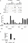
Figure 6. Relish Is Displaced from the Attacin-A Promoter by the Repressosome
(A) Left panel: soluble chromatin extracts were prepared from SL2 cells with (15 min or 8 h) or without (0 min) LPS/PGN treatment, and immunoprecipitated with antibodies against Relish, Jun, or Stat92E as described in Figure 3. Right panel: double ChIP assays. The precipitates obtained from the first ChIP of cells with (15 min) or without LPS/PGN treatment were analyzed separately in a second ChIP with the antibodies indicated at the top (second ChIP). The amounts of Attacin-A promoter fragments co-precipitated with the indicated antibody are shown. (B) The transcript levels of each target gene are shown under Luciferase, Relish, Jra, Stat92E, or Dsp1 knock-down conditions with LPS/PGN treatment (10 μg/ml; 1h). The averages and standard deviations of triplicates assays are shown.
Figure 7
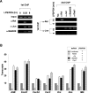
Figure 7. Up-Regulation of Relish Target Genes in Repressosome Mutant Flies during Bacterial Infection
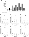
(A) Transcript levels of LPS/PGN-induced Attacin-A in mutant flies. Wild type (w1118) and mutant flies were infected with E. coli, and the levels of the Attacin-A transcript were measured by real time PCR analysis 24 h after infection. Total RNA from groups of five flies was pooled for the analysis. The averages and standard deviations of three independent assays are shown. Attacin-A was not expressed without bacterial infection in any of the mutants (unpublished data). Abbreviations for the mutant flies are as follows: w, w1118; R, Rel/Rel; S/d, UAS-shStat92E/+; da-Gal4/+; S/d/R, UAS-shStat92E/+; da-Gal4/Rel; S/h, UAS-hs-shStat92E/UAS-hs-shStat92E; S/h/R, UAS-hs-shStat92E/+; Rel/+; J, Jra1A109/CyO; J/R, Jra1A109/+; Rel/+; D, Dsp1[EP355]/Dsp1[EP355]; D/R, Dsp1[EP355]/Y; Rel/+. (B–G) Bacterial clearance assays. Wild type and mutants were injected with the same number of E. coli, and the number of live bacteria inside each injected fly was measured as described in Materials and Methods and represented by a dot on the graph. The bars represent mean of colony forming unit (cfu). The abbreviations used are as in (A). p-values were calculated by Student's t-test. *p < 0.01. **p < 0.05. ***p > 0.2.
Figure 8
Figure 8. Lower Survival Rate Caused by Mis-Regulation of Repressosome Complex in Flies
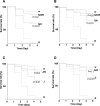
Survival of various mutant flies after bacterial infection. Three-d-old wild-type and mutant flies were infected with E. coli and their survival was measured each day after infection. Survival curves are plotted as Kaplan-Meier plots. Statistical significance is tested using log-rank analysis with MedCare software. The abbreviations used are as in Figure 7.

Comment in

Similar articles

Cited by

References

    1. Janeway CA, Medzhitov R. Innate immune recognition. Annu Rev Immunol. 2002;20:197–216. - PubMed
    1. Kimbrell DA, Beutler B. The evolution and genetics of innate immunity. Nat Rev Genet. 2001;2:256–267. - PubMed
    1. Kim T, Kim Y-J. Overview of innate immunity in Drosophila . J Biochem Mol Biol. 2005;38:121–127. - PubMed
    1. Silverman N, Maniatis T. NF-κB signaling pathways in mammalian and insect innate immunity. Genes Dev. 2001;15:2321–2342. - PubMed
    1. Hedengren M, Asling B, Dushay MS, Ando I, Ekengren S, et al. Relish, a central factor in the control of humoral, but not cellular immunity in Drosophila . Mol Cell. 1999;4:827–837. - PubMed

Publication types

MeSH terms