

Review
doi: 10.1172/JCI33099.
MicroRNAs: powerful new regulators of heart disease and provocative therapeutic targets
Affiliations
- PMID: 17786230
- PMCID: PMC1952642
- DOI: 10.1172/JCI33099
Item in Clipboard
Review
MicroRNAs: powerful new regulators of heart disease and provocative therapeutic targets
J Clin Invest.
2007 Sep.
Abstract
MicroRNAs act as negative regulators of gene expression by inhibiting the translation or promoting the degradation of target mRNAs. Recent studies have revealed key roles of microRNAs as regulators of the growth, development, function, and stress responsiveness of the heart, providing glimpses of undiscovered regulatory mechanisms and potential therapeutic targets for the treatment of heart disease.
Figures
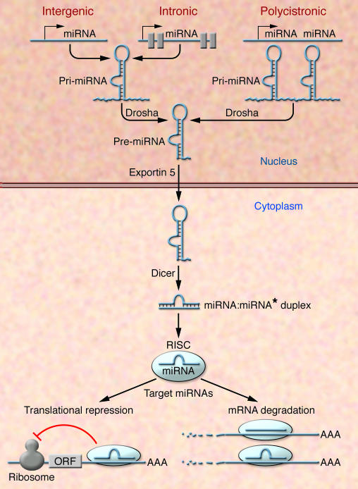
The primary transcripts of miRNAs, called pri-miRNAs, are transcribed as individual miRNA genes, from introns of protein-coding genes, or from polycistronic transcripts. The RNase Drosha further processes the pri-miRNA into 70–100 nucleotide, hairpin-shaped precursors, called pre-miRNA, which are exported from the nucleus by exportin 5. In the cytoplasm, the pre-miRNA is cleaved by Dicer into an miRNA:miRNA* duplex. Assembled into the RISC, the mature miRNA negatively regulates gene expression by either translational repression or mRNA degradation, which is dependent on sequence complementarity between the miRNA and the target mRNA. ORF, open reading frame.
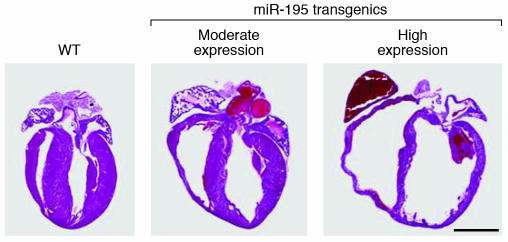
H&E sections of 2-week-old wild-type and transgenic mice expressing miR-195 under control of the αMHC promoter. In transgenic as compared with wild-type mice, moderate levels of miR-195 expression (26-fold) cause cardiac hypertrophy, and higher levels of expression (29-fold) cause dilated cardiomyopathy with ventricular dilatation and wall thinning. Reproduced with permission from Proceedings of the National Academy of Sciences of the United States of America (8). Scale bar: 2 mm.

Stress signals drive a pathological remodeling response of the adult heart, which is characterized by hypertrophy, ventricular dilatation, and fibrosis. miR-208 is required for stress-dependent remodeling and miR-195, which is induced by cardiac stress, is sufficient to drive cardiac remodeling. miR-133 appears to play a negative role in cardiac hypertrophy (5, 6, 8).

(A) Schematic diagram of a heart following thoracic aortic banding (TAB). (B) Sections of hearts of approximately 3-month-old wild-type and miR-208–/– mice are shown following sham operation or TAB for 21 days. High-magnification views of the ventricular wall are shown at the bottom. Trichrome staining identifies fibrosis in blue. Note that hypertrophy and fibrosis are diminished in miR208–/– mice compared with wild-type following TAB. Scale bars: 2 mm (top); 20 μm (bottom). Reproduced with permission from Science (5).
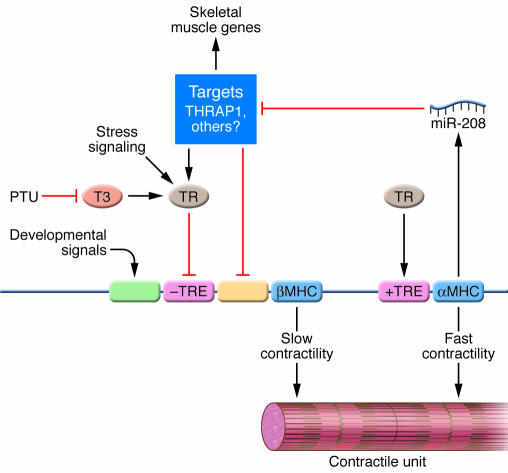
αMHC and βMHC promote fast and slow contractility, respectively. TRs act through positive and negative TREs to activate and repress expression of the αMHC and βMHC genes, respectively, which are linked. Propylthiouracil (PTU) prevents T3 biosynthesis, resulting in hypothyroidism and upregulation of βMHC due to loss of the repressive action of the TR on the negative TRE. Stress signals also activate βMHC expression, at least in part through the TR. Developmental signals drive βMHC expression before birth through separate regulatory elements. miR-208, encoded by the αMHC gene, negatively regulates mRNA targets encoding THRAP1 and other negative regulators of βMHC expression. miR-208 also represses an activator of fast skeletal muscle genes. Modified with permission from Science (5).
Similar articles
-
MicroRNAs: novel regulators in cardiac development and disease.Cardiovasc Res. 2008 Sep 1;79(4):562-70. doi: 10.1093/cvr/cvn137. Epub 2008 May 29. Cardiovasc Res. 2008. PMID: 18511432 Review.
-
[Potential role of microRNAs in human diseases and the exploration on design of small molecule agents].Yao Xue Xue Bao. 2007 Nov;42(11):1115-21. Yao Xue Xue Bao. 2007. PMID: 18300464 Review. Chinese.
-
MicroRNA epigenetic alterations: predicting biomarkers and therapeutic targets in human diseases.Clin Genet. 2008 Oct;74(4):307-15. doi: 10.1111/j.1399-0004.2008.01075.x. Epub 2008 Aug 18. Clin Genet. 2008. PMID: 18713257 Review.
-
MicroRNAs: novel regulators in the hallmarks of human cancer.Cancer Lett. 2009 Nov 28;285(2):116-26. doi: 10.1016/j.canlet.2009.04.031. Epub 2009 May 22. Cancer Lett. 2009. PMID: 19464788 Review.
-
Toward microRNA-based therapeutics for heart disease: the sense in antisense.Circ Res. 2008 Oct 24;103(9):919-28. doi: 10.1161/CIRCRESAHA.108.183426. Circ Res. 2008. PMID: 18948630 Free PMC article. Review.
Cited by
-
Heart structure-specific transcriptomic atlas reveals conserved microRNA-mRNA interactions.PLoS One. 2013;8(1):e52442. doi: 10.1371/journal.pone.0052442. Epub 2013 Jan 2. PLoS One. 2013. PMID: 23300973 Free PMC article.
-
Transcription factor FoxO1, the dominant mediator of muscle wasting in chronic kidney disease, is inhibited by microRNA-486.Kidney Int. 2012 Aug;82(4):401-11. doi: 10.1038/ki.2012.84. Kidney Int. 2012. PMID: 22475820 Free PMC article.
-
MiR-30-regulated autophagy mediates angiotensin II-induced myocardial hypertrophy.PLoS One. 2013;8(1):e53950. doi: 10.1371/journal.pone.0053950. Epub 2013 Jan 9. PLoS One. 2013. PMID: 23326547 Free PMC article.
-
MicroRNA Regulatory Pathways in the Control of the Actin-Myosin Cytoskeleton.Cells. 2020 Jul 9;9(7):1649. doi: 10.3390/cells9071649. Cells. 2020. PMID: 32660059 Free PMC article. Review.
-
Horizontal transfer of microRNAs: molecular mechanisms and clinical applications.Protein Cell. 2012 Jan;3(1):28-37. doi: 10.1007/s13238-012-2003-z. Epub 2012 Feb 9. Protein Cell. 2012. PMID: 22314808 Free PMC article. Review.
References
-
- Hoffman J.I. Incidence of congenital heart disease: II. Prenatal incidence. Pediatr. Cardiol. 1995;16:155–165. - PubMed
-
- Rosamond W., et al. Heart disease and stroke statistics--2007 update: a report from the American Heart Association Statistics Committee and Stroke Statistics Subcommittee. Circulation. 2007;115:e69–e171. - PubMed
-
- Yang B., et al. The muscle-specific microRNA miR-1 regulates cardiac arrhythmogenic potential by targeting GJA1 and KCNJ2. Nat. Med. 2007;13:486–491. - PubMed
-
- Zhao Y., et al. Dysregulation of cardiogenesis, cardiac conduction, and cell cycle in mice lacking miRNA-1-2. Cell. 2007;129:303–317. - PubMed
-
- van Rooij E., et al. Control of stress-dependent cardiac growth and gene expression by a microRNA. Science. 2007;316:575–579. - PubMed
Publication types
MeSH terms
Substances
LinkOut - more resources
Full Text Sources
Other Literature Sources
Medical
