. 2007 Aug;27(2):281-95.
doi: 10.1016/j.immuni.2007.07.010.

Inflammation directs memory precursor and short-lived effector CD8(+) T cell fates via the graded expression of T-bet transcription factor

Affiliations

Inflammation directs memory precursor and short-lived effector CD8(+) T cell fates via the graded expression of T-bet transcription factor

Nikhil S Joshi et al. Immunity. 2007 Aug.

Abstract

As acute infections resolve, effector CD8(+) T cells differentiate into interleukin-7 receptor(lo) (IL-7R(lo)) short-lived effector cells (SLECs) and IL-7R(hi) memory precursor effector cells (MPECs) capable of generating long-lived memory CD8(+) T cells. By using another SLEC marker, KLRG1, we found that KLRG1(hi) effector cells began appearing early during infection and were committed to downregulating IL-7R. Unlike IL-7R(hi) MPECs, KLRG1(hi) IL-7R(lo) SLECs relied on IL-15, but IL-15 could not sustain their long-term maintenance or homeostatic turnover. The decision between SLEC and MPEC fates was regulated by the amount of inflammatory cytokines (i.e., IL-12) present during T cell priming. According to the amount of inflammation, a gradient of T-bet was created in which high T-bet expression induced SLECs and low expression promoted MPECs. These results elucidate a mechanism by which the innate immune system sets the relative amounts of a lineage-determining transcription factor in activated CD8(+) T cells and, correspondingly, regulates their memory cell potential.


Figures

Figure 1
Figure 1
KLRG1hi IL-7Rlo effector CD8 T cells are short-lived and require IL-15 for survival. (A) IL-7Rhi and IL-7Rlo Thy1.1+ effector CD8 T cells from P14 chimeric mice were compared on day 7 of LCMV infection using Affymetrix GeneChips. Table shows the fold increase in expression for selected NK receptors (IL-7Rlo > IL-7Rhi cells). (B and C) Analysis of MPEC and SLEC subsets following LCMV infection of P14 chimeric mice. (B) Plots are gated on Thy1.1+ P14 T cells and show expression of KLRG1 and IL-7R in blood over time. (C) Line graphs show KLRG1hi IL-7Rlo (■) and KLRG1lo IL-7Rhi (▲) P14 CD8 T cell numbers in the spleen, liver, lung, inguinal lymph node (LN) and total from all tissues. The magnitude of contraction between days 8−40 and 8−75 is indicated. (D) CFSE-labeled P14 memory CD8 T cells from day ∼40 pi were transferred into naïve mice and then analyzed for CFSE and KLRG1 expression 4−6 weeks later. (E) wt and IL-15−/− mice were infected with LCMV and Db GP33−41 MHC class I tetramer+ CD8 T cells were analyzed for KLRG1 expression 8, 15 and 30 days pi. Similar data were observed for NP396−404-specific CD8 T cells (data not shown). (F) KLRG1hi IL-7Rlo or KLRG1lo IL-7Rhi P14 CD8 T cells were sorted day 8 pi and transferred in equal numbers into wt or IL-15−/− recipients for 10−15 days. Bar graph shows the number of donor cells recovered from IL-15−/− recipients normalized to the number recovered from wt recipients.
Figure 2
Figure 2
KLRG1 marks effector CD8 T cells committed to an SLEC fate. (A and B) P14 chimeric mice were infected with LCMV and on days 4−8 pi the Thy1.1+ P14 effector CD8 T cells were analyzed for expression of (A) KLRG1 and (B) KLRG1 and IL-7R. (B) Bottom row, histograms show IL-7R expression on KLRG1hi (filled) or KLRG1lo (open) P14 CD8 T cells. IL-7R MFI is shown (KLRG1lo/KLRG1hi). (C) Bar graph shows IL-7R mRNA levels (normalized to the ribosomal gene L9) in the indicated cell populations measured by real-time PCR. **=p<0.001. (D) Day 5 pi KLRG1hi and KLRG1lo P14 CD8 T cells were sorted and transferred in equal number back into day 5 LCMV infected recipients and analyzed 4 days post transfer (pt) for KLRG1 and IL-7R expression.
Figure 3
Figure 3
Phenotypic and functional comparisons between KLRG1hi and KLRG1lo effector CD8 T cells. (A) Histograms show expression of the indicated proteins on KLRG1hi (filled) and KLRG1lo (open) P14 CD8 T cells on days 5 and 8 pi. The MFI of KLRG1lo/KLRG1hi cells is shown. (B) In vivo CTL assay comparing day 5 and 8 KLRG1hi (black) and KLRG1lo (white) P14 effector CD8 T cells. The percent killing over 4 hrs was normalized to the on Effector:Target (E:T) ratio. (C) Splenocytes from day 5 and 8 LCMV infected P14 chimeric were stimulated with GP33−41 peptide and analyzed for IFNγ and IL-2 production by intracellular cytokine staining. Histograms show IFNγ (left and center) and IL-2 (right) production by KLRG1hi (filled) and KLRG1lo (open) Thy1.1+ CD8 P14 CD8 T cells. IL-2 plots are gated on IFNγ-producing cells. Similar results were found in endogenous LCMV-specific CD8 T cells stimulated with NP396−404, GP33−41 and GP276−284. Note, KLRG1 expression does not change during 5 hr stimulation (data not shown). (D) Histograms show the percent of KLRG1hi (top) and KLRG1lo cells (bottom) P14 CD8 T cells in S/G2/M phases of the cell cycle on day 5 pi using 7-AAD.
Figure 4
Figure 4
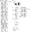
Levels of inflammation regulate KLRG1hi IL-7Rlo SLEC formation. (A) P14 chimeric mice were infected with LM-GP33 and 1 day pi were either left untreated (No Rx, black bars) or treated with Ampicillin (Amp Rx, white bars). FACS plots show KLRG1 and IL-7R expression and bar graphs show the number of total or KLRG1lo IL-7Rhi MPECs on day 7 pi. (B) P14 chimeric mice were infected with rVVhp −0, −17, or −19 (see text) and 8 days pi Thy1.1+ P14 CD8 T cells were analyzed for KLRG1 and IL-7R expression. (C) P14 chimeric mice were concurrently immunized with DC-33 and varying doses of Listeria (not expressing GP33−41). FACS plots show KLRG1 and IL-7R expression on day 7 P14 effector CD8 T cells. (D) Purified naive Thy1.1+ P14 CD8 T cells were stimulated with GP33−41 peptide-loaded cells ± CpG ODN or the indicated cytokines for 24−48h and then transferred into naïve recipients. Thy1.1+ P14 CD8 T cells were analyzed for KLRG1 and IL-7R expression 5−6 days pt. (E and F) Naïve wt or T-bet−/− P14 CD8 T cells were stimulated as in (D) with IL-12 or IFNγ or both (E) or decreasing concentrations of IL-12 (F). (E) Histograms show T-bet expression in wt (line) or T-bet−/− (shaded) Thy1.1+ CD44hi CD8 T cells and the T-bet MFI is indicated. (F) Line graph shows the MFI of T-bet with either peptide alone (dashed), the indicated concentration of IL-12 (solid), or T-bet−/− P14 CD8 T cells + IL-12 (gray). Data in graph is representative of 3 independent experiments.
Figure 5
Figure 5
T-bet expression is necessary and sufficient for development of KLRG1hi SLECs. (A) wt or T-bet−/− mice or (B) wt mice containing ∼1x10⁴ wt or T-bet−/− P14 CD8 T cells were infected with LCMV and analyzed 7−8 days later for IL-7R and KLRG1 expression on GP33−41-specific CD8 T cells (A) or Thy1.1+ P14 CD8 splenocytes (B). Similar data to (A) were observed with NP396−404-specific CD8 T cells (data not shown). (C) Bar graph compares the total combined number of endogenous GP33−41 and NP396−404-specific CD8 T cells between wt (black) or T-bet−/− (white) animals on day 8 of LCMV infection. Note, similar numbers of IL-7Rhi effector cells in wt and T-bet−/− animals. **=p<0.001, *=p<0.01. (D) As in Figure 4D, wt or T-bet−/− Thy1.1+ P14 CD8 T cells were stimulated with IL-12, transferred into naïve recipients and analyzed for KLRG1 and IL-7R expression 5−6 days later. (E) wt P14 CD8 T cells were transduced with control (MSCV) or T-bet-expressing RVs, transferred into naïve recipients, and analyzed 5 or 30+ days later for KLRG1 and IL-7R expression on Thy1.1+ GFP+ CD8 T cells. (F) wt or T-bet−/− P14 CD8 T cells were transduced with MSCV or T-bet RVs and transferred into recipients that were subsequently infected with LCMV. Seven days later, Thy1.1+ GFP+ splenocytes were analyzed for IL-7R and KLRG1 expression.
Figure 6
Figure 6
T-bet functions in both MPECs and SLECs according to an expression gradient. (A) Naïve and day 8 KLRG1hi IL-7Rlo SLECs or KLRG1lo IL-7Rhi MPECs were sorted and examined by Western blotting T-bet and GRp94 levels. (B) wt and T-bet−/− P14 CD8 T cells were analyzed 5, 8 and 30 days pi for CD122 expression. (C) T-bet+/+, T-bet+/− or T-bet−/− P14 CD8 T cells were analyzed 7 and 30 days pi for KLRG1 and IL-7R expression and T-bet expression (bottom histogram plot). (D) T-bet+/+, T-bet+/− or T-bet−/− P14 memory CD8 T cells were analyzed 30 days pi for CD122 expression (E) T-bet−/− P14 CD8 T cells were transduced with T-bet RV or one expressing T-bet fused to the estrogen receptor (T-bet:ER) and transferred into mice subsequently infected with LCMV. Mice were treated with 0 – 8 mg of Tamoxifen (Tm) during infection and on day 7 pi, GFP+ donor splenocytes were analyzed for expression of KLRG1 and IL-7R.
Figure 7
Figure 7
Model of SLEC and MPEC development during acute viral infection. Naive CD8 T cells are IL-7Rhi, CD122lo (IL-2/15βR), KLRG1neg and T-betneg and are IL-7 dependent. Early during infection, most effector CD8 T cells become CD122hi and downregulate IL-7R to an intermediate-to-low level, but expression of T-bet and KLRG1 is set depending on their exposure to inflammatory cytokines (e.g. IL-12). Effector CD8 T cells that are exposed to lower levels of inflammation express less T-bet (light blue cells) and begin to upregulate IL-7R to become KLRG1lo IL-7Rhi MPECs (turquoise cells). Effector CD8 T cells that encounter higher levels of inflammatory cytokines express relatively more T-bet and KLRG1 (dark blue cells), stably repress IL-7R and consequentially become KLRG1hi IL-7Rlo SLECs. SLECs become IL-15 dependent, however, IL-15 alone cannot support their long-term persistence or homeostatic turnover and they decline over time. In contrast, MPECs remain dually responsive to IL-7 and IL-15 and preferentially develop into long-lived memory CD8 T cells that can self-renew.

Comment in

Similar articles

Cited by

References

    1. Agnello D, Lankford CS, Bream J, Morinobu A, Gadina M, O'Shea JJ, Frucht DM. Cytokines and transcription factors that regulate T helper cell differentiation: new players and new insights. J Clin Immunol. 2003;23:147–161. - PubMed
    1. Ahmed R, Gray D. Immunological memory and protective immunity: understanding their relation. Science. 1996;272:54–60. - PubMed
    1. Bachmann MF, Beerli RR, Agnellini P, Wolint P, Schwarz K, Oxenius A. Long-lived memory CD8+ T cells are programmed by prolonged antigen exposure and low levels of cellular activation. Eur J Immunol. 2006;36:842–854. - PubMed
    1. Bachmann MF, Schwarz K, Wolint P, Meijerink E, Martin S, Manolova V, Oxenius A. Cutting edge: distinct roles for T help and CD40/CD40 ligand in regulating differentiation of proliferation-competent memory CD8+ T cells. J Immunol. 2004;173:2217–2221. - PubMed
    1. Badovinac VP, Haring JS, Harty JT. Initial T Cell Receptor Transgenic Cell Precursor Frequency Dictates Critical Aspects of the CD8+ T Cell Response to Infection. Immunity. 2007;26:827–841. - PMC - PubMed

Publication types

MeSH terms