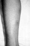
Mucocutaneous lesions of Behcet's disease
- PMID: 17722228
- PMCID: PMC2628050
- DOI: 10.3349/ymj.2007.48.4.573
Mucocutaneous lesions of Behcet's disease
Abstract
Behcet's disease is particularly prevalent in "Silk Route" populations, but it has a global distribution. The diagnosis of the disease is based on clinical criteria as there is as yet no pathognomonic test, and mucocutaneous lesions, which figure prominently in the presentation and diagnosis, may be considered the diagnostic hallmarks. Among the internationally accepted criteria, painful oral and genital ulcers, cutaneous vasculitic lesions and reactivity of the skin to needle prick or injection (the pathergy reaction) are considered hallmarks of Behcet's disease, and often precede other manifestations. Their recognition may permit earlier diagnosis and treatment, with salutary results. This paper describes the various lesions that constitute the syndrome and focuses on those that may be considered characteristic.
Figures


Similar articles
-
Behçet's disease: treatment of mucocutaneous lesions.Clin Exp Rheumatol. 2005 Jul-Aug;23(4):532-9. Clin Exp Rheumatol. 2005. PMID: 16095126 Review.
-
Behçet's disease: A comprehensive review with a focus on epidemiology, etiology and clinical features, and management of mucocutaneous lesions.J Dermatol. 2016 Jun;43(6):620-32. doi: 10.1111/1346-8138.13381. Epub 2016 Apr 14. J Dermatol. 2016. PMID: 27075942 Review.
-
Conjunctival ulcers in Behçet's disease.Ophthalmology. 2003 Jun;110(6):1137-41. doi: 10.1016/S0161-6420(03)00265-3. Ophthalmology. 2003. PMID: 12799237
-
[Behçet's disease in a Dutch boy with painful skin lesions].Ned Tijdschr Geneeskd. 2003 May 3;147(18):873-7. Ned Tijdschr Geneeskd. 2003. PMID: 12756879 Dutch.
-
Histopathologic study of cutaneous lesions in Behçet's syndrome.J Dermatol. 1990 Jun;17(6):333-41. doi: 10.1111/j.1346-8138.1990.tb01653.x. J Dermatol. 1990. PMID: 2384635
Cited by
-
Behcet's Disease Presenting as Pancolonic Ulcerations in a Patient With Kikuchi-Fujimoto Disease: A Rare Gastrointestinal Manifestation.ACG Case Rep J. 2024 Aug 30;11(9):e01455. doi: 10.14309/crj.0000000000001455. eCollection 2024 Sep. ACG Case Rep J. 2024. PMID: 39221232 Free PMC article.
-
The clinical manifestations and treatment outcomes of Behçet's disease: A single-center experience.Health Sci Rep. 2024 Jul 24;7(7):e2238. doi: 10.1002/hsr2.2238. eCollection 2024 Jul. Health Sci Rep. 2024. PMID: 39055614 Free PMC article.
-
Investigation of optic nerve function in Behçet's patients with optic disc hyperfluorescence in fundus fluorescein angiography.Int Ophthalmol. 2024 Feb 13;44(1):76. doi: 10.1007/s10792-024-02927-y. Int Ophthalmol. 2024. PMID: 38351422
-
Matrix Metalloproteinase-2 and -3 Levels in Patients with Behçet's Disease and Implication for the Presence of Vascular Aneurysm or Neurologic Involvement.Eur J Rheumatol. 2023 Jul;10(3):101-106. doi: 10.5152/eurjrheum.2023.23007. Eur J Rheumatol. 2023. PMID: 37681256 Free PMC article.
-
Update on the Diagnosis of Behçet's Disease.Diagnostics (Basel). 2022 Dec 23;13(1):41. doi: 10.3390/diagnostics13010041. Diagnostics (Basel). 2022. PMID: 36611332 Free PMC article. Review.
References
-
- Sakane T, Takeno M, Suzuki N, Inaba G. Behçet's disease. N Engl J Med. 1999;341:1284–1291. - PubMed
-
- Ehrlich GE, Kajani M, Schwartz IR, McAlack RF. Further studies of platelet rosettes around granulocytes in Behçet's syndrome. Inflammation. 1975;1:223–225. - PubMed
-
- Lehner T. Immunopathogenesis of Behçet's disease. Ann Med Interne (Paris) 1999;150:483–487. - PubMed
-
- Alpsoy E, Yılmaz E, Coşkun M, Savaş A, Yeğin O. HLA antigens and linkage disequilibrium patterns in Turkish Behçet's patients. J Dermatol. 1998;25:158–162. - PubMed
-
- Alpsoy E, Çayırlı C, Er H, Yılmaz E. The levels of plasma interleukin-2 and soluble interleukin-2R in Behçet's disease: as a marker of disease activity. J Dermatol. 1998;25:513–516. - PubMed
Publication types
MeSH terms
LinkOut - more resources
Full Text Sources
Medical

