. 2007 Oct;171(4):1334-41.
doi: 10.2353/ajpath.2007.070199. Epub 2007 Aug 23.

Sex hormones induce direct epithelial and inflammation-mediated oxidative/nitrosative stress that favors prostatic carcinogenesis in the noble rat

Affiliations

Sex hormones induce direct epithelial and inflammation-mediated oxidative/nitrosative stress that favors prostatic carcinogenesis in the noble rat

Neville N C Tam et al. Am J Pathol. 2007 Oct.

Abstract

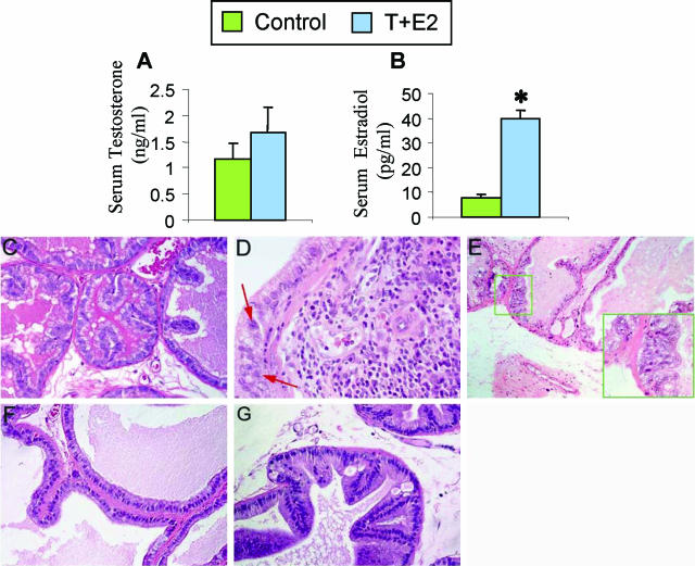
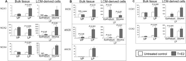
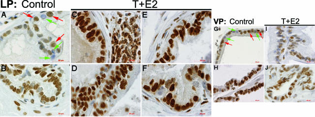
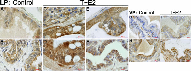
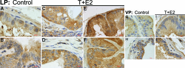
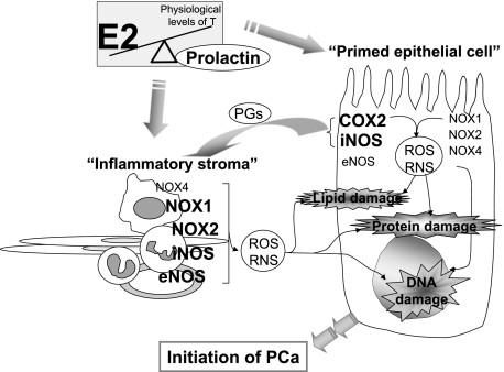
Oxidative and nitrosative stress have been implicated in prostate carcinogenesis, but the cause(s) of redox imbalance in the gland remains poorly defined. We and others have reported that administration of testosterone plus 17beta-estradiol to Noble rats for 16 weeks induces dysplasia and stromal inflammation of the lateral prostate (LP) but not the ventral prostate. Here, using laser capture microdissected specimens, we found that the combined hormone regimen increased the expression of mRNA of specific members of NAD(P)H oxidase (NOX-1, NOX-2, and NOX4), nitric-oxide synthase [NOS; inducible NOS and endothelial NOS], and cyclooxygenase (COX-2) in the LP epithelium and/or its adjacent inflammatory stroma. Accompanying these changes was the accumulation of 8-hydroxy-2'-deoxyguanosine, 4-hydroxynonenal protein adducts, and nitrotyrosine, primarily in the LP epithelium, suggesting that NOX, NOS, and COX may mediate hormone-induced oxidative/nitrosative stress in epithelium. We concluded that the oxidative/nitrosative damage resulting from the testosterone-plus-17beta-estradiol treatment is not solely derived from stromal inflammatory lesions but likely also originates from the epithelium per se. In this context, the up-regulation of COX-2 from epithelium represents a potential mechanism by which the hormone-initiated epithelium might induce inflammatory responses. Thus, we link alterations in the hormonal milieu with oxidative/nitrosative/inflammatory damage to the prostate epithelium that promotes carcinogenesis.


Figures

Figure 1
Figure 1
T+E2 co-treatment results in changes in the sex hormone milieu (A and B) and a lobe-specific induction of stromal inflammatory response and focal epithelial dysplastic lesions in LPs (C–E), but not in VPs (F and G), of treated rats. After a 16-week implantation of Silastic capsules containing T and E2, serum levels of T and E2 were determined by radioimmunoassay. A: Serum T levels of treated rats were not significantly different from those of untreated control rats. B: Serum E2 levels of treated rats were significantly (*P < 0.05) increased by four- to fivefold compared with levels in untreated control rats. Data represent the mean ± SEM of values for at least five animals. C: Normal control LP. Epithelial tubules are lined by a single layer of cuboidal secretory cells; the stroma comprises primarily a periacinar layer of smooth muscle cells and interstitial fibroblasts. Note the scarcity of inflammatory cells in the interstitial stroma. D: T+E2-treated LP. Dysplastic epithelium associated with stromal inflammation is shown. Infiltrating inflammatory cells accumulate in the stroma juxtaposed to the prostatic epithelium exhibiting a continuum of normal morphology to dysplasia, as evidenced by the presence of pleomorphic nuclei (red arrows), the earliest recognizable sign of preneoplasia. E: T+E2-treated LP. Epithelial dysplasia with no inflammatory cell infiltration in the adjacent stroma. The inset illustrates a dysplastic lesion characterized by deranged epithelial cells with pleomorphic nuclei and loss of cell polarity. F: Normal control VP. Glandular acini are composed of columnar epithelial cells surrounded by a thin layer of smooth muscles and fibroblasts. Inflammatory cells are rarely detected in the interstitial stroma. G: T+E2-treated VP. A representative region of the VP shows no induction of epithelial dysplasia and stromal inflammatory response.
Figure 2
Figure 2
Aberrant up-regulation of (A) O2·-generating NOX, (B) ·NO-producing NOS, and (C) proinflammatory COX genes in the LP in which epithelial and stromal compartments show differential response to dual exposure to T + E2. Total RNA was extracted from each half of VP or LP lobes, which are referred to as the “bulk tissues” panel (left) or from the microdissected epithelial and stromal cells in LPs’ cryosections, which represent the “LCM-derived cells” panel (right). The results of real-time quantitative PCR analyses of target-gene abundance are expressed in mRNA copy numbers that have been normalized for 1 million copies of RPL19, an endogenous housekeeping gene. “ND” denotes an undetectable gene. In the iNOS panel of B, the copy numbers represented by both columns of VPs (control and T+E2 treatment) are multiplied by 100 for the sake of visualization. Each column represents the average value from four or five animals, and error bars represent SEM.
Figure 3
Figure 3
Immunolocalization of 8-OHdG in the LP and VP after T+E2 co-treatment. A and B: Untreated control LP. Negatively stained nuclei (green arrows) are often found interspersed with positive nuclei (red arrows) in the glandular epithelium (A). Relatively strong nuclear staining in the ductal epithelium and stroma is shown (B). C: T+E2-treated LP. A representative mild dysplastic tubule closely associated with stromal inflammatory infiltration is shown. Both epithelial cells and inflammatory cells exhibit a strong nuclear immunoreactivity. D: T+E2-treated LP. High-grade dysplastic epithelia exhibit intense staining in nuclei. Note that inflammatory infiltration is not evident in the area shown. E: T+E2-treated LP. A remarkable increase in nuclear 8-OHdG immunostaining is seen both in a nondysplastic (apparently morphologically normal) acinus. F: T+E2-treated LP. A representative prostatic duct shows strong and uniform 8-OHdG staining in the epithelium. G and H: Untreated control VP. Numerous negatively stained nuclei (green arrows) are mingled with weakly positive nuclei (red arrows) in the acinar epithelium (G), whereas strong nuclear staining is found in the ductal epithelium (H). I and J: T+E2-treated VP. No remarkable increase of the 8-OHdG staining in acini (I) and ducts is seen (J). Scale bars = 20 μm.
Figure 4
Figure 4
Immunolocalization of 4-HNE protein adducts in the LP and VP after T+E2 co-treatment. A and B: Untreated control LP. Positive immunostaining is observed in the acinar (A) and ductal epithelium (B). C: T+E2-treated LP. A noticeable increase in immunoreactivity of 4-HNE protein adducts in a dysplastic gland closely associated with stromal inflammatory infiltration is shown. D: T+E2-treated LP. A representative high-grade dysplastic gland, which lacks the induction of stromal inflammatory response, exhibits intense immunostaining in the epithelium. E: T+E2-treated LP. A morphologically normal gland shows intense staining in the epithelium. Note the sparse staining in the adjacent stroma with no association with stromal inflammation. F: T+E2-treated LP. A modest increase in immunostaining is seen in the ductal epithelium. G and H: Untreated control VP. Weak and sporadic immunostaining is detected in acini (G), whereas relatively strong staining is found in ducts (H). I and J: T+E2-treated VP. No apparent increase of the staining in the glandular (I) and ductal (J) regions is seen. Scale bars = 20 μm.
Figure 5
Figure 5
Immunolocalization of nitrotyrosine in the LP and the VP after T+E2 co-treatment. A and B: Untreated control LP. Positive immunostaining is localized predominantly to the glandular (A) and ductal (B) epithelium. C: T+E2-treated LP. A representative dysplastic acinar region shows a remarkable increase in nitrotyrosine staining in the epithelium and the interstitial inflammatory stroma. D: T+E2-treated LP. High-grade dysplastic epithelium with minimal association with stromal inflammation shows a strong and uniform nitrotyrosine staining. Note the generally weak staining in the interstitial stroma. E: T+E2-treated LP. Intense nitrotyrosine immunoreactivity in a nondysplastic gland is shown. Note the sparse staining in the adjacent stroma without inflammatory infiltration. F: T+E2-treated LP. There is a modest increase of nitrotyrosine staining in the ductal epithelium. G and H: Untreated control VP. Immunostaining for nitrotyrosine in the acinar epithelium (G) is generally weak or negative. A representative duct (H) shows conspicuous staining in the epithelium. I and J: T+E2-treated VP. There is only a mild elevation of nitrotyrosine staining in acini (I) and ducts (J). Scale bars = 20 μm.
Figure 6
Figure 6
Proposed working model of sex hormone-induced oxidative/nitrosative stress that favors initiation of prostate cancer. An altered circulating hormone milieu (ie, increased ratio of E2 to T, and the E2-induced hyperprolactinemia) can directly induce oxidative/nitrosative damages to macromolecules (DNA, lipids, and proteins) of prostatic epithelial cells by the in situ induction of iNOS and COX-2 expression. In parallel, the “hormone-primed” epithelium, with elevated COX-2 expression, can promote inflammatory cell recruitment and infiltration to the stroma by the release of proinflammatory prostaglandins. The resulting reactive inflammatory stroma, which expresses high levels of NOX1, NOX2, iNOS, and eNOS, could in turn exacerbate the epithelial OS/NS-related injury in a reciprocal manner. Thus, we propose that these multifaceted, hormone-driven cellular events may create a vicious double-feed forward cycles of dysregulated ROS/RNS metabolism and inflammatory activation between the initiated epithelium and the inflammatory stroma that could ultimately lead to a pro-cancer microenvironment in the tissue. The hormonal actions on OS/NS are discussed in detail in the Discussion. The “inflammatory stroma” comprises primarily inflammatory cell infiltrates, fibroblasts, smooth muscle cells, and endothelium. PG, prostaglandins.

Similar articles

Cited by

References

    1. Bostwick DG, Alexander EE, Singh R, Shan A, Qian J, Santella RM, Oberley LW, Yan T, Zhong W, Jiang X, Oberley TD. Antioxidant enzyme expression and reactive oxygen species damage in prostatic intraepithelial neoplasia and cancer. Cancer. 2000;89:123–134. - PubMed
    1. Oberley TD, Zhong W, Szweda LI, Oberley LW. Localization of antioxidant enzymes and oxidative damage products in normal and malignant prostate epithelium. Prostate. 2000;44:144–155. - PubMed
    1. Fang FC. Antimicrobial reactive oxygen and nitrogen species: concepts and controversies. Nat Rev Microbiol. 2004;2:820–832. - PubMed
    1. Lambeth JD. NOX enzymes and the biology of reactive oxygen. Nat Rev Immunol. 2004;4:181–189. - PubMed
    1. Arbiser JL, Petros J, Klafter R, Govindajaran B, McLaughlin ER, Brown LF, Cohen C, Moses M, Kilroy S, Arnold RS, Lambeth JD. Reactive oxygen generated by Nox1 triggers the angiogenic switch. Proc Natl Acad Sci USA. 2002;99:715–720. - PMC - PubMed

Publication types

MeSH terms