

The surface protein Srr-1 of Streptococcus agalactiae binds human keratin 4 and promotes adherence to epithelial HEp-2 cells
- PMID: 17709412
- PMCID: PMC2168289
- DOI: 10.1128/IAI.00717-07
The surface protein Srr-1 of Streptococcus agalactiae binds human keratin 4 and promotes adherence to epithelial HEp-2 cells
Abstract
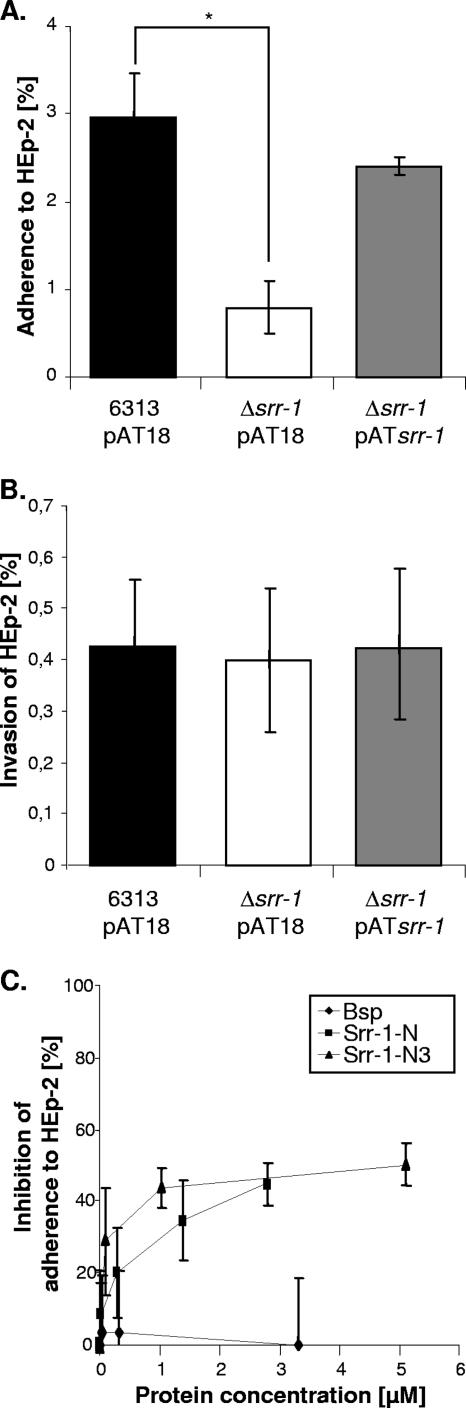
Streptococcus agalactiae is frequently the cause of bacterial sepsis and meningitis in neonates. In addition, it is a commensal bacterium that colonizes the mammalian gastrointestinal tract. During its commensal and pathogenic lifestyles, S. agalactiae colonizes and invades a number of host compartments, thereby interacting with different host proteins. In the present study, the serine-rich repeat protein Srr-1 from S. agalactiae was functionally investigated. Immunofluorescence microscopy showed that Srr-1 was localized on the surface of streptococcal cells. The Srr-1 protein was shown to interact with a 62-kDa protein in human saliva, which was identified by matrix-assisted laser desorption ionization-time-of-flight analysis as human keratin 4 (K4). Immunoblot and enzyme-linked immunosorbent assay experiments allowed us to narrow down the K4 binding domain in Srr-1 to a region of 157 amino acids (aa). Furthermore, the Srr-1 binding domain of K4 was identified in the C-terminal 255 aa of human K4. Deletion of the srr-1 gene in the genome of S. agalactiae revealed that this gene plays a role in bacterial binding to human K4 and that it is involved in adherence to epithelial HEp-2 cells. Binding to immobilized K4 and adherence to HEp-2 cells were restored by introducing the srr-1 gene on a shuttle plasmid into the srr-1 mutant. Furthermore, incubation of HEp-2 cells with the K4 binding domain of Srr-1 blocked S. agalactiae adherence to epithelial cells in a dose-dependent fashion. This is the first report describing the interaction of a bacterial protein with human K4.
Figures


Similar articles
-
The novel fibrinogen-binding protein FbsB promotes Streptococcus agalactiae invasion into epithelial cells.Infect Immun. 2004 Jun;72(6):3495-504. doi: 10.1128/IAI.72.6.3495-3504.2004. Infect Immun. 2004. PMID: 15155657 Free PMC article.
-
Structure of KRT4 binding domain of Srr-1 from Streptococcus agalactiae reveals a novel β-sheet complementation.Int J Biol Macromol. 2015 Apr;75:97-105. doi: 10.1016/j.ijbiomac.2014.12.048. Epub 2015 Jan 17. Int J Biol Macromol. 2015. PMID: 25603146
-
Rga is a regulator of adherence and pilus formation in Streptococcus agalactiae.Microbiology (Reading). 2011 Aug;157(Pt 8):2319-2327. doi: 10.1099/mic.0.044933-0. Epub 2011 Feb 17. Microbiology (Reading). 2011. PMID: 21330442
-
Cellular interactions by LPxTG-anchored pneumococcal adhesins and their streptococcal homologues.Cell Microbiol. 2011 Feb;13(2):186-97. doi: 10.1111/j.1462-5822.2010.01560.x. Epub 2010 Dec 28. Cell Microbiol. 2011. PMID: 21199258 Review.
-
Surface proteins of Streptococcus agalactiae and horizontal gene transfer.Int J Med Microbiol. 2004 Sep;294(2-3):169-75. doi: 10.1016/j.ijmm.2004.06.018. Int J Med Microbiol. 2004. PMID: 15493827 Review.
Cited by
-
Membrane trafficking of the bacterial adhesin GspB and the accessory Sec transport machinery.J Biol Chem. 2019 Feb 1;294(5):1502-1515. doi: 10.1074/jbc.RA118.005657. Epub 2018 Dec 4. J Biol Chem. 2019. PMID: 30514759 Free PMC article.
-
Molecular dissection of the secA2 locus of group B Streptococcus reveals that glycosylation of the Srr1 LPXTG protein is required for full virulence.J Bacteriol. 2009 Jul;191(13):4195-206. doi: 10.1128/JB.01673-08. Epub 2009 Apr 24. J Bacteriol. 2009. PMID: 19395494 Free PMC article.
-
The Two Distinct Types of SecA2-Dependent Export Systems.Microbiol Spectr. 2019 May;7(3):10.1128/microbiolspec.psib-0025-2018. doi: 10.1128/microbiolspec.PSIB-0025-2018. Microbiol Spectr. 2019. PMID: 31215505 Free PMC article. Review.
-
Surface proteins involved in the adhesion of Streptococcus salivarius to human intestinal epithelial cells.Appl Microbiol Biotechnol. 2018 Mar;102(6):2851-2865. doi: 10.1007/s00253-018-8794-y. Epub 2018 Feb 13. Appl Microbiol Biotechnol. 2018. PMID: 29442170 Free PMC article.
-
Streptococcus oralis Neuraminidase Modulates Adherence to Multiple Carbohydrates on Platelets.Infect Immun. 2017 Feb 23;85(3):e00774-16. doi: 10.1128/IAI.00774-16. Print 2017 Mar. Infect Immun. 2017. PMID: 27993975 Free PMC article.
References
-
- Altschul, S. F., W. Gish, W. Miller, E. W. Myers, and D. J. Lipman. 1990. Basic local alignment search tool. J. Mol. Biol. 215:403-410. - PubMed
-
- Areschoug, T., M. Stalhammar-Carlemalm, I. Karlsson, and G. Lindahl. 2002. Streptococcal beta protein has separate binding sites for human factor H and IgA-Fc. J. Biol. Chem. 277:12642-12648. - PubMed
-
- Arrecubieta, C., M. H. Lee, A. Macey, T. J. Foster, and F. D. Lowy. 2007. SdrF, a Staphylococcus epidermidis surface protein, binds type I collagen. J. Biol. Chem. 282:18767-18776. - PubMed
-
- Balter, S., C. G. Whitney, and A. Schuchat. 2000. Epidemiology of group B streptococcal infections, p. 154-162. In V. A. Fischetti, R. P. Novick, J. J. Ferretti, D. A. Portnoy, and J. I. Rood (ed.), Gram-positive pathogens. American Society for Microbiology, Washington, DC.
Publication types
MeSH terms
Substances
LinkOut - more resources
Full Text Sources
Molecular Biology Databases
