. 2007 Aug 13;178(4):595-610.
doi: 10.1083/jcb.200703002.

An in vitro nuclear disassembly system reveals a role for the RanGTPase system and microtubule-dependent steps in nuclear envelope breakdown

Affiliations

An in vitro nuclear disassembly system reveals a role for the RanGTPase system and microtubule-dependent steps in nuclear envelope breakdown

Petra Mühlhäusser et al. J Cell Biol. .

Abstract

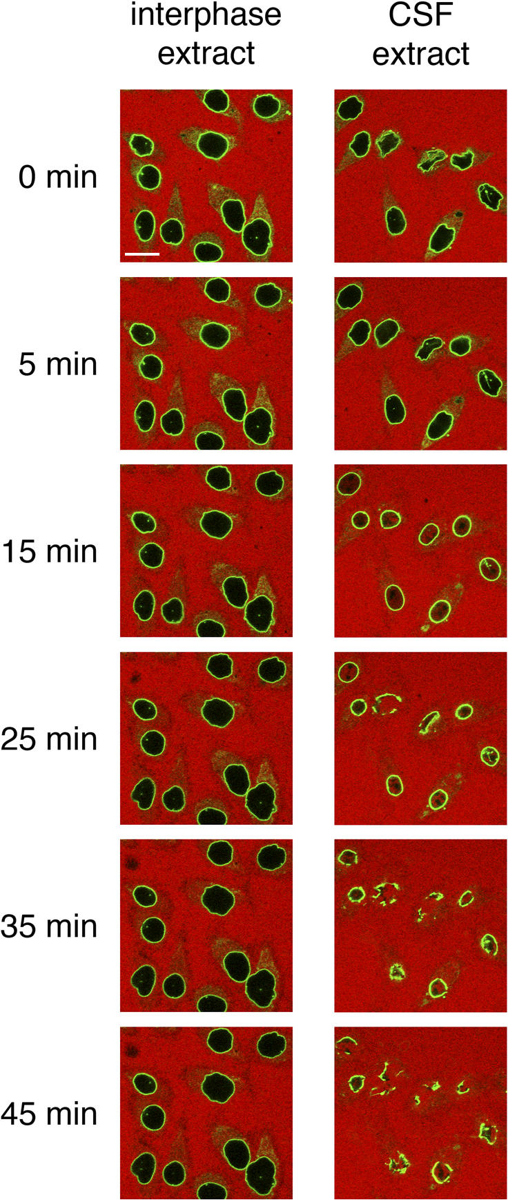
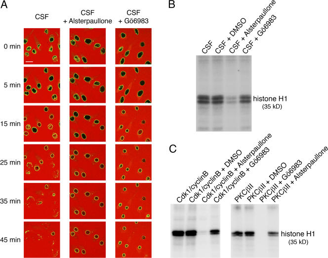
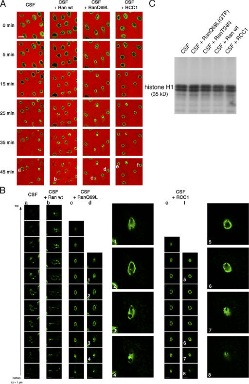
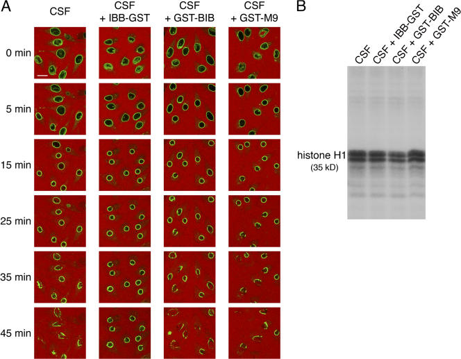
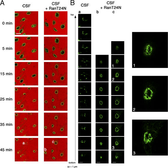
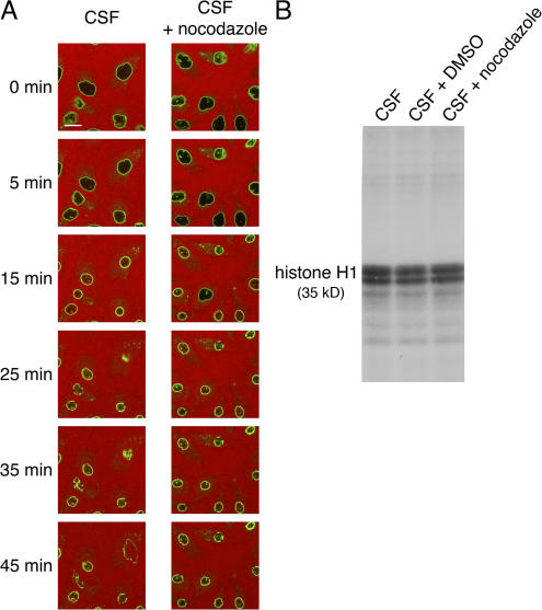
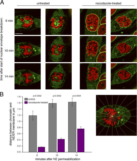
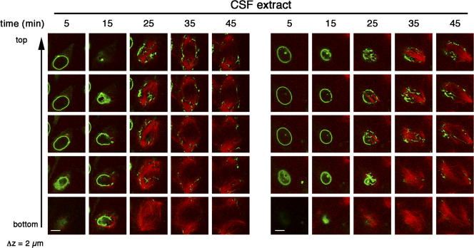
During prophase, vertebrate cells disassemble their nuclear envelope (NE) in the process of NE breakdown (NEBD). We have established an in vitro assay that uses mitotic Xenopus laevis egg extracts and semipermeabilized somatic cells bearing a green fluorescent protein-tagged NE marker to study the molecular requirements underlying the dynamic changes of the NE during NEBD by live microscopy. We applied our in vitro system to analyze the role of the Ran guanosine triphosphatase (GTPase) system in NEBD. Our study shows that high levels of RanGTP affect the dynamics of late steps of NEBD in vitro. Also, inhibition of RanGTP production by RanT24N blocks the dynamic rupture of nuclei, suggesting that the local generation of RanGTP around chromatin may serve as a spatial cue in NEBD. Furthermore, the microtubule-depolymerizing drug nocodazole interferes with late steps of nuclear disassembly in vitro. High resolution live cell imaging reveals that microtubules are involved in the completion of NEBD in vivo by facilitating the efficient removal of membranes from chromatin.


Figures

Figure 1.
Figure 1.
Incubation of HeLa cell nuclei in mitotic egg extracts results in nuclear disassembly. Time course of in vitro nuclear disassembly. HeLa cells stably expressing GFP-LAP2β (green) were grown on coverslips to ∼50% confluency. The cells were permeabilized and incubated in interphase (left) or mitotic (CSF; right) Xenopus egg extract. Extracts were supplemented with an energy-regenerating system and 250 μg/ml of 155-kD TRITC-labeled dextran (red) to allow for the monitoring of changes in NE permeability. Nuclear disassembly was followed by time-lapse confocal laser-scanning microscopy. Bar, 20 μm.
Figure 2.
Figure 2.
Efficient in vitro nuclear disassembly depends on Cdk1 and PKC activity. (A) Permeabilized GFP-LAP2β (green)–expressing HeLa cells were incubated in CSF extracts containing an energy-regenerating system and a 155-kD TRITC-labeled dextran (red). Extracts were either untreated (solvent control; left) or treated with 200 μM of the kinase inhibitors alsterpaullone (Cdk1 inhibitor; middle) or Gö6983 (PKC inhibitor; right) for 10 min at RT. Nuclear disassembly was monitored by time-lapse confocal laser-scanning microscopy. The relative fluorescence intensity inside nuclei (n > 18) was quantified, and an increase of >10% inside nuclei was used as a threshold. Dextran influx into nuclei incubated in untreated CSF extract and alsterpaullone-treated or Gö6983-treated extracts occurred after 15 min (±4 min) and 22 min (±3 min) or 12 min (±2 min), respectively. (B) Kinase activity of untreated and inhibitor-treated CSF extracts was assayed using histone H1 as substrate. Final concentrations of alsterpaullone and Gö6983 were 200 μM. (C) The specificity of the inhibitors used was tested using recombinant Cdk1–cyclin B1 and PKCβII. Bar, 20 μm.
Figure 3.
Figure 3.
Nuclear import is not required for the initiation of NEBD. (A) To block nucleocytoplasmic transport, permeabilized GFP-LAP2β (green)–expressing HeLa cells were preincubated with 15 μM of recombinant Impβ45–462 (right). Nuclei were then incubated in CSF extracts supplemented with an energy- regenerating system and a 155-kD TRITC-labeled dextran (red). In vitro nuclear breakdown was monitored by time-lapse confocal laser-scanning microscopy. The relative fluorescence intensity inside nuclei (n > 18) was quantified as in Fig. 2. Dextran influx into nuclei of control cells or cells preincubated with Impβ45–462 occurred after 12 min (±3 min) and 11 min (±4 min), respectively. (B) High magnification confocal sectioning of disassembled nuclei from A (indicated by letters) was performed using a 63× objective and a 4× zoom. Z-step width is 1 μm. On the right side, representative magnifications of selected frames from the z stacks (indicated by numbers) are shown. Bars (A), 20 μm; (B) 10 μm.
Figure 4.
Figure 4.
Inhibition of NE rupture in vitro by excess of RanGTP in CSF extracts. (A) Permeabilized GFP-LAP2β (green)–expressing HeLa cells were incubated in untreated CSF extract or CSF extracts supplemented with 20 μM of either recombinant Ran wild type (wt), recombinant RanQ69L(GTP), or RCC1. CSF extracts had been preincubated with the recombinant proteins for 10 min at RT before the disassembly reaction. Nuclear breakdown was monitored in the presence of 155-kD TRITC-labeled dextran (red) by time-lapse confocal laser-scanning microscopy. The relative fluorescence intensity inside nuclei (n > 18) was quantified as in Fig. 2. Dextran influx into nuclei incubated in untreated CSF extract or extracts incubated with Ran wild type, RanQ69L(GTP), or RCC1 occurred after 13 min (±4 min), 11 min (±5 min), 11 min (±4 min), or 15 min (±3 min), respectively. (B) High magnification confocal sectioning of disassembled nuclei from A (indicated by letters) was performed using a 63× objective and a 4× zoom. Z-step width is 1 μm. Representative magnifications of selected frames from the z stacks (indicated by numbers) are shown for RanQ69L and RCC1 addition. (C) Kinase activity of untreated CSF extract and CSF extract supplemented with 20 μM of recombinant RanQ69L(GTP), RanT24N, Ran wild type, or RCC1 was assayed using histone H1 as substrate. Bars (A), 20 μm; (B) 10 μm.
Figure 5.
Figure 5.
The IBB of importin α inhibits the late steps of NEBD in vitro. (A) Permeabilized GFP-LAP2β (green)–expressing HeLa cells were incubated in untreated CSF extract (left) or CSF extract supplemented with recombinant IBB-GST, GST-BIB, or GST-M9 (20 μM each; preincubated in the extract for 10 min at RT before the disassembly reaction). Nuclear disassembly was monitored in the presence of 155-kD TRITC-labeled dextran (red) by time-lapse confocal microscopy. (B) Kinase activity of untreated CSF extract and CSF extract supplemented with 20 μM recombinant IBB-GST, GST-BIB, or GST-M9 was assayed using histone H1 as substrate. Bar, 20 μm.
Figure 6.
Figure 6.
Inhibition of RCC1 activity by RanT24N interferes with final steps of NEBD in vitro. (A) Nuclei of semipermeabilized GFP-LAP2β (green)–expressing HeLa cells were preloaded with either Ran wild-type (left) or RanT24N (right). Then, cells were incubated in CSF extract supplemented with either 20 μM Ran wild-type (left) or RanT24N (right), an energy-regenerating system, and a 155-kD TRITC-labeled dextran (red). NEBD was monitored by time-lapse confocal laser-scanning microscopy. The relative fluorescence intensity inside nuclei (n > 14) was quantified as in Fig. 2. Dextran influx into nuclei of control cells or cells preincubated with RanT24N occurred after 16 min (±3 min) and 19 min (±4 min), respectively. (B) High magnification confocal sectioning of disassembled nuclei from A (indicated by letters) was performed using a 63× objective and a 4× zoom. Z-step width is 1 μm. On the right, representative magnifications of selected frames from the z stacks (indicated by numbers) are shown. Bars (A), 20 μm; (B) 10 μm.
Figure 7.
Figure 7.
Microtubules contribute to NE rupture in vitro. (A) Permeabilized GFP-LAP2β (green)–expressing HeLa cells were incubated in untreated CSF extract or CSF extract containing 10 μM nocodazole (preincubated for 10 min at RT before the disassembly reaction). Nuclear disassembly was monitored in the presence of 155-kD TRITC-labeled dextran (red) by time-lapse confocal microscopy. (B) Kinase activity of untreated CSF extract and CSF extract supplemented with 10 μM nocodazole, or the corresponding amount of DMSO was assayed using histone H1 as substrate. Bar, 20 μm.
Figure 8.
Figure 8.
Microtubule-dependent removal of the NE/ER membrane network during mitosis in HeLa cells. (A) HeLa cells stably expressing GFP-LAP2β (green) and H2B-mRFP (red) were transiently transfected with the plasmid coding for mPlum-GST-M9. Early prophase cells, which were either untreated or treated with 1 μg/ml nocodazole, were chosen, and the efflux of mPlum-GST-M9 out of the nuclei was monitored by live confocal microscopy. The increase in NE permeability judged by a 1.3-fold increase of the fluorescence intensity of mPlum-GST-M9 in the cytoplasm (not depicted) was defined as the start of NEBD (time = 0 min). Stacks of four confocal images were obtained after 6, 10, and 14 min. (B) Distances between the chromatin and the NE/ER membrane were determined in the four slices of each stack along 20 lines of a radial grid (interval of lines = 18°) emanating from the center of the chromatin mass. Distances were determined for nine untreated cells and nine cells treated with nocodazole. Mean distances and SEM (error bars) are indicated. P-values were calculated using a directional Mann-Whitney test (α = 0.01; n = 9). Bar, 10 μm.
Figure 9.
Figure 9.
Microtubule-dependent disintegration of the NE in vitro. GFP-LAP2β (green)–expressing HeLa cells were permeabilized and incubated with CSF extract supplemented with 2 μg rhodamine-labeled tubulin (red) and an energy-regenerating system. NEBD and microtubule polymerization were monitored by confocal laser-scanning microscopy. Five sections with a z-step width of 2 μm were taken at the indicated times. Bars, 10 μm.

Similar articles

Cited by

References

    1. Beaudouin, J., D. Gerlich, N. Daigle, R. Eils, and J. Ellenberg. 2002. Nuclear envelope breakdown proceeds by microtubule-induced tearing of the lamina. Cell. 108:83–96. - PubMed
    1. Buendia, B., J.C. Courvalin, and P. Collas. 2001. Dynamics of the nuclear envelope at mitosis and during apoptosis. Cell. Mol. Life Sci. 58:1781–1789. - PMC - PubMed
    1. Busson, S., D. Dujardin, A. Moreau, J. Dompierre, and J.R. De Mey. 1998. Dynein and dynactin are localized to astral microtubules and at cortical sites in mitotic epithelial cells. Curr. Biol. 8:541–544. - PubMed
    1. Campbell, R.E., O. Tour, A.E. Palmer, P.A. Steinbach, G.S. Baird, D.A. Zacharias, and R.Y. Tsien. 2002. A monomeric red fluorescent protein. Proc. Natl. Acad. Sci. USA. 99:7877–7882. - PMC - PubMed
    1. Carazo-Salas, R.E., G. Guarguaglini, O.J. Gruss, A. Segref, E. Karsenti, and I.W. Mattaj. 1999. Generation of GTP-bound Ran by RCC1 is required for chromatin-induced mitotic spindle formation. Nature. 400:178–181. - PubMed

Publication types

Substances