. 2007 Sep;5(9):e220.
doi: 10.1371/journal.pbio.0050220.

Three-dimensional analysis of a viral RNA replication complex reveals a virus-induced mini-organelle

Affiliations

Three-dimensional analysis of a viral RNA replication complex reveals a virus-induced mini-organelle

Benjamin G Kopek et al. PLoS Biol. 2007 Sep.

Abstract

Positive-strand RNA viruses are the largest genetic class of viruses and include many serious human pathogens. All positive-strand RNA viruses replicate their genomes in association with intracellular membrane rearrangements such as single- or double-membrane vesicles. However, the exact sites of RNA synthesis and crucial topological relationships between relevant membranes, vesicle interiors, surrounding lumens, and cytoplasm generally are poorly defined. We applied electron microscope tomography and complementary approaches to flock house virus (FHV)-infected Drosophila cells to provide the first 3-D analysis of such replication complexes. The sole FHV RNA replication factor, protein A, and FHV-specific 5-bromouridine 5'-triphosphate incorporation localized between inner and outer mitochondrial membranes inside approximately 50-nm vesicles (spherules), which thus are FHV-induced compartments for viral RNA synthesis. All such FHV spherules were outer mitochondrial membrane invaginations with interiors connected to the cytoplasm by a necked channel of approximately 10-nm diameter, which is sufficient for ribonucleotide import and product RNA export. Tomographic, biochemical, and other results imply that FHV spherules contain, on average, three RNA replication intermediates and an interior shell of approximately 100 membrane-spanning, self-interacting protein As. The results identify spherules as the site of protein A and nascent RNA accumulation and define spherule topology, dimensions, and stoichiometry to reveal the nature and many details of the organization and function of the FHV RNA replication complex. The resulting insights appear relevant to many other positive-strand RNA viruses and support recently proposed structural and likely evolutionary parallels with retrovirus and double-stranded RNA virus virions.


Conflict of interest statement

Competing interests. The authors have declared that no competing interests exist.

Figures

Figure 1
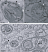
Figure 1. FHV Protein A Is Localized in Virus-Induced Mitochondrial Spherules
(A and B) Examples of anti-protein A immunogold labeling of mitochondrial spherules in FHV-infected cells. Cells were fixed in 4% paraformaldehyde and 0.2% glutaraldehyde, postfixed in 0.1% osmium, dehydrated and embedded in LR Gold resin, sectioned and placed on nickel-grids, immunostained with protein A antisera and a secondary antibody conjugated to ultrasmall gold particles, and silver-enhanced. The mitochondrial matrix, cytoplasm (Cyt) and examples of the many spherules (S) in the mitochondrial intermembrane space (IMS) are labeled for reference. White arrowheads indicate gold particles at a mitochondrial membrane but not directly over a spherule. Black arrowheads indicate gold particles in the cytoplasm. As illustrated in panel A, islands of cytoplasm surrounded by a mitochondrial ring are seen frequently in EM sections of FHV-infected mitochondria. (C) A schematic, based on 3-D tomographic analysis shown below, of how such cytoplasmic islands are generated by EM sectioning of the frequently cup-shaped, FHV-modified mitochondria [see panel (B)]. The bottom image in (C) further shows how some planes of sectioning give rise to “vesicle packet” structures seen in Figures 2C and 3D below. White, cytoplasm; blue, space between outer and inner mitochondrial membranes; yellow, matrix.
Figure 2
Figure 2. Association of Viral RNA Synthesis with FHV Spherules
(A and B) Examples of incorporated BrUTP immunogold labeling of mitochondrial spherules from cells infected with FHV. Isolated mitochondria were incubated for 1 h at 28 °C with a transcription mix that included BrUTP. (C) Localization of RNA synthesis to spherules in FHV-infected cells into which BrUTP was introduced by transfection. Length of labeling period was 15 min. Samples were prepared for EM and immunogold labeling as in Figure 1, except that anti-BrU antibody was used instead of anti-protein A. See Figure 1C for an explanation of the nonstandard mitochondrial morphologies seen in the upper left and lower right of (C). The arrowheads and labeling of mitochondria, spherules, and cytoplasm are as in Figure 1. In interpreting the immunogold localization, note that the primary-secondary antibody complex linking the gold particles to their target epitopes may span up to 20 nm.
Figure 3
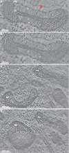
Figure 3. EMT Reconstructions of Mitochondria from FHV-Infected Drosophila Cells
Labels denote outer mitochondrial membrane (OM) and inner mitochondrial membrane (IM). (A and B) Slices through a tomographic reconstruction showing FHV-induced spherule rearrangements of a mitochondrion (see Video S1 to view the entire z-series). (B) is a slice that is ∼15 nm down the z-axis (perpendicular to the plane of the image) from the slice in (A). White arrowheads indicate the necks that connect spherules to the OM. Asterisks mark two spherules that appear to be free vesicles in (A) but are shown to have necked connections to the outer membrane in (B). A red arrow marks the ∼10-nm channel connecting a representative spherule interior to the cytoplasm. This red arrow also corresponds to the same spherule and connection as the red arrow in Figure 4B–4C. (C and D) Images from another tomogram that are displaced from each other in the z-axis by ∼150 nm. Note the change in morphology of mitochondrion 1 where spherules that appear to be connected to the outer mitochondrial membrane in (C) appear as a vesicle packet in (D).
Figure 4
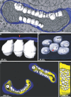
Figure 4. 3-D Maps of FHV-Modified Mitochondria
Blue indicates outer mitochondrial membrane, white indicates FHV spherules, yellow indicates inner mitochondrial membrane. (A) Merged image of a 3-D map of the outer membrane and spherules of the mitochondrion from Figure 3A and 3B and a slice of the tomogram showing the electron density map from which it was derived. (B) A portion of the map in (A) showing a close-up view of the connections between the outer mitochondrial membrane and the spherules. The red arrow marks that same spherule as the one depicted by the red arrow in Figure 3A. (C) A 90 ° rotation of (B) showing the channels that connect the spherule interiors to the cytoplasm. The outer membrane has been made translucent to show the spherules behind it. Again, the red arrow corresponds to the red arrows in Figures 3A and 4B. (D) 3-D maps of the mitochondria shown in Figure 3C. (E) A view of mitochondrion 2 from (D) that has been rotated and has had the outer membrane removed to show the range of spherule sizes.
Figure 5
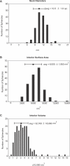
Figure 5. Spherule Dimensions
Distribution of spherule neck channel diameters (A), interior surface areas (B), and interior volumes (C). The data shown represents measurements of 150 (A) and 175 (B and C) individual spherules.
Figure 6
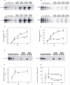
Figure 6. Measurements of the number of molecules of FHV (+)RNAs and (−)RNAs and protein A per cell in FHV-infected Drosophila cells
(A and B) Cells were harvested and counted at the time points indicated above the figure, and total RNA was extracted. An amount of RNA corresponding to 1.0 × 10⁵, 1.0 × 10⁴, or 1.0 × 10³ cell equivalents was loaded as indicated above each lane and subjected to Northern blot hybridization with radio-labeled probes specific for the detection of (+)RNA1 (top panel of A), (−)RNA1 (top panel of B), (+)RNA2 (bottom panel of A), or (−)RNA2 (bottom panel of B). Specific signals are indicated by the arrowheads. (C) Graph of the number of molecules of (+)RNA1 (diamond) and (+)RNA2 (squares) per cell over a 24-h time course. (D) Graph of the number of molecules of (−)RNA1 and (−)RNA2 per cell over a 24-h time course. (E) An aliquot of the same cells harvested for the RNA analysis at the indicated time points was lysed in Laemmli sample buffer and subjected to immunoblot analysis with polyclonal antisera for protein A. Protein A levels were measured by comparison with the signal intensities derived from known amounts of purified protein A. (F) Graph of the number of molecules of protein A per cell over a 24-h time course. (G) Graph of the ratios of protein A versus (−)RNA1 (diamonds) and (−)RNA2 during the course of infection. The 24 hpi data shown represent the mean and standard deviation for three independent experiments. The data shown for 4, 8, and 12 hpi represent the mean and standard deviation for two independent experiments.
Figure 7
Figure 7. Modeling of FHV Transmembrane Protein A into a Spherule
(A) 3-D map of a single spherule where half of the membrane has been removed to show the interior. As in Figure 4, the spherule membrane is white and the contiguous outer mitochondrial membrane is blue. (B) Schematic of likely protein A organization within a spherule. Based on the average density of globular proteins, FHV protein A (112 kDa) is modeled in as a green sphere of ∼7 nm in diameter. Based on the average of ∼100 protein A molecules per spherule (Table 1), the figure shows the potential packing arrangement of 50 protein A molecules in half a spherule. The spheres are shown lining the interior surface area of the spherule membrane because protein A is a transmembrane protein [38]. (C) Schematic representation of the structure and organization of the FHV RNA replication complex. Protein A (ptn A; green spheres) forms a shell within the mitochondrial membrane spherule within which RNA synthesis occurs (N = N terminus; C = C terminus). Protein A is also shown as possibly extending into the spherule neck, since it may be a determinant of the relatively constant 10-nm diameter neck. As noted in Figure 1 (white arrowheads), a small fraction of protein A may reside on the outer mitochondrial membrane external to spherules. The diagram shows (+)RNA synthesis (red arrow) from (−)RNA templates (black segmented line), which is the predominant form of FHV RNA synthesis throughout all but the earliest phases of FHV infection (Figure 6A–6D).

Similar articles

Cited by

References

    1. van Regenmortel MHV, editor. Virus taxonomy. San Diego: Academic Press; 2000. 1162
    1. Carette JE, van Lent J, MacFarlane SA, Wellink J, van Kammen A. Cowpea mosaic virus 32- and 60-kilodalton replication proteins target and change the morphology of endoplasmic reticulum membranes. J Virol. 2002;76:6293–6301. - PMC - PubMed
    1. Egger D, Wolk B, Gosert R, Bianchi L, Blum HE, et al. Expression of hepatitis C virus proteins induces distinct membrane alterations including a candidate viral replication complex. J Virol. 2002;76:5974–5984. - PMC - PubMed
    1. Gosert R, Kanjanahaluethai A, Egger D, Bienz K, Baker SC. RNA replication of mouse hepatitis virus takes place at double-membrane vesicles. J Virol. 2002;76:3697–3708. - PMC - PubMed
    1. Kujala P, Ikaheimonen A, Ehsani N, Vihinen H, Auvinen P, et al. Biogenesis of the Semliki Forest virus RNA replication complex. J Virol. 2001;75:3873–3884. - PMC - PubMed

Publication types