Interactions of the G quartet forming semaphorin 3F RNA with the RGG box domain of the fragile X protein family
- PMID: 17693432
- PMCID: PMC2018618
- DOI: 10.1093/nar/gkm581
Interactions of the G quartet forming semaphorin 3F RNA with the RGG box domain of the fragile X protein family
Abstract
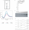
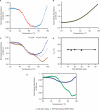
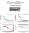
Fragile X syndrome, the most common cause of inherited mental retardation, is caused by the transcriptional silencing of the fmr1 gene due to an unstable expansion of a CGG trinucleotide repeat and its subsequent hypermethylation in its 5' UTR. This gene encodes for the fragile X mental retardation protein (FMRP), an RNA-binding protein that has been shown to use its RGG box domain to bind to G quartet-forming RNA. In this study, we performed a detailed analysis of the interactions between the FMRP RGG box domain and one of its proposed RNA targets, human semaphorin 3F (S3F) RNA by using biophysical methods such as fluorescence, UV and circular dichroism spectroscopy. We show that this RNA forms a G quartet-containing structure, which is recognized with high affinity and specificity by the FMRP RGG box. In addition, we analyzed the interactions of human S3F RNA with the RGG box and RG cluster of the two FMRP autosomal paralogs, the FXR1P and FXR2P. We found that this RNA is bound with high affinity and specificity only by the FXR1P RGG box, but not by the FXR2P RG cluster. Both FMRP and FXR1P RGG box are able to unwind the G quartet structure of S3F RNA, however, the peptide concentrations required in this process are very different: a ratio of 1:6 RNA:FMRP RGG box versus 1:2 RNA:FXR1P RGG box.
Figures


Similar articles
-
Thermodynamics of the fragile X mental retardation protein RGG box interactions with G quartet forming RNA.Biochemistry. 2006 Jul 11;45(27):8319-30. doi: 10.1021/bi060209a. Biochemistry. 2006. PMID: 16819831
-
Analysis of the Fragile X mental retardation protein isoforms 1, 2 and 3 interactions with the G-quadruplex forming semaphorin 3F mRNA.Mol Biosyst. 2012 Feb;8(2):642-9. doi: 10.1039/c1mb05322a. Epub 2011 Dec 1. Mol Biosyst. 2012. PMID: 22134704
-
Fragile X mental retardation protein recognition of G quadruplex structure per se is sufficient for high affinity binding to RNA.Mol Biosyst. 2008 Dec;4(12):1212-9. doi: 10.1039/b812537f. Epub 2008 Oct 27. Mol Biosyst. 2008. PMID: 19396385
-
Biology of the fragile X mental retardation protein, an RNA-binding protein.Biochem Cell Biol. 1999;77(4):331-42. Biochem Cell Biol. 1999. PMID: 10546896 Review.
-
The fragile X mental retardation protein, FMRP, recognizes G-quartets.Ment Retard Dev Disabil Res Rev. 2004;10(1):49-52. doi: 10.1002/mrdd.20008. Ment Retard Dev Disabil Res Rev. 2004. PMID: 14994288 Review.
Cited by
-
Aberrant astrocyte protein secretion contributes to altered neuronal development in multiple models of neurodevelopmental disorders.Nat Neurosci. 2022 Sep;25(9):1163-1178. doi: 10.1038/s41593-022-01150-1. Epub 2022 Aug 30. Nat Neurosci. 2022. PMID: 36042312 Free PMC article.
-
The Human Fragile X Mental Retardation Protein Inhibits the Elongation Step of Translation through Its RGG and C-Terminal Domains.Biochemistry. 2020 Oct 13;59(40):3813-3822. doi: 10.1021/acs.biochem.0c00534. Epub 2020 Sep 29. Biochemistry. 2020. PMID: 32945655 Free PMC article.
-
The new (dis)order in RNA regulation.Cell Commun Signal. 2016 Apr 6;14:9. doi: 10.1186/s12964-016-0132-3. Cell Commun Signal. 2016. PMID: 27048167 Free PMC article. Review.
-
Therapeutic Strategies in Fragile X Syndrome: From Bench to Bedside and Back.Neurotherapeutics. 2015 Jul;12(3):584-608. doi: 10.1007/s13311-015-0355-9. Neurotherapeutics. 2015. PMID: 25986746 Free PMC article. Review.
-
Crystal structure reveals specific recognition of a G-quadruplex RNA by a β-turn in the RGG motif of FMRP.Proc Natl Acad Sci U S A. 2015 Sep 29;112(39):E5391-400. doi: 10.1073/pnas.1515737112. Epub 2015 Sep 15. Proc Natl Acad Sci U S A. 2015. PMID: 26374839 Free PMC article.
References
-
- O’Donnell WT, Warren ST. A decade of molecular studies of fragile x syndrome. Annu. Rev. Neurosci. 2002;25:315–338. - PubMed
-
- Jin P, Warren ST. Understanding the molecular basis of fragile X syndrome. Hum. Mol. Gen. 2000;6:901–908. - PubMed
-
- Ashley CT, Wilkinson KD, Reines D, Warren ST. FMR1 protein: conserved RNP family domains and selective RNA binding. Science. 1993;262:563–566. - PubMed
-
- Antar LN, Bassell GJ. Sunrise at the synapse: the FMRP mRNP shaping the synaptic interface. Neuron. 2003;37:555–558. - PubMed

