Comparative Study
. 2007 Aug 1;27(31):8395-404.
doi: 10.1523/JNEUROSCI.2478-07.2007.

Opposite regulation of oligodendrocyte apoptosis by JNK3 and Pin1 after spinal cord injury

Affiliations
Comparative Study

Opposite regulation of oligodendrocyte apoptosis by JNK3 and Pin1 after spinal cord injury

Qi Ming Li et al. J Neurosci. .

Abstract

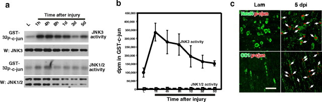
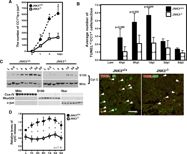
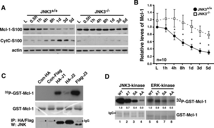
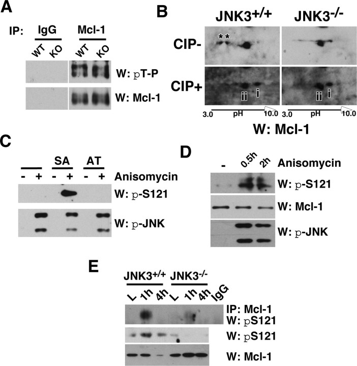
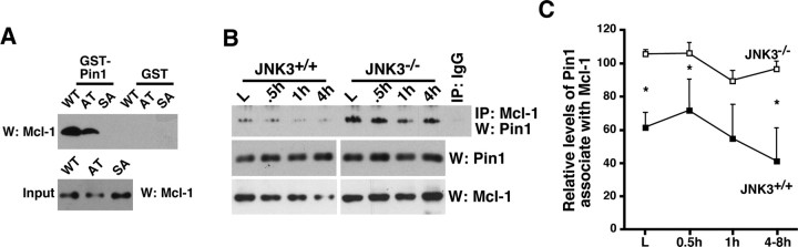
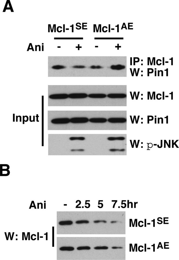
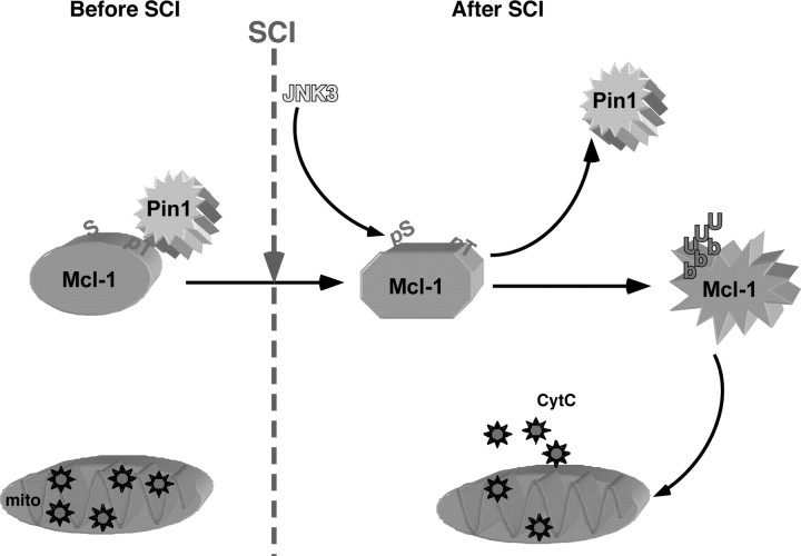
Although oligodendrocytes undergo apoptosis after spinal cord injury, molecular mechanisms responsible for their death have been unknown. We report that oligodendrocyte apoptosis is regulated oppositely by c-Jun N-terminal kinase 3 (JNK3) and protein interacting with the mitotic kinase, never in mitosis A I (Pin1), the actions of which converge on myeloid cell leukemia sequence-1 (Mcl-1). Activated after injury, JNK3 induces cytochrome c release by facilitating the degradation of Mcl-1, the stability of which is maintained in part by Pin1. Pin1 binds Mcl-1 at its constitutively phosphorylated site, Thr163Pro, and stabilizes it by inhibiting ubiquitination. After injury JNK3 phosphorylates Mcl-1 at Ser121Pro, facilitating the dissociation of Pin1 from Mcl-1. JNK3 thus induces Mcl-1 degradation by counteracting the protective binding of Pin1. These results are confirmed by the opposing phenotypes observed between JNK3-/- and Pin1-/- mice: oligodendrocyte apoptosis and cytochrome c release are reduced in JNK3-/- but elevated in Pin1-/- mice. This report thus unveils a mechanism by which cytochrome c release is under the opposite control of JNK3 and Pin1, regulators for which the activities are intricately coupled.


Figures

Figure 1.
Figure 1.
JNK3 is the major kinase activated after spinal cord injury. A, B, JNK3 and JNK1/2 activity was measured in immunoprecipitation/kinase assays by using GST-c-Jun as the substrate. For the specificity of JNK3 immunoprecipitation, see supplemental Figure 1 (available at www.jneurosci.org as supplemental material). Note that JNK1/2 activities are at least 500-fold lower than JNK3 activity, based on the radioactivity counts on GST-c-Jun. n = 3. The error bars represent the range of the disintegrations per minute counts. C, The p-c-Jun immunoreactivity, a measure of JNK activation, was detected in oligodendrocytes and neurons after injury. Arrows point to p-c-Jun+ cells. Scale bar, 30 μm.
Figure 2.
Figure 2.
JNK3 is necessary for mitochondria-mediated apoptosis of oligodendrocytes after spinal cord injury. A, Reduction in the number of p-c-Jun+ cells among CC1+ oligodendrocytes in JNK3−/− mice as compared with the number in JNK3+/+ mice. p-c-Jun immunoreactivity among CC1+ cells was quantified in JNK3+/+ and JNK3−/− mice. The error bars represent SEM. Asterisks represent the time points at which the two genotypes showed statistical difference (Student's t test, p < 0.05). B, Top, Oligodendrocyte apoptosis after spinal cord injury is attenuated in JNK3−/− mice compared with JNK3+/+ mice. Adult JNK3+/+ and JNK3−/− mice were subjected to T9 hemisection. At the indicated time every fourth coronal section was processed for TUNEL and CC1 immunostaining; cells that were positive for both stainings were counted (p values, Student's t test). The error bars represent SEM. B, Bottom, A representative picture of TUNEL+/CC1+ cells. Arrowheads point to TUNEL+/CC1+ cells. Scale bar, 30 μm. C, Top, JNK3 is necessary for cytC release from the mitochondria after spinal cord injury. The spinal cord lysates were processed for mitochondrial fractionation by using sucrose gradients. C, Bottom, As controls for fractionation, the mitochondrial, S100, and nuclear fractions were analyzed in a Western blot with a mitochondrial marker, COX-IV, a cytosolic marker, RhoGDI, and a nuclear marker, c-Jun. D, Quantification of the cytC levels in the S100 fraction from JNK3+/+ and JNK3−/− mice. Asterisks represent the time points at which the two genotypes showed statistical difference (Student's t test; p < 0.02).
Figure 3.
Figure 3.
JNK3 regulates Mcl-1 stability after injury. A, Mcl-1 protein levels change most dramatically beginning 4 h after injury in JNK3+/+ mice, coinciding with cytC release. In JNK3−/− mice, the Mcl-1 levels remain elevated. Actin control is shown; the control for fractionation is shown in Figure 2C. B, Quantification of the relative Mcl-1 levels at different time points after injury. Asterisks represent the time points at which the two genotypes show statistical difference (Student's t test, p < 0.05). C, JNK3 phosphorylates Mcl-1 in vitro. JNK1, 2, or 3 was immunoprecipitated from 293T cells and subjected to kinase assays, using GST-Mcl-1 as the substrate. The asterisks in the stained gel represent IgG. As a control, the same lysates were subjected to immunoprecipitation and blotted for JNK Western. The experiments were repeated three to four times with similar results. D, JNK3 phosphorylates Mcl-1 at both S121P and T163P, whereas ERK phosphorylates Mcl-1 at T163P in vitro. GST-Mcl-1 proteins bearing single and double mutations as indicated were used as substrates in kinase reactions. The bottom panels show the amount of GST-Mcl-1 protein in each lane as Coomassie-stained controls. The asterisk in the stained gel represents IgG. The experiments were repeated three to four times with similar results.
Figure 4.
Figure 4.
JNK3 phosphorylates Mcl-1 at S121 in vivo after injury. A, Mcl-1 was phosphorylated at T163P in the uninjured spinal cord regardless of JNK3 genotype. The uninjured lysates were subjected to immunoprecipitation with the control IgG or Mcl-1 antibody and probed in a Western blot with pThr-Pro antibody. The same blot was stripped and reprobed for Mcl-1 as a control. B, JNK3 phosphorylated Mcl-1 at an additional site after injury in vivo. Injury lysates collected at 1 dpi were subjected to two-dimensional electrophoresis and subsequent Western blot with Mcl-1, with and without CIP treatment. Note that Mcl-1 has two additional phosphorylated bands in JNK3+/+ but not in JNK3−/− mice without CIP treatment (asterisks), and the distance between unphosphorylated and partially phosphorylated Mcl-1 is greater in JNK3+/+ than in JNK3−/− mice after CIP treatment (compare the distance between i and ii). C, D, Specificity of the pS121P-Mcl-1 antibody. 293T cells were transfected with Mcl-1SA and Mcl-1AT mutants (C) or not (D). To phosphorylate the endogenous Mcl-1 or the mutants, we treated 293T cells with 50 ng/ml anisomycin for 30 min or 2 h. To inhibit degradation of phosphorylated Mcl-1, we also added 10 μm MG132 to cells before anisomycin treatment. Note that pS121P-Mcl-1 antibody detected Mcl-1SA, but not the Mcl-1AT mutant, and only when JNK was activated (C) and, similarly, the endogenous Mcl-1 only after anisomycin treatment (D). E, JNK3 phosphorylated Mcl-1 at S121P after injury. The injury lysates were subjected to immunoprecipitation with the control IgG or Mcl-1 antibody and probed in Westerns with the pS121P-Mcl-1 antibody. As controls, direct Western blots with pS121P-Mcl-1 and Mcl-1 antibodies also are shown. All of the experiments were repeated two to three times with similar results.
Figure 5.
Figure 5.
Pin1 binding to Mcl-1 inhibits ubiquitination and degradation of Mcl-1. A, Pin1 binds Mcl-1 at T163P. 293T cells were transfected with Mcl-1 wild type and mutants and were treated with 50 ng/ml anisomycin for 30 min to activate the endogenous JNK pathway. Lysates then were subjected to pulldown assays with GST or GST-Pin1. The Mcl-1 input controls are shown also. B, Pin1 binds Mcl-1 in vivo, and its binding to Mcl-1 decreases with injury more rapidly in the wild-type mice as compared with JNK3−/− mice. The lysates were immunoprecipitated with Mcl-1 or the control IgG, and the bound Pin1 was probed. The levels of Pin1 and Mcl-1 are shown as controls. C, Quantification of the amount of Pin1 bound to Mcl-1 (n = 3). Asterisks represent the time points at which the two genotypes showed a statistical difference (Student's t test, p < 0.05). The difference at 1 h after injury was p = 0.08 (Student's t test).
Figure 6.
Figure 6.
Pin1 is necessary for inhibiting cytC release, at least in part by regulating Mcl-1 stability in vivo. A, CytC not only is released constitutively in the basal state but also increases additionally after injury in Pin1−/− mice. B, Quantification of cytC release at different time points after injury. C, Quantification of the relative Mcl-1 levels at different time points after injury. B, C, Asterisks represent the time points at which the two genotypes showed a statistical difference (Student's t test, p < 0.05). D, Apoptosis of oligodendrocytes increases in Pin1−/− mice after spinal cord injury. The error bars represent SEM.
Figure 7.
Figure 7.
Pin1 inhibits ubiquitination of Mcl-1. A, Pin1 inhibits the extent of Mcl-1 ubiquitination in a dose-dependent manner. At 1 d after transfection, 293T cells were treated with 10 μm MG132 to stop Mcl-1 degradation; 6 h later the lysates were subjected to immunoprecipitation with Mcl-1 and probed for ubiquitin with HA antibody. Ub-Mcl-1 indicates ubiquitinated Mcl-1. As controls, Mcl-1 and Pin1 Westerns are shown also. B, Pin1 binding regulates Mcl-1 ubiquitination. The T163A mutant that no longer binds Pin1 becomes ubiquitinated even in the presence of an increasing amount of Pin1. The experiments were performed similarly to those in A. C, Mcl-1 degradation after cycloheximide treatment is delayed in the presence of excess Pin1. 293T cells were transfected with the indicated constructs and treated with 50 μg/ml cycloheximide for the indicated amount of time. All of the experiments were repeated three to four times with similar results.
Figure 8.
Figure 8.
JNK3-dependent phosphorylation of Mcl-1 at S121P is necessary for Pin1 to dissociate from Mcl-1. A, The Pin1 binding to Mcl-1 fails to decrease with the S121A mutant. Mcl-1SE and Mcl-1AE mutants were introduced to 293T cells, which subsequently were subjected to 250 ng/ml anisomycin treatment. Anisomycin blocks protein synthesis and activates JNK at that concentration. The amount of bound Pin1 to Mcl-1 mutants was assessed. Controls for JNK activation after anisomycin treatment are shown as p-JNK Western blots along with Mcl-1 and Pin1 input controls. B, Anisomycin-induced Mcl-1 degradation is delayed in the Mcl-1AE mutant when compared with the Mcl-1SE mutant. 293T cells were transfected with the indicated constructs and treated with 250 ng/ml anisomycin for the indicated period of time. All of the experiments were repeated four to five times with similar results.
Figure 9.
Figure 9.
A model of JNK3 action in vivo. In the absence of injury, Mcl-1 is maintained in its phosphorylated state at T163P, which recruits Pin1 to bind. Pin1 binding inhibits Mcl-1 ubiquitination and degradation. With the Mcl-1 protein level elevated, cytC remains in the mitochondria (mito). With injury (SCI), however, JNK3 is activated and phosphorylates Mcl-1 at S121P, which results in a conformational shift in Mcl-1, thereby facilitating Pin1 to dissociate. With Pin1 removed from Mcl-1, Mcl-1 undergoes ubiquitination and subsequent degradation. Degradation of Mcl-1 leads to changes in the dynamics of Bcl-2 molecule interactions, leading to cytC release into the cytosol.

Similar articles

Cited by

References

    1. Beattie MS, Harrington AW, Lee R, Kim JY, Boyce SL, Longo FM, Bresnahan JC, Hempstead BL, Yoon SO. ProNGF induces p75-mediated death of oligodendrocytes following spinal cord injury. Neuron. 2002;36:375–386. - PMC - PubMed
    1. Becker EB, Bonni A. Pin1 mediates neural-specific activation of the mitochondrial apoptotic machinery. Neuron. 2006;49:655–662. - PubMed
    1. Behrens A, Sibilia M, Wagner EF. Amino-terminal phosphorylation of c-Jun regulates stress-induced apoptosis and cellular proliferation. Nat Genet. 1999;21:326–329. - PubMed
    1. Benn SC, Woolf CJ. Adult neuron survival strategies—slamming on the brakes. Nat Rev Neurosci. 2004;5:686–700. - PubMed
    1. Brecht S, Kirchhof R, Chromik A, Willesen M, Nicolaus T, Raivich G, Wessig J, Waetzig V, Goetz M, Claussen M, Pearse D, Kuan CY, Vaudano E, Behrens A, Wagner E, Flavell RA, Davis RJ, Herdegen T. Specific pathophysiological functions of JNK isoforms in the brain. Eur J Neurosci. 2005;21:363–377. - PubMed

Publication types

MeSH terms

Substances