. 2008 Jan;38(1):78-87.
doi: 10.1165/rcmb.2007-0174OC. Epub 2007 Jul 26.

Plasminogen activation induced pericellular fibronectin proteolysis promotes fibroblast apoptosis

Affiliations

Plasminogen activation induced pericellular fibronectin proteolysis promotes fibroblast apoptosis

Jeffrey C Horowitz et al. Am J Respir Cell Mol Biol. 2008 Jan.

Abstract

Apoptosis of fibroblasts/myofibroblasts is a critical event in the resolution of tissue repair responses; however, mechanisms for the regulation of (myo)fibroblast apoptosis/survival remain unclear. In this study, we demonstrate counter-regulatory interactions between the plasminogen activation system and transforming growth factor-beta1 (TGF-beta1) in the control of fibroblast apoptosis. Plasmin treatment induced fibroblast apoptosis in a time- and dose-dependent manner in association with proteolytic degradation of extracellular matrix proteins, as detected by the release of soluble fibronectin peptides. Plasminogen, which was activated to plasmin by fibroblasts, also induced fibronectin proteolysis and fibroblast apoptosis, both of which were blocked by alpha2-antiplasmin but not by inhibition of matrix metalloproteinase activity. TGF-beta1 protected fibroblasts from apoptosis induced by plasminogen but not from apoptosis induced by exogenous plasmin. The protection from plasminogen-induced apoptosis conferred by TGF-beta1 is associated with the up-regulation of plasminogen activator-1 (PAI-1) expression and inhibition of plasminogen activation. Moreover, lung fibroblasts from mice genetically deficient in PAI-1 lose the protective effect of TGF-beta1 against plasminogen-induced apoptosis. These findings support a novel role for the plasminogen activation system in the regulation of fibroblast apoptosis and a potential role of TGF-beta1/PAI-1 in promoting (myo)fibroblast survival in chronic fibrotic disorders.


Figures
<b>Figure 1.</b>
Figure 1.
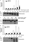
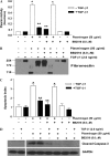
Plasmin induces fibroblast apoptosis in association with fibronectin proteolysis. (A) IMR-90 fibroblasts were cultured to 60% confluence and growth arrested for 24 hours before treatment with plasmin (doses indicated) for 16 hours. Equal amounts of protein from the cell culture supernatant were subjected to SDS-PAGE and Western immunoblotting for fibronectin. (B) Similarly cultured IMR-90 fibroblasts were treated with plasmin (0.5 U/ml) ± TGF-β1 (2.0 ng/ml) and cell culture supernatants were assessed for fibronectin. (C) IMR-90 fibroblasts were cultured to 60% confluence in a 96-well plate, growth arrested in serum-free media for 24 hours, and treated with plasmin for 16 hours. Apoptosis was assessed by enzyme-linked immunosorbent assay (ELISA) for ssDNA. *P < 0.001 compared with control (n = 4). (D) IMR-90 fibroblasts cultured as above were treated with plasmin (0.1 U/ml) for the times noted and whole cell lysates were assessed for cleaved caspase-3 by SDS-PAGE and Western immunoblotting. Membranes were stripped and probed for GAPDH. (E) IMR-90 fibroblasts were treated with plasmin ± TGF-β1 (2.0 ng/ml) for 16 hours, and lysates were assessed for cleaved caspase-3 by SDS-PAGE and Western immunoblotting. Membranes were stripped and probed for GAPDH. Data are representative of at least three independent experiments.
<b>Figure 2.</b>
Figure 2.
TGF-β1 inhibits plasminogen activation and fibronectin proteolysis in normal human lung fibroblasts. (A) IMR-90 fibroblasts were cultured and growth arrested for 24 hours in serum-free media without phenol red before treatment with exogenous plasminogen without (open bars) or with (solid bars) TGF-β1 (2.0 ng/ml) for 16 hours. The cell culture supernatants were assessed for plasmin activity as described in Materials and Methods. *P < 0.001 versus untreated control. **P < 0.001 compared with the equivalent plasminogen dose without TGF-β1. n = 4 per condition. (B) IMR-90 fibroblasts were treated with plasminogen at the indicated doses for 16 hours and cell culture supernatants were assessed for fibronectin by SDS-PAGE and Western immunoblotting. (C) IMR-90 fibroblasts were treated with plasminogen ± TGF-β1 (2.0 ng/ml) for 16 hours, and cell culture supernatants were assessed for fibronectin by SDS-PAGE and Western immunoblotting. (D) IMR-90 fibroblasts were treated with plasminogen (50 μg/ml) for the time periods indicated in the absence (open bars) or presence (solid bars) of TGF-β1 (2.0 ng/ml) and cell culture supernatants were assessed for plasmin activity. *P < 0.001 compared with untreated control. **P < 0.001 compared with equivalent time-point without TGF-β1. n = 4 per condition. (E) Similarly cultured IMR-90 cells were treated with plasminogen (50 μg/ml) for the indicated times and cell culture supernatants were assessed for fibronectin by SDS-PAGE and Western immunoblotting. Data are representative of at least three independent experiments.
<b>Figure 3.</b>
Figure 3.
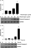
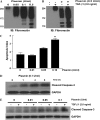
Plasminogen-induced fibroblast apoptosis is dose- and time-dependent. IMR-90 fibroblasts were cultured in a 96-well plate or in 35-mm dishes to 60% confluence and growth arrested for 24 hours before treatment with plasminogen. (A) Fibroblasts were treated with plasminogen for 18 hours and apoptosis was assessed via ELISA for ssDNA (n = 4; * P < 0.001 versus control). (B) Fibroblasts were treated with plasminogen (doses indicated) for 18 hours and cell lysates were assessed for cleaved caspase-3 by Western immunoblotting. The blot was then stripped and probed for GAPDH. (C) Fibroblasts in a 96-well plate were treated with plasminogen (50 μg/ml) and apoptosis was assessed by ELISA for ssDNA (n = 4; *P < 0.001 versus time 0). (D) Fibroblasts were treated with plasminogen (50 μg/ml) and whole cell lysates were assessed for cleaved caspase-3 by Western immunoblotting. The blots were stripped and re-probed for GAPDH. All data represent experiments performed at least three times.
<b>Figure 4.</b>
Figure 4.
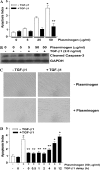
TGF-β1 inhibits plasminogen-induced fibroblast apoptosis. (A and B) IMR-90 fibroblasts were cultured and treated with plasminogen ± TGF-β1 (2.0 ng/ml) for 18 hours. (A) Apoptosis was assessed by ELISA for ssDNA. n = 4; *P < 0.001 versus control, **P < 0.001 versus plasminogen without TGF-β1. (B) Apoptosis was assessed by Western immunoblotting for cleaved caspase-3. The blot was then stripped and probed for GAPDH. (C) IMR-90 fibroblasts were treated with/without plasminogen (50 μg/ml) in the presence/absence of TGF-β1 (2 ng/ml) for 18 hours and visualized by microscopy. Representative photomicrographs are shown at ×20 power. (D) IMR-90 fibroblasts were treated with/without plasminogen (50 μg/ml) in the presence/absence of TGF-β1 given as a co-treatment or delayed for the times noted. Apoptosis was assessed by ELISA for ssDNA 18 hours after the plasminogen treatment. *P < 0.001 versus untreated control, **P < 0.001 versus compared with plasminogen treatment alone.
<b>Figure 5.</b>
Figure 5.
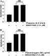
Blockade of the type 1 TGF-β receptor does not increase plasminogen- or plasmin-mediated fibroblast apoptosis. (A) IMR-90 fibroblasts were treated with (A) plasmin (0.5 U/ml) or (B) plasminogen (50 μg/ml) in the presence/absence of the ALK-5 inhibitor SB431542 (1.0 μM). Apoptosis was assessed by ELISA for ssDNA after 18 hours. *P < 0.001 versus untreated controls (n = 4 per condition, and the experiment was repeated three times).
<b>Figure 6.</b>
Figure 6.
Plasminogen-induced fibroblast apoptosis is not mediated by MMPs. IMR-90 fibroblasts were treated with/without plasminogen (50 μg/ml) ± TGF-β1 (2.0 ng/ml) and/or the broad-spectrum MMP inhibitor BB2516 (5.0 μM) for 18 hours. (A) Cell culture supernatants were assessed for plasmin activity. n = 4; *P < 0.001 versus controls, **P < 0.001 versus plasminogen alone. (B) Cell culture supernatants were assessed for fibronectin by Western immunoblotting. (C) Apoptosis was assessed using ELISA for ssDNA. n = 4; *P < 0.001 versus control, **P < 0.001 versus plasminogen. (D) Apoptosis was also assessed by Western immunoblotting of whole cell lysates for cleaved caspase-3. Blots were stripped and probed for GAPDH. All data represent experiments performed at least three times.
<b>Figure 7.</b>
Figure 7.
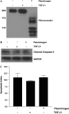
Fibronectin proteolytic peptides in conditioned media do not induce fibroblast apoptosis. IMR-90 fibroblasts were treated with/without plasminogen (50 μg/ml) or TGF-β1 (2 ng/ml) for 18 hours. (A) Cell culture supernatants were assessed for fibronectin degradation peptides by Western immunoblotting. (B and C) α2-antiplasmin was added to the culture supernatants to neutralize residual plasmin activity. This conditioned media was then used to treat growth-arrested IMR-90 fibroblasts pre-treated with suramin (300 μM). After 18 hours, apoptosis was assessed by (B) Western immunoblotting for cleaved caspase 3 or by (C) ELISA for ssDNA.
<b>Figure 8.</b>
Figure 8.
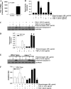
PAI-1 expression is required for TGF-β1 inhibition of plasminogen-induced fibroblast apoptosis. (A) IMR-90 fibroblasts were treated ± TGF-β1 (2.0 ng/ml) for 18 hours. PAI-1 RNA was assessed via Affymetrix microarray gene chip analysis. n = 3; P = 0.001. (B) IMR-90 fibroblasts were treated with plasminogen (50 μg/ml) ± TGF-β1 (2.0 ng/ml) or exogenous PAI-1 (22.5 ng/ml) for 18 hours, and cell culture supernatants were assessed for plasmin activity. n = 2; *P < 0.001 versus control. **P < 0.001 versus plasminogen. (C) Whole cell lysates from IMR-90 fibroblasts were assessed for cleaved caspase-3 by Western immunoblotting, and the blots were stripped and probed for GAPDH. (D) Primary lung fibroblasts isolated from wild-type (open bars) and PAI-1−/− mice (solid bars) were cultured to 60% confluence and treated with plasminogen (50 μg/ml) ± TGF-β1 (5.0 ng/ml) for 18 hours. Cell culture supernatants were assessed for plasmin activity. n = 2; *P < 0.001 versus untreated control, **P < 0.001 versus plasminogen alone; #P < 0.001 versus wild-type + plasminogen. (E) Primary lung fibroblasts from wild-type and PAI-1−/− mice were treated with plasminogen (50 μg/ml) ± TGF-β1 (5.0 ng/ml) for 18 hours and whole cell lysates were assessed for cleaved caspase-3. Membranes were stripped and probed for GAPDH. (F) Primary lung fibroblasts from wild-type (open bars) and PAI-1−/− (solid bars) mice were treated with plasminogen (50 μg/ml) ± TGF-β1 (5.0 ng/ml) or exogenous PAI-1 (22.5 ng/ml) for 18 hours and apoptosis was assessed by ELISA for ssDNA. n = 4; *P < 0.01 versus untreated control; **P < 0.001 versus plasminogen treated. Data represent three independent experiments.
<b>Figure 9.</b>
Figure 9.
Proposed mechanism of plasminogen-mediated apoptosis and the effect of TGF-β1. Fibroblasts secrete a fibronectin-rich pericellular matrix and receive adhesion-mediated signals that regulate cell phenotype. Pericellular matrix–generated signals may include biomechanical and tensile forces that promote or sustain pro-survival signals. Fibroblasts express plasminogen activators (uPA and/or tPA), which activate plasminogen in the cellular microenvironment to plasmin, which then mediates fibronectin proteolysis and fibroblast apoptosis. The specific mechanism linking pericellular fibronectin proteolysis to fibroblast apoptosis has not been determined. The pro-fibrotic cytokine, TGF-β1, induces the secretion of PAI-1, which blocks plasminogen activation, fibronectin proteolysis, and fibroblast apoptosis.

Similar articles

Cited by

References

    1. Tomasek JJ, Gabbiani G, Hinz B, Chaponnier C, Brown RA. Myofibroblasts and mechano-regulation of connective tissue remodelling. Nat Rev Mol Cell Biol 2002;3:349–363. - PubMed
    1. Desmouliere A, Redard M, Darby I, Gabbiani G. Apoptosis mediates the decrease in cellularity during the transition between granulation tissue and scar. Am J Pathol 1995;146:56–66. - PMC - PubMed
    1. Hinz B, Phan SH, Thannickal VJ, Galli A, Bochaton-Piallat ML, Gabbiani G. The myofibroblast: one function, multiple origins. Am J Pathol 2007;170:1807–1816. - PMC - PubMed
    1. Thannickal VJ, Horowitz JC. Evolving concepts of apoptosis in idiopathic pulmonary fibrosis. Proc Am Thorac Soc 2006;3:350–356. - PMC - PubMed
    1. Gabbiani G. The myofibroblast in wound healing and fibrocontractive diseases. J Pathol 2003;200:500–503. - PubMed

Publication types

MeSH terms