

Downregulation of Wnt signaling is a trigger for formation of facultative heterochromatin and onset of cell senescence in primary human cells
- PMID: 17643369
- PMCID: PMC2698096
- DOI: 10.1016/j.molcel.2007.05.034
Downregulation of Wnt signaling is a trigger for formation of facultative heterochromatin and onset of cell senescence in primary human cells
Abstract
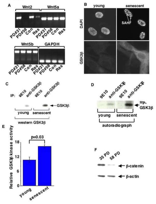
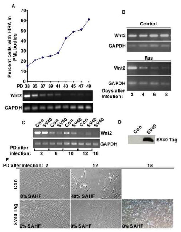
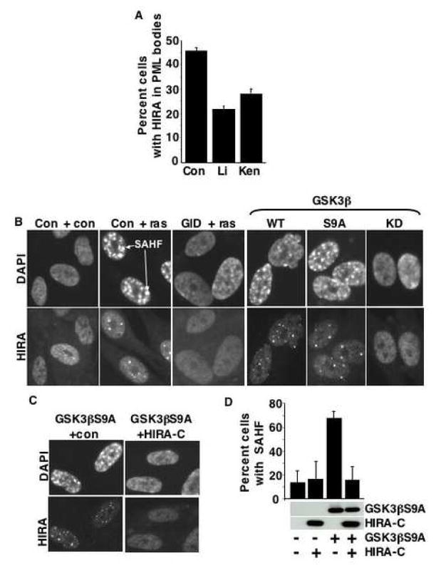
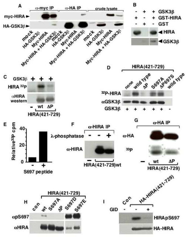
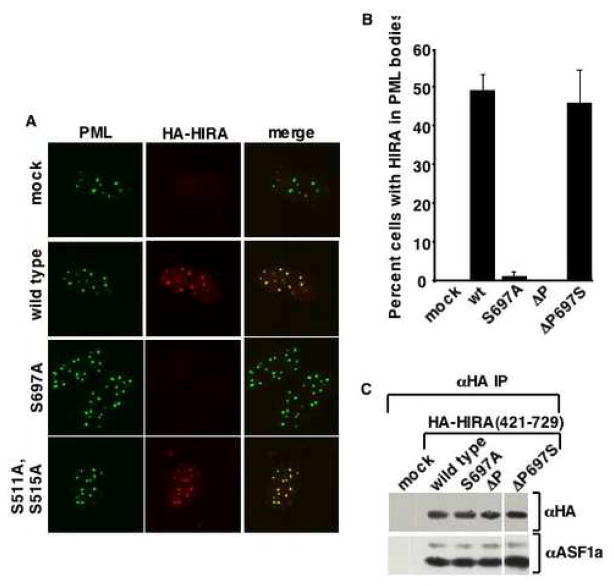
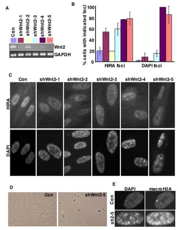
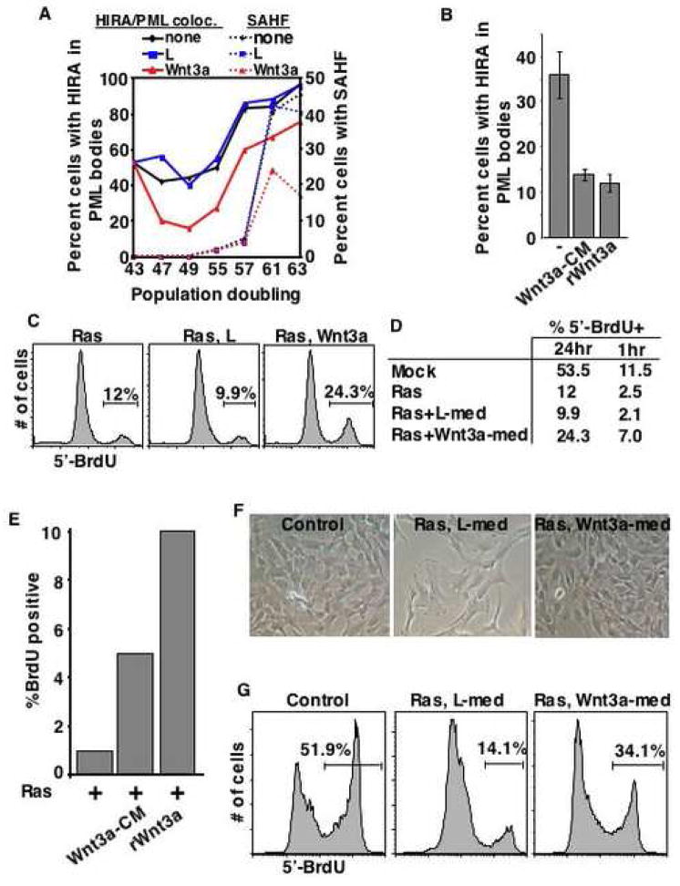
Cellular senescence is an irreversible proliferation arrest of primary cells and an important tumor suppression process. Senescence is often characterized by domains of facultative heterochromatin, called senescence-associated heterochromatin foci (SAHF), which repress expression of proliferation-promoting genes. Formation of SAHF is driven by a complex of histone chaperones, HIRA and ASF1a, and depends upon prior localization of HIRA to PML nuclear bodies. However, how the SAHF assembly pathway is activated in senescent cells is not known. Here we show that expression of the canonical Wnt2 ligand and downstream canonical Wnt signals are repressed in senescent human cells. Repression of Wnt2 occurs early in senescence and independently of the pRB and p53 tumor suppressor proteins and drives relocalization of HIRA to PML bodies, formation of SAHF and senescence, likely through GSK3beta-mediated phosphorylation of HIRA. These results have major implications for our understanding of both Wnt signaling and senescence in tissue homeostasis and cancer progression.
Figures
Similar articles
-
Definition of pRB- and p53-dependent and -independent steps in HIRA/ASF1a-mediated formation of senescence-associated heterochromatin foci.Mol Cell Biol. 2007 Apr;27(7):2452-65. doi: 10.1128/MCB.01592-06. Epub 2007 Jan 22. Mol Cell Biol. 2007. PMID: 17242198 Free PMC article.
-
Human UBN1 is an ortholog of yeast Hpc2p and has an essential role in the HIRA/ASF1a chromatin-remodeling pathway in senescent cells.Mol Cell Biol. 2009 Feb;29(3):758-70. doi: 10.1128/MCB.01047-08. Epub 2008 Nov 24. Mol Cell Biol. 2009. PMID: 19029251 Free PMC article.
-
Formation of MacroH2A-containing senescence-associated heterochromatin foci and senescence driven by ASF1a and HIRA.Dev Cell. 2005 Jan;8(1):19-30. doi: 10.1016/j.devcel.2004.10.019. Dev Cell. 2005. PMID: 15621527
-
Remodeling of chromatin structure in senescent cells and its potential impact on tumor suppression and aging.Gene. 2007 Aug 1;397(1-2):84-93. doi: 10.1016/j.gene.2007.04.020. Epub 2007 May 1. Gene. 2007. PMID: 17544228 Free PMC article. Review.
-
Chromatin maintenance and dynamics in senescence: a spotlight on SAHF formation and the epigenome of senescent cells.Chromosoma. 2014 Oct;123(5):423-36. doi: 10.1007/s00412-014-0469-6. Epub 2014 May 27. Chromosoma. 2014. PMID: 24861957 Review.
Cited by
-
Wnt Signaling Is Deranged in Asthmatic Bronchial Epithelium and Fibroblasts.Front Cell Dev Biol. 2021 Mar 15;9:641404. doi: 10.3389/fcell.2021.641404. eCollection 2021. Front Cell Dev Biol. 2021. PMID: 33791298 Free PMC article.
-
Wnt signaling in regulation of biological functions of the nurse cell harboring Trichinella spp.Parasit Vectors. 2016 Sep 2;9(1):483. doi: 10.1186/s13071-016-1770-4. Parasit Vectors. 2016. PMID: 27589866 Free PMC article.
-
Perturbation of METTL1-mediated tRNA N7- methylguanosine modification induces senescence and aging.Nat Commun. 2024 Jul 8;15(1):5713. doi: 10.1038/s41467-024-49796-8. Nat Commun. 2024. PMID: 38977661 Free PMC article.
-
Dual regulation of TERT activity through transcription and splicing by DeltaNP63alpha.Aging (Albany NY). 2008 Dec 9;1(1):58-67. doi: 10.18632/aging.100003. Aging (Albany NY). 2008. PMID: 20157588 Free PMC article.
-
Identification of ribonucleotide reductase M2 as a potential target for pro-senescence therapy in epithelial ovarian cancer.Cell Cycle. 2014;13(2):199-207. doi: 10.4161/cc.26953. Epub 2013 Oct 29. Cell Cycle. 2014. PMID: 24200970 Free PMC article.
References
-
- Ahuja D, Saenz-Robles MT, Pipas JM. SV40 large T antigen targets multiple cellular pathways to elicit cellular transformation. Oncogene. 2005;24:7729–7745. - PubMed
-
- Bartkova J, Rezaei N, Liontos M, Karakaidos P, Kletsas D, Issaeva N, Vassiliou LV, Kolettas E, Niforou K, Zoumpourlis VC, et al. Oncogene-induced senescence is part of the tumorigenesis barrier imposed by DNA damage checkpoints. Nature. 2006;444:633–637. - PubMed
-
- Braig M, Lee S, Loddenkemper C, Rudolph C, Peters AH, Schlegelberger B, Stein H, Dorken B, Jenuwein T, Schmitt CA. Oncogene-induced senescence as an initial barrier in lymphoma development. Nature. 2005;436:660–665. - PubMed
Publication types
MeSH terms
Substances
Grants and funding
LinkOut - more resources
Full Text Sources
Other Literature Sources
Research Materials
Miscellaneous
