

Overexpression of PDGF-BB decreases colorectal and pancreatic cancer growth by increasing tumor pericyte content
- PMID: 17641778
- PMCID: PMC1913488
- DOI: 10.1172/JCI31334
Overexpression of PDGF-BB decreases colorectal and pancreatic cancer growth by increasing tumor pericyte content
Abstract
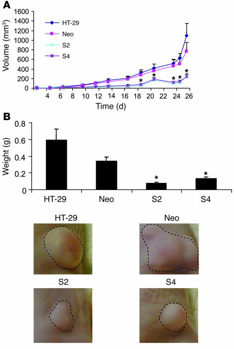
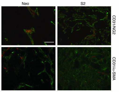
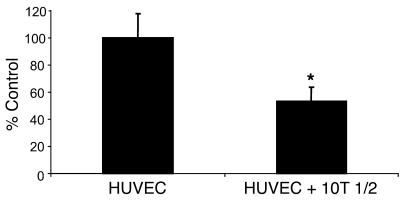
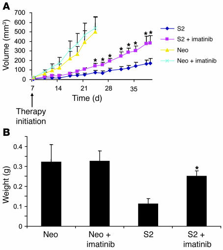
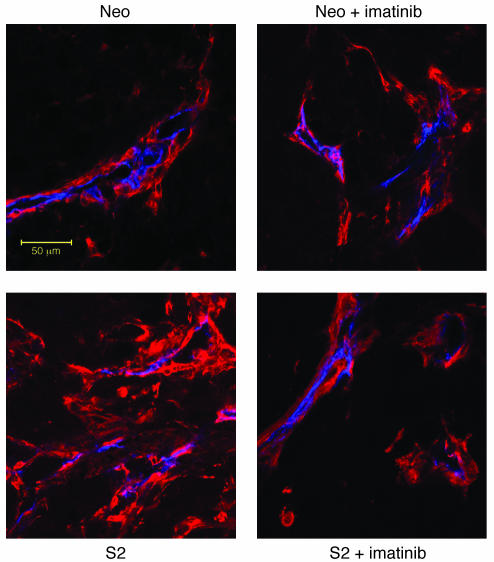
We hypothesized that overexpression of PDGF-BB in colorectal cancer (CRC) and pancreatic cancer cells would result in increased pericyte coverage of ECs in vivo, rendering the tumor vasculature more resistant to antiangiogenic therapy. We stably transfected the cDNA for the PDGF-B into HT-29 human CRC and FG human pancreatic cancer cells. Surprisingly, when HT-29 or FG parental and transfected cells were injected into mice (subcutaneously and orthotopically), we observed marked inhibition of tumor growth in the PDGF-BB-overexpressing clones. In the PDGF-BB-overexpressing tumors, we observed an increase in pericyte coverage of ECs. Treatment of PDGF-BB-overexpressing tumors with imatinib mesylate (PDGFR inhibitor) resulted in increased growth and decreased total pericyte content compared with those in untreated PDGF-BB-overexpressing tumors. In vitro studies demonstrated the ability of VSMCs to inhibit EC proliferation by approximately 50%. These data show that increasing the pericyte content of the tumor microenvironment inhibits the growth of angiogenesis-dependent tumors. Single-agent therapy targeting PDGF receptor must be used with caution in tumors when PDGFR is not the target on the tumor cell itself.
Figures

Similar articles
-
Overexpression of platelet-derived growth factor-BB increases tumor pericyte content via stromal-derived factor-1alpha/CXCR4 axis.Cancer Res. 2009 Aug 1;69(15):6057-64. doi: 10.1158/0008-5472.CAN-08-2007. Epub 2009 Jul 7. Cancer Res. 2009. PMID: 19584297
-
Inhibition of platelet-derived growth factor receptor phosphorylation by STI571 (Gleevec) reduces growth and metastasis of human pancreatic carcinoma in an orthotopic nude mouse model.Clin Cancer Res. 2003 Dec 15;9(17):6534-44. Clin Cancer Res. 2003. PMID: 14695158
-
Identification of a subset of pericytes that respond to combination therapy targeting PDGF and VEGF signaling.Int J Cancer. 2007 Dec 15;121(12):2606-14. doi: 10.1002/ijc.22999. Int J Cancer. 2007. PMID: 17691110
-
PDGF receptors as targets in tumor treatment.Adv Cancer Res. 2007;97:247-74. doi: 10.1016/S0065-230X(06)97011-0. Adv Cancer Res. 2007. PMID: 17419949 Review.
-
Imatinib and prostate cancer: lessons learned from targeting the platelet-derived growth factor receptor.Expert Opin Investig Drugs. 2013 Jun;22(6):787-94. doi: 10.1517/13543784.2013.787409. Epub 2013 Apr 1. Expert Opin Investig Drugs. 2013. PMID: 23540855 Review.
Cited by
-
Differential cytokine and chemokine expression after ablation vs. resection in colorectal cancer liver metastasis.Surg Open Sci. 2024 Jan 14;18:29-34. doi: 10.1016/j.sopen.2024.01.005. eCollection 2024 Mar. Surg Open Sci. 2024. PMID: 38318321 Free PMC article.
-
Inhibition of platelet-derived growth factor receptor synergistically increases the pharmacological effect of tamoxifen in estrogen receptor α positive breast cancer.Oncol Lett. 2021 Apr;21(4):294. doi: 10.3892/ol.2021.12555. Epub 2021 Feb 17. Oncol Lett. 2021. PMID: 33732370 Free PMC article.
-
Recruitment and retention: factors that affect pericyte migration.Cell Mol Life Sci. 2014 Jan;71(2):299-309. doi: 10.1007/s00018-013-1432-z. Epub 2013 Aug 4. Cell Mol Life Sci. 2014. PMID: 23912898 Free PMC article. Review.
-
VEGF-A/VEGFR-2 and FGF-2/FGFR-1 but not PDGF-BB/PDGFR-β play important roles in promoting immature and inflammatory intraplaque angiogenesis.PLoS One. 2018 Aug 20;13(8):e0201395. doi: 10.1371/journal.pone.0201395. eCollection 2018. PLoS One. 2018. PMID: 30125282 Free PMC article.
-
A combined score of clinical factors and serum proteins can predict time to recurrence in high grade serous ovarian cancer.Gynecol Oncol. 2019 Mar;152(3):574-580. doi: 10.1016/j.ygyno.2018.12.015. Epub 2018 Dec 18. Gynecol Oncol. 2019. PMID: 30578005 Free PMC article.
References
-
- Ellis L.M. A targeted approach for antiangiogenic therapy of metastatic human colon cancer. Am. Surg. 2003;69:3–10. - PubMed
-
- Jung Y.D., et al. The role of the microenvironment and intercellular cross-talk in tumor angiogenesis. Semin. Cancer Biol. 2002;12:105–112. - PubMed
-
- von Tell D., Armulik A., Betsholtz C. Pericytes and vascular stability. Exp. Cell Res. 2006;312:623–629. - PubMed
-
- Gerhardt H., Betsholtz C. Endothelial-pericyte interactions in angiogenesis. Cell Tissue Res. 2003;314:15–23. - PubMed
-
- Sims D.E. Diversity within pericytes. Clin. Exp. Pharmacol. Physiol. 2000;27:842–846. - PubMed
Publication types
MeSH terms
Substances
LinkOut - more resources
Full Text Sources
Other Literature Sources
Medical
Miscellaneous
