

Common molecular signature in SOD1 for both sporadic and familial amyotrophic lateral sclerosis
- PMID: 17636119
- PMCID: PMC1941502
- DOI: 10.1073/pnas.0705044104
Common molecular signature in SOD1 for both sporadic and familial amyotrophic lateral sclerosis
Abstract
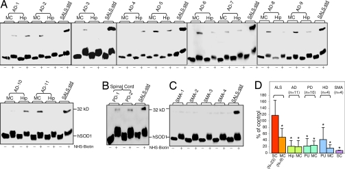
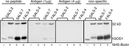
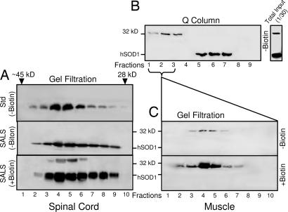
Amyotrophic lateral sclerosis (ALS) is a devastating motor neuron degenerative disease whose etiology and pathogenesis remain poorly understood. Most cases of ALS ( approximately 90%) are sporadic (SALS), occurring in the absence of genetic associations. Approximately 20% of familial ALS (FALS) cases are due to known mutations in the copper, zinc superoxide dismutase (SOD1) gene. Molecular evidence for a common pathogenesis of SALS and FALS has remained elusive. Here we use covalent chemical modification to reveal an attribute of spinal cord SOD1 common to both SOD1-linked FALS and SALS, but not present in normal or disease-affected tissues from other neurodegenerative diseases, including Alzheimer's, Parkinson's, and Huntington's diseases and spinal muscular atrophy, a non-ALS motor neuron disease. Biotinylation reveals a 32-kDa, covalently cross-linked SOD1-containing protein species produced not only in FALS caused by SOD1 mutation, but also in SALS. These studies use chemical modification as a novel tool for the detection of a disease-associated biomarker. Our results identify a shared molecular event involving a known target gene and suggest a common step in the pathogenesis between SALS and FALS.
Conflict of interest statement
Conflict of interest statement: V.R.L. is a founder and J.L. a consultant to Prosetta Corporation, an early stage biotechnology company.
Figures


Similar articles
-
FUS-immunoreactive inclusions are a common feature in sporadic and non-SOD1 familial amyotrophic lateral sclerosis.Ann Neurol. 2010 Jun;67(6):739-48. doi: 10.1002/ana.22051. Ann Neurol. 2010. PMID: 20517935 Free PMC article.
-
Pathological TDP-43 distinguishes sporadic amyotrophic lateral sclerosis from amyotrophic lateral sclerosis with SOD1 mutations.Ann Neurol. 2007 May;61(5):427-34. doi: 10.1002/ana.21147. Ann Neurol. 2007. PMID: 17469116
-
TDP-43 immunoreactivity in neuronal inclusions in familial amyotrophic lateral sclerosis with or without SOD1 gene mutation.Acta Neuropathol. 2007 May;113(5):535-42. doi: 10.1007/s00401-007-0206-9. Epub 2007 Feb 27. Acta Neuropathol. 2007. PMID: 17333220
-
Recent advances in research on neuropathological aspects of familial amyotrophic lateral sclerosis with superoxide dismutase 1 gene mutations: neuronal Lewy body-like hyaline inclusions and astrocytic hyaline inclusions.Histol Histopathol. 1999 Jul;14(3):973-89. doi: 10.14670/HH-14.973. Histol Histopathol. 1999. PMID: 10425565 Review.
-
SOD1 in neurotoxicity and its controversial roles in SOD1 mutation-negative ALS.Adv Biol Regul. 2016 Jan;60:95-104. doi: 10.1016/j.jbior.2015.10.006. Epub 2015 Oct 31. Adv Biol Regul. 2016. PMID: 26563614 Review.
Cited by
-
DNA Damage and Chromatin Rearrangement Work Together to Promote Neurodegeneration.Mol Neurobiol. 2025 Jan;62(1):1282-1290. doi: 10.1007/s12035-024-04331-0. Epub 2024 Jul 8. Mol Neurobiol. 2025. PMID: 38977621 Free PMC article. Review.
-
T cell-microglial dialogue in Parkinson's disease and amyotrophic lateral sclerosis: are we listening?Trends Immunol. 2010 Jan;31(1):7-17. doi: 10.1016/j.it.2009.09.003. Epub 2009 Oct 31. Trends Immunol. 2010. PMID: 19879804 Free PMC article. Review.
-
Nonamyloid aggregates arising from mature copper/zinc superoxide dismutases resemble those observed in amyotrophic lateral sclerosis.J Biol Chem. 2010 Dec 31;285(53):41701-11. doi: 10.1074/jbc.M110.113696. Epub 2010 Oct 25. J Biol Chem. 2010. PMID: 20974846 Free PMC article.
-
Cu, Zn-superoxide dismutase 1 (SOD1) is a novel target of Puromycin-sensitive aminopeptidase (PSA/NPEPPS): PSA/NPEPPS is a possible modifier of amyotrophic lateral sclerosis.Mol Neurodegener. 2011 May 7;6:29. doi: 10.1186/1750-1326-6-29. Mol Neurodegener. 2011. PMID: 21548977 Free PMC article.
-
Proteostasis and movement disorders: Parkinson's disease and amyotrophic lateral sclerosis.Cold Spring Harb Perspect Biol. 2011 Oct 1;3(10):a007500. doi: 10.1101/cshperspect.a007500. Cold Spring Harb Perspect Biol. 2011. PMID: 21844169 Free PMC article. Review.
References
-
- Rosen DR, Siddique T, Patterson D, Figlewicz DA, Sapp P, Hentati A, Donaldson D, Goto J, O'Regan JP, Deng HX, et al. Nature. 1993;362:59–62. - PubMed
-
- Cleveland DW, Rothstein JD. Nat Rev Neurosci. 2001;2:806–819. - PubMed
-
- Reaume AG, Elliott JL, Hoffman EK, Kowall NW, Ferrante RJ, Siwek DF, Wilcox HM, Flood DG, Beal MF, Brown RH, Jr, et al. Nat Genet. 1996;13:43–47. - PubMed
-
- Bruijn LI, Miller TM, Cleveland DW. Annu Rev Neurosci. 2004;27:723–749. - PubMed
-
- Valentine JS, Doucette PA, Potter SZ. Annu Rev Biochem. 2004;74:563–593. - PubMed
Publication types
MeSH terms
Substances
Grants and funding
LinkOut - more resources
Full Text Sources
Other Literature Sources
Medical
Miscellaneous
